. 2012;7(9):e44639.
doi: 10.1371/journal.pone.0044639. Epub 2012 Sep 11.

Multi-scale biomechanical remodeling in aging and genetic mutant murine mitral valve leaflets: insights into Marfan syndrome

Affiliations

Multi-scale biomechanical remodeling in aging and genetic mutant murine mitral valve leaflets: insights into Marfan syndrome

Russell A Gould et al. PLoS One. 2012.

Abstract

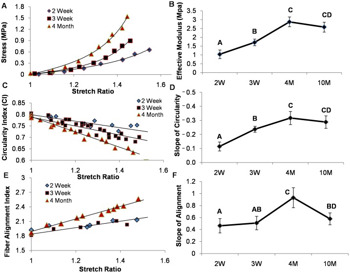
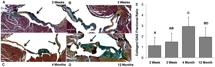
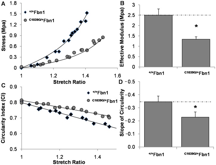
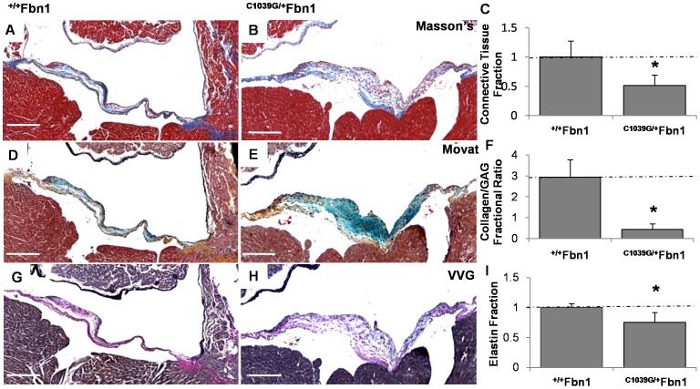
Mitral valve degeneration is a key component of the pathophysiology of Marfan syndrome. The biomechanical consequences of aging and genetic mutation in mitral valves are poorly understood because of limited tools to study this in mouse models. Our aim was to determine the global biomechanical and local cell-matrix deformation relationships in the aging and Marfan related Fbn1 mutated murine mitral valve. To conduct this investigation, a novel stretching apparatus and gripping method was implemented to directly quantify both global tissue biomechanics and local cellular deformation and matrix fiber realignment in murine mitral valves. Excised mitral valve leaflets from wild-type and Fbn1 mutant mice from 2 weeks to 10 months in age were tested in circumferential orientation under continuous laser optical imaging. Mouse mitral valves stiffen with age, correlating with increases in collagen fraction and matrix fiber alignment. Fbn1 mutation resulted in significantly more compliant valves (modulus 1.34 ± 0.12 vs. 2.51 ± 0.31 MPa, respectively, P<.01) at 4 months, corresponding with an increase in proportion of GAGs and decrease in elastin fraction. Local cellular deformation and fiber alignment change linearly with global tissue stretch, and these slopes become more extreme with aging. In comparison, Fbn1 mutated valves have decoupled cellular deformation and fiber alignment with tissue stretch. Taken together, quantitative understanding of multi-scale murine planar tissue biomechanics is essential for establishing consequences of aging and genetic mutations. Decoupling of local cell-matrix deformation kinematics with global tissue stretch may be an important mechanism of normal and pathological biomechanical remodeling in valves.


Conflict of interest statement

Competing Interests: The authors have declared that no competing interests exist.

Figures

Figure 1
Figure 1. Meso-scale uniaxial tensile device.
(A) Experimental stage controls the opposite co-linear translation of two cylindrical elastomeric beams. Tissue is mounted on the top surface of the posts using filter paper. The assembly is fully submerged in a buffered saline bath, and able to be imaged continuously under an upright confocal system. (B) Silicon post defection (v), post separation (s), and horizontal translation (d) were used for the biomechanical analysis. (C) Fabricated device implements a screw-driven wedge design for separation of the two linear rail guides at micro-meter resolution. (D) Detailed view of the silicon-post setup located inside the water bath. Distance between the posts is approximately 500 um.
Figure 2
Figure 2. Macro and micro-scale tissue analysis.
(A–D) Macro scale valve deformation at varying stretch ratios (λ) viewed at 10× under confocal microscopy (Green: 5-DTAF stained matrix fibers, Red: vital dye labeled cell bodies). Stars denote centroid location of the cantilever posts. (E–H) Micro-scale cellular deformation of the same group of cells viewed during the same test at 40X under confocal microscopy. Arrows denote tracked morphology of individual cells. (I–L) Micro-scale fiber alignment from the same region viewed during the same test at 40× under confocal microscopy. Fiber un-crimping and alignment is clearly visible as stretch progresses left to right.
Figure 3
Figure 3. Temporal biomechanical analysis of C57/B6J mitral valves.
(A) Representative stress-strain curves of mitral valves loaded in the circumferential direction (not to failure). (B) Stress-strain data were fit to an exponential Fung model, from which coefficients were used to determine effective modulus. (C) Representative circularity index curves as defined by the ratio of 4*Pi*(Area/Perimeter2). (D) Circularity-index curves were modeled as a linear fit and the negative slope was used for comparison. (E) Representative fiber-alignment curves were defined by the Fourier-Transform of collagen alignment and summed within ten degrees of the image horizontal, which was parallel to the tissue and loading direction. (F) Fiber-alignment data were fit to a linear model, the negative slope of which was used for comparison. Error bars show ±SD, n ≥6 for each condition. Groups that do not share letters are significantly different from each other according to a one-way ANOVA with Tukey’s post hoc (p≤0.05).
Figure 4
Figure 4. Temporal histological examination of C57/B6J mitral valves.
(A) 2 week old murine mitral valves contain similar fractional amounts of collagen and GAGs, with the majority of collagen near the attachment zones but mostly undefined architecture (Movat’s stain: yellow/orange = collagen, green/blue = GAGs). (B) More collagen relative to GAGs is present in 3 week old MV, with matrix stratification developing (Arrows). (C) At 4 months, murine mitral valves have nearly 3 times the amount of collagen to GAGs, are significantly more compact, and have well defined atrialis/fibrosa strata (Arrows). (D) At 12 months, murine mitral valves have significantly less collagen to GAGs, with dramatically increased thickening and reduced structural organization. (E) Digital quantification of matrix composition using color thresholding. Data compared as the ratio of collagen to GAGs within each valve leaflet. Magnification, ×4. Scale bars: 200 µm. Error bars show ±SD, n ≥3 valves per time point. Bars that do not share any letters are significantly different according to a one-way ANOVA with Tukey’s post hoc (p≤0.05).
Figure 5
Figure 5. Biomechanical analysis of +/+ Fbn1 and C1039G/+ Fbn1 at 4 months.
(A) Representative stress-strain responses of mitral valves loaded in the circumferential direction. (B) Stress-strain data were fit to an exponential Fung model, coefficients of which were used to determine effective modulus. (C) Representative stretch induced cell shape changes responses defined by the circularity index (CI) = 4*Pi*(Area/Perimeter2). (D) Circularity-index data were modeled as a linear fit, the negative slope of which was used for comparison. Error bars show ±SD, n ≥6 valves per condition. Asterisks signify statistical differences according to a Student’s t-test (p≤0.05).
Figure 6
Figure 6. Differences between +/+ Fbn1 and C1039G/+ Fbn1 mitral valve matrix composition at 4 months.
(A–B) Masson’s Trichrome reveals a reduction in the fractional amount of connective tissue in the marfan mitral valve compared to wildtype. (C) Digital quantification of the connective tissue composition using color thresholding, with blue regions denoting connective tissue. (D–F) Movat’s stain reveals over 3-fold reduction in the fractional amount of collagen compared to GAGs in the Marfan mitral valve relative to the wildtype (yellow-collagen, blue-GAGs). (G–I) Verhoeff’s–van Gieson (VVG) stain reveals a significant reduction in the fractional amount of elastin in the Marfan mitral valve compared to the wildtype (purple/black-elastin). Magnification, ×4. Scale bars: 200 µm. Error bars show ±SD, n ≥3 valves per condition. Asterisks denote statistical differences according to a Student’s t-test (p≤0.05).

Similar articles

Cited by

References

    1. Pyeritz RE (2000) The Marfan syndrome. Annu Rev Med 51: 481–510. - PubMed
    1. Dietz HC, Loeys B, Carta L, Ramirez F (2005) Recent progress towards a molecular understanding of Marfan syndrome. Am J Med Genet C Semin Med Genet 139C: 4–9. - PubMed
    1. Freed LA, Levy D, Levine RA, Larson MG, Evans JC, et al. (1999) Prevalence and clinical outcome of mitral-valve prolapse. N Engl J Med 341: 1–7. - PubMed
    1. Judge DP, Dietz HC (2008) Therapy of Marfan syndrome. Annu Rev Med 59: 43–59. - PubMed
    1. Mendelson K, Schoen FJ (2006) Heart valve tissue engineering: concepts, approaches, progress, and challenges. Ann Biomed Eng 34: 1799–1819. - PMC - PubMed

Publication types