. 2012;7(9):e44798.
doi: 10.1371/journal.pone.0044798. Epub 2012 Sep 12.

Apoptosis induced by knockdown of uPAR and MMP-9 is mediated by inactivation of EGFR/STAT3 signaling in medulloblastoma

Affiliations

Apoptosis induced by knockdown of uPAR and MMP-9 is mediated by inactivation of EGFR/STAT3 signaling in medulloblastoma

Ramaprasada Rao Kotipatruni et al. PLoS One. 2012.

Expression of concern in

Abstract

Background: Medulloblastoma is a highly invasive cancer of central nervous system diagnosed mainly in children. Matrix metalloproteinase-9 (MMP-9) and urokinase plasminogen activator receptor (uPAR) are over expressed in several cancers and well established for their roles in tumor progression. The present study is aimed to determine the consequences of targeting these molecules on medulloblastoma progression.

Methodology/principal findings: Radiation is one of the foremost methods applied for treating cancer and considerable evidence showed that radiation elevated uPAR and MMP-9 expression in medulloblastoma cell. Therefore efforts are made to target these molecules in non-irradiated and irradiated medulloblastoma cells. Our results showed that siRNA-mediated knockdown of uPAR and MMP-9, either alone or in combination with radiation modulated a series of events leading to apoptosis. Down regulation of uPAR and MMP-9 inhibited the expression of anti-apoptotic molecules like Bcl-2, Bcl-xL, survivin, XIAP and cIAPI; activated BID cleavage, enhanced the expression of Bak and translocated cyctochrome C to cytosol. Capsase-3 and -9 activities were also increased in uPAR- and MMP-9-downregulated cells. The apoptosis induced by targeting MMP-9 and uPAR was initiated by inhibiting epidermal growth factor receptor (EGFR) mediated activation of STAT3 and NF-κB related signaling molecules. Silencing uPAR and MMP-9 inhibited DNA binding activity of STAT3 and also reduced the recruitment of STAT3 protein at the promoter region of Bcl-2 and survivin genes. Our results suggest that inhibiting uPAR and MMP-9 reduced the expression of anti-apoptotic molecules by inactivating the transcriptional activity of STAT3. In addition, treating pre-established medulloblastoma with siRNAs against uPAR and MMP-9 both alone or in combination with radiation suppressed uPAR, MMP-9, EGFR, STAT3 expression and induced Bak activation leading to apoptosis.

Conclusion/significance: Taken together, our results illustrated that RNAi mediated targeting of uPAR and MMP-9 might have therapeutic potential against medulloblastoma.


Conflict of interest statement

Competing Interests: The authors have declared that no competing interests exist.

Figures

Figure 1
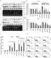
Figure 1. Transfection with pU, pM and pUM in combination with radiation specifically down regulates expression of uPAR and MMP-9 and induces apoptosis.
Daoy and D283 cell lines were transfected with either transfection reagent alone (control, Con), pSV (scrambled vector) or gene specific shRNA’s as described in materials and methods. A) RT-PCR analysis and western blotting was carried to determine the expression levels of uPAR and MMP-9 in cell transfected with reagent, pSV, pU-, pM- and pUM-transfected Daoy and D283 cells (with and without radiation, 8 Gy). The experiments were repeated three times and representative images were shown. The immunoblots were stripped and re-probed with GAPDH as a loading control. Semi-quantitative RT-PCR analysis was carried out to detect mRNA levels of uPAR and MMP-9 using specific primers. B) Protein band and PCR amplicon intensities were quantified by densitometry analysis using ImageJ software (National Institutes of Health). The levels of uPAR and MMP-9 protein were normalized to GAPDH levels in mock-transfected cells. Columns: mean of triplicate experiments; bars: s.d.; *p<0.01, significant difference from pSV-transfected cells. C) 72 hrs after Transfection (with or without radiation), the cells were trypsinized and analyzed by flow cytometry to measure the number of apoptotic TUNEL-positive cells using the APO-BrdU TUNEL Assay kit. The percent apoptotic cells from each treatment are represented and the mean ± s.d. from three separate experiments was represented; *p<0.05 and ** p<0.01 were considered significant compared to pSV-transfected cells and pSV+IR treated cells, respectively. D) Apoptotic cells were also quantified using an Annexin V assay followed by FACS analysis. Results are reported as the percent of cells (minimum 10,000 analyzed) that were Annexin V-positive. Gating was based on positive and negative control cells. Representative FACS data of Daoy and D283 are shown (n = 3).
Figure 2
Figure 2. uPAR and MMP-9 gene silencing induced mitochondrial apoptosis critically depends on Bak activation and cytochrome C release into cytosol in Daoy and D283 cells.
A) 72 hrs after transfecting Daoy and D283 cells with pU, pM and pUM (with or without radiation, 8 Gy), the cells were collected and analyzed by western blotting with the indicated antibodies. The membrane was reprobed with GAPDH to confirm equal protein loading. B) Sub-cellular fractions were prepared from the transfected cells as described in materials and methods. Cytochrome C release from mitochondria into the cytosol and mitochondrial Bak levels were determined by Western blotting of mitochondrial and cytosolic protein extracts. Membranes were re-probed with α-tubulin (cytosolic marker) or COX IV (mitochondrial marker). C) Mitochondrial membrane potential damage was evaluated using MitoLight green (Millipore). To determine the initiation of mitochondrial apoptosis in shRNA transfected Daoy and D283 cells, we carried out FACS analysis of mitochondrial membrane potential by sorting cells as described in materials and methods. Graphs represents percent number of cells showing reduced mitochondrial membrane potential in Daoy and D283 cell transfected with shuPAR, shMMP9 and shuPAR-MMP9 (treated with and without radiation). Each experiment is repeated 3 times * p<0.05 were statistically significant compared to pSV-transfected cells. A total of 10,000 cells were sorted per treatment.
Figure 3
Figure 3. Transcriptional silencing of uPAR and MMP-9 activates caspase-3, caspase-9 and PARP.
Activity of caspase-3 and caspase-9 in Daoy and D283 cells was determined using A) caspase-3 and B) caspase-9 colorimetric activity kits. Total cell lysates collected from transfected cells (with and with radiation) were collected, incubated with the respective peptide substrate, and treated with caspase conjugate p-nitroaniline (Ac-DEVD-pNA). Further, activity was measured at 405 nm using a microplate reader. Y-axis shows the percent activity of the respective caspase by normalizing the control to 100%. Bars represent the means ± s.d. from three independent experiments. * p<0.01 and ** p<0.05 were statistically significant compared to control and pSV-transfected cells. C) Immunoblot analysis of proteins isolated from cells treated with pU, pM and pUM (with and without radiation) was carried out to determine the activities of caspase-3, caspase-9 and PARP cleavage. D) The above immunoblots were stripped and re-probed with survivin, XIAP and cIAP1 specific antibodies, to determine the expression levels of inhibitory proteins.
Figure 4
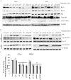
Figure 4. Silencing uPAR and MMP-9 inhibits nuclear levels and activation of STAT3 and NF-κB p65 (Rel-A). A)
Total cell lysates were evaluated by immunoblotting to determine the expression of NFκB p65 (Rel-A), IκBα, total and phosphorylated forms of EGFR and STAT3. Uniform loading of the respective protein was confirmed by re-probing the membrane with β-actin antibody. B) Nuclear levels of phosphorylated STAT3 and Rel-A were determined by analyzing the nuclear extracts isolated from transfected cells by western blotting. C–D) Nuclear extracts were prepared from cells transfected with pU, pM and pUM (with and with radiation), and the DNA binding activity of the nuclear extracts to C) STAT3 and D) NFκB p65 probe was determined using Electrophorotic Mobility gel Shift Assay. All experiments were repeated three times.
Figure 5
Figure 5. uPAR and MMP-9 activates EGFR and STAT3; Inhibition of STAT3 and NF-κB 65 induces apoptosis in Daoy and D283 cells.
To determine possible cross-talk between STAT3 and Rel-A during apoptosis, each gene was downregulated using specific siRNA followed by determination of the nuclear levels of STAT3 and Rel-A in both Daoy and D283 cells. A) Medulloblastoma cells transfected with full-length uPAR expressing plasmid or/and incubated with recombinant MMP-9 (25 ηg/ml), were either blocked with EGFR neutralizing antibody or non-specific isotype IgG to determine the role of the extracellular proteases in activation of EGFR and STAT3. Western blot analysis was carried out on cell lysates isolated from Daoy and D283 to determine the levels of phosphorylated EGFR and STAT3. B) Western blot analysis was carried out on the nuclear extracts isolated from STAT3-downregulated cells. Membranes were probed with phosphorylated forms of STAT3 and Rel-A. C) Similarly, nuclear extracts isolated from Rel-A downregulated cells were evaluated by Western blotting to detect phosphorylated forms of Rel-A and STAT3 in the nucleus. D) Chromatin immunoprecipitation assay was carried out with the nuclear extracts isolated from Day and D283 cells transfected with either STAT3 siRNA or pUM plasmid. Chromatin was immunopreciptated with STAT3 and analyzed by PCR using primers specific for Bcl-2 promoter region to determine the STAT3 recruitment at Bcl-2 promoter sequences. Chromatin immunoprecipitated with isotype IgG was used as negative control. Input DNA was confirmed by amplifying the Bcl-2 promoter form the chromatin aliquot collected prior to immunoprecipitation step.
Figure 6
Figure 6. Inhibition of uPAR and MMP-9 induces apoptosis in vivo.
Daoy cells were implanted intracranially in nude mice, which were treated with control pSV or pUM (150 µg) either alone or in combination with radiation as described in Materials and Methods. A–B) Paraffin-embedded brain tumor sections from mice that received control shRNA or pUM either alone and combination of radiation were analyzed by immunohistochmeical analysis using antibodies for A) uPAR and B) MMP-9 followed by treatment with secondary antibody conjugated with Alexa Fluor 594 and 488 fluorescent tagged. C) Induction of apoptosis in pUM-treated tumors was confirmed by carrying out TdT-mediated nick end labeling (TUNEL) assay. The tissues were analyzed for apoptosis using fluorescence microscopy. D) The relative number of TUNEL-positive cells was counted and represented graphically. Bars represent the means ± s.d. from three independent experiments, * p<0.05. E) Immunoflourscence analysis of paraffin-embedded brain sections to detect the levels of NF-κB p65 and Stat3 in pUM and pSV treated mice (with and without radiation). After incubating with the anti Rel-A and Stat3 antibodies for overnight, the tissue sections were washed and incubated in secondary antibody conjugated with Alexa Fluor 594 and 488 fluorescent tagged, respectively. Representative images from three independent experiments were shown in each panel.

Similar articles

Cited by

References

    1. Rood BR, MacDonald TJ, Packer RJ (2004) Current treatment of medulloblastoma: recent advances and future challenges. Semin Oncol 31: 666–675. - PubMed
    1. Rossi A, Caracciolo V, Russo G, Reiss K, Giordano A (2008) Medulloblastoma: from molecular pathology to therapy. Clin Cancer Res 14: 971–976. - PMC - PubMed
    1. Crawford JR, MacDonald TJ, Packer RJ (2007) Medulloblastoma in childhood: new biological advances. Lancet Neurol 6: 1073–1085. - PubMed
    1. Rao JS (2003) Molecular mechanisms of glioma invasiveness: the role of proteases. Nat Rev Cancer 3: 489–501. - PubMed
    1. Murphy G, Docherty AJ (1992) The matrix metalloproteinases and their inhibitors. Am J Respir Cell Mol Biol 7: 120–125. - PubMed

Publication types

MeSH terms