. 2012;7(9):e44824.
doi: 10.1371/journal.pone.0044824. Epub 2012 Sep 11.

Immune complex signatures of patients with active and inactive SLE revealed by multiplex protein binding analysis on antigen microarrays

Affiliations

Immune complex signatures of patients with active and inactive SLE revealed by multiplex protein binding analysis on antigen microarrays

Krisztián Papp et al. PLoS One. 2012.

Abstract

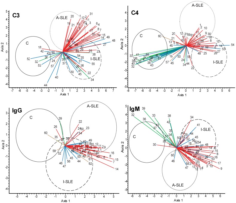
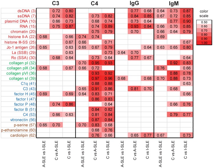
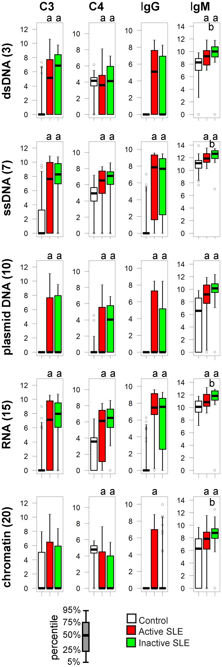
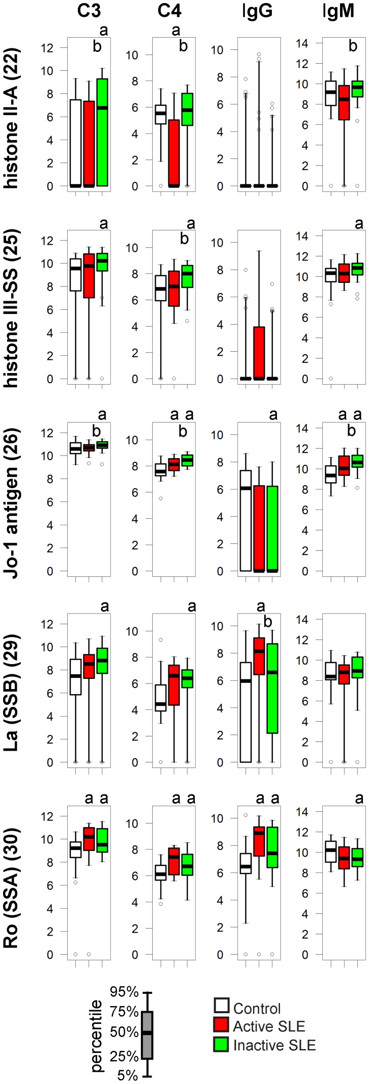
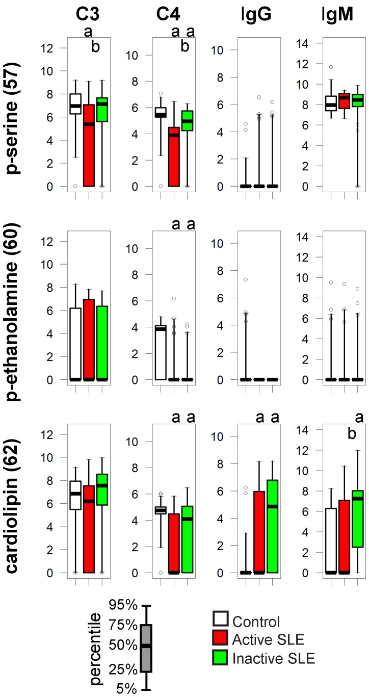
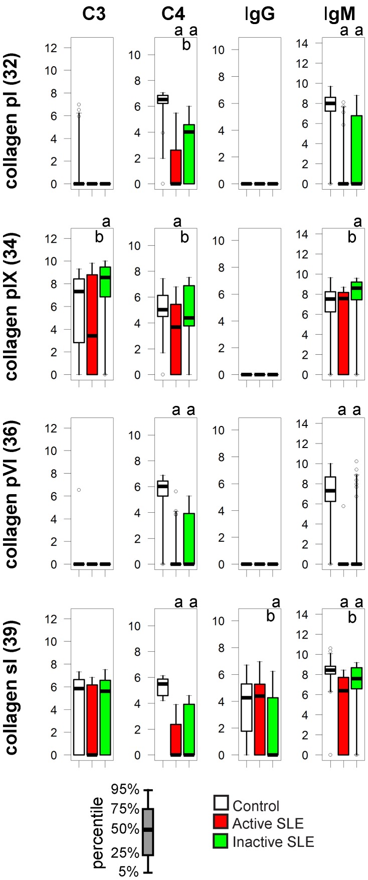
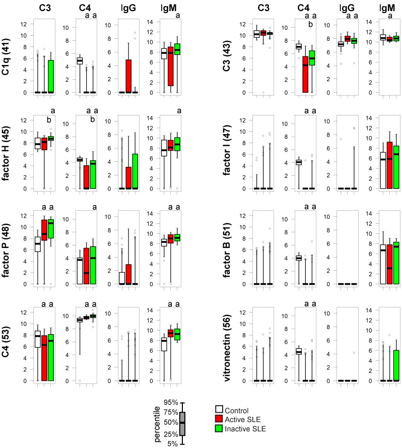
Systemic lupus erythematosus is characterized by dysfunctional clearance of apoptotic debris and the development of pathogenic autoantibodies. While the complement system is also involved in the disease no attempt has been made to generate a comprehensive view of immune complex formation from various autoantigens. We increased the complexity of autoantibody profiles by measuring the binding of two complement proteins, C3 and C4, in addition to two antibody classes, IgG and IgM, to a collection of autoantigens. These complement components covalently bind to those microarray features where antibodies and other serum components induce complement activation. Using this technology, we compared functional serum antibody profiles of control subjects (n = 31) and patients with lupus erythematosus (n = 61) in the active (n = 22) and inactive (n = 39) phase of the disease. Multivariate analysis was applied to identify contributions of binding data on 25 antigens to the discrimination of the study groups. Receiver operating characteristic analysis was used to portray the discriminative property of each measured parameter for each antigen in pairwise group comparisons. Complement C3 and C4 deposition increased on autoantibody targets in spite of the decreased serum complement concentrations, and decreased on other autoantigens, demonstrating the imbalance of complement function in patients with lupus erythematosus. Our observations confirmed previously known markers of disease and showed that C3 and C4 deposition data were at least as powerful as Ig binding data in separating the study groups.


Conflict of interest statement

Competing Interests: The authors have read the journal's policy and have the following conflicts: Eötvös Loránd University and Hungarian Academy of Sciences owns a pending patent on antigen microarray-based complement activation measurements[MEASUREMENT OF COMPLEMENT ACTIVATION PRODUCTS ON ANTIGEN ARRAYS; US2010075864 (A1); The invention employs the functional complement system in the biological sample tested, thereby the information gained relates to antigen recognition properties and functional consequences in the organism from which the sample was taken and relies on the ability of antigen recognition molecules, primarily antibodies to activate the complement system in the sample tested, upon binding to elements of an antigen array.]. PV, ZS, KP and J. Prechl are employed by Diagnosticum Ltd, the company licensing rights of the mentioned patent. Diagnosticum Ltd develops serological assays utilizing the technology described by the patent. This does not alter the authors' adherence to all the PLoS ONE policies on sharing data and materials.

Figures

Figure 1
Figure 1. Canonical variates analysis (CVA) of binding data.
Binding data of C3, C4, IgG and IgM were used to generate a canonical space, defined by axis 1 and axis 2, from the discriminant functions derived through eigenanalysis. Coordinates of observations for the three study groups (control, C; active SLE, A-SLE; inactive SLE, I-SLE) in this canonical space are enclosed within the ellipsoids with a 95% confidence. Vectors represent correlations of variables (antigens) with the canonical axes and are superimposed on the ordination of observations: the length and directionality of these vectors offer a possibility to evaluate the relative influence of antigens upon the separation of groups. For example, the vector of C3 binding data of antigen 8 separates group A-SLE and I-SLE from group C but not A-SLE and I-SLE from each other. Antigen groups are color-coded: all nuclear materials (nucleic acids and nuclear proteins), red; collagens, green; complement, blue; lipids, brown. The complete list of antigens shown here is found in Table 2.
Figure 2
Figure 2. Discriminative properties of antibody binding and complement deposition.
The three study groups (control, C; active SLE, A-SLE; inactive SLE, I-SLE) were compared in every pair using ROC analysis for each of the four measured proteins. AreaUnder the Curve (AUC) values for antigen binding events that were found to have significant (p<0.05) discriminative properties are shown in the form of a heat map. Antigen dilution points with the highest summarized AUC values are shown.
Figure 3
Figure 3. Alterations of immune complex composition on nucleic acid antigens.
A. Bar charts depict the reactivity profile of the three study groups, four components of immune complexes shown separately. Scale of y axis is relative fluorescence units. ap<0.05 compared to control group, bp<0.05 comparing active to inactive SLE Empty circles represent outliers: samples exceeding the upper or lower quartile values with 1.5-times the interquartile distance.
Figure 4
Figure 4. Alterations of immune complex composition on nuclear protein antigens.
See Figure 3. for details.
Figure 5
Figure 5. Alterations of immune complex composition on cardiolipin.
See Figure 3. for details.
Figure 6
Figure 6. Alterations of immune complex composition on collagens.
See Figure 3. for details.
Figure 7
Figure 7. Alterations of IC composition on complement proteins.
See Figure 3. For details.

References

    1. Hochberg MC (1997) Updating the American College of Rheumatology revised criteria for the classification of systemic lupus erythematosus. Arthritis Rheum 40: 1725. - PubMed
    1. Smeenk R, Brinkman K, van den Brink H, Termaat RM, Berden J, et al. (1990) Antibodies to DNA in patients with systemic lupus erythematosus. Their role in the diagnosis, the follow-up and the pathogenesis of the disease. Clin Rheumatol 9: 100–110. - PubMed
    1. Liu CC, Ahearn JM (2009) The search for lupus biomarkers. Best Pract Res Clin Rheumatol 23: 507–523. - PMC - PubMed
    1. Sturfelt G, Truedsson L (2005) Complement and its breakdown products in SLE. Rheumatology (Oxford) 44: 1227–1232. - PubMed
    1. Truedsson L, Bengtsson AA, Sturfelt G (2007) Complement deficiencies and systemic lupus erythematosus. Autoimmunity 40: 560–566. - PubMed

Publication types