. 2012;7(9):e44945.
doi: 10.1371/journal.pone.0044945. Epub 2012 Sep 12.

Phosphoinositide 3-kinase C2β regulates RhoA and the actin cytoskeleton through an interaction with Dbl

Affiliations

Phosphoinositide 3-kinase C2β regulates RhoA and the actin cytoskeleton through an interaction with Dbl

Karolina Błajecka et al. PLoS One. 2012.

Abstract

The regulation of cell morphology is a dynamic process under the control of multiple protein complexes acting in a coordinated manner. Phosphoinositide 3-kinases (PI3K) and their lipid products are widely involved in cytoskeletal regulation by interacting with proteins regulating RhoGTPases. Class II PI3K isoforms have been implicated in the regulation of the actin cytoskeleton, although their exact role and mechanism of action remain to be established. In this report, we have identified Dbl, a Rho family guanine nucleotide exchange factor (RhoGEF) as an interaction partner of PI3KC2β. Dbl was co-immunoprecipitated with PI3KC2β in NIH3T3 cells and cancer cell lines. Over-expression of Class II phosphoinositide 3-kinase PI3KC2β in NIH3T3 fibroblasts led to increased stress fibres formation and cell spreading. Accordingly, we found high basal RhoA activity and increased serum response factor (SRF) activation downstream of RhoA upon serum stimulation. In contrast, the dominant-negative form of PI3KC2β strongly reduced cell spreading and stress fibres formation, as well as SRF response. Platelet-derived growth factor (PDGF) stimulation of wild-type PI3KC2β over-expressing NIH3T3 cells strongly increased Rac and c-Jun N-terminal kinase (JNK) activation, but failed to show similar effect in the cells with the dominant-negative enzyme. Interestingly, epidermal growth factor (EGF) and PDGF stimulation led to increased extracellular signal-regulated kinase (Erk) and Akt pathway activation in cells with elevated wild-type PI3KC2β expression. Furthermore, increased expression of PI3KC2β protected NIH3T3 from detachment-dependent death (anoikis) in a RhoA-dependent manner. Taken together, these findings suggest that PI3KC2β modulates the cell morphology and survival through a specific interaction with Dbl and the activation of RhoA.


Conflict of interest statement

Competing Interests: The authors have declared that no competing interests exist.

Figures

Figure 1
Figure 1. PI3KC2β interacts with Dbl GEF for Rho GTPases.
(A) Lysates of NIH3T3 cells stably overexpressing wild-type Glu-PI3KC2β (NIH3T3-C2β-WT) were immunoprecipitated (IP) with an anti-C2β, anti-Glu (EE) tag, anti-Dbl or control IgG antibody. PI3KC2β interaction with the endogenous Dbl, as well as Grb2 and Src was detected by immunoblotting (IB) with the indicated antibodies. (B) Complex of endogenous PI3KC2β and Dbl was immunoprecipitated from the whole cell extract (WCE) of the parental NIH3T3 cells and IMR5 neuroblastoma human cancer cells with anti-Dbl, anti-C2β and control IgG antibody. Samples were subjected to western blot analysis with the indicated antibodies. In the case of IMR5 cells Triton 0.5% and Brij96 1% lysis buffers were used, while NIH3T3 cells were lysed with Triton 1%. Asterisk indicates PI3KC2β. (C) Interaction between the PI3KC2β and Dbl was examined by the pull-down (PD) of PI3KC2β GST-fused N-terminal and C2 C-terminal domains in HEK293 cells transfected with HA-proto-Dbl (Dbl), pcDNA3 empty vector (V) or untransfected control (-). Complex formation was shown by immunoblotting with anti-Dbl antibody. Asterisk indicates ectopically expressed (HA-tag) Dbl. (D) Interaction between the Dbl and Grb2 was examined by the GST-Grb2 pull-down in HA-proto-Dbl (Dbl) transfected and untransfected (-) HEK293 cells. Pull-down samples were subjected to western blot analysis with indicated antibodies. All experiments were repeated at least two times with similar results.
Figure 2
Figure 2. PI3KC2β binds with high affinity to the spectrin- and PH- Dbl domain.
(A) Schematic representation of Dbl mutant constructs, which were kind gift of Prof. Danny Manor from the School of Medicine at Case Western Reserve University, Cleveland, USA. (B) COS-1 cells co-transfected with Myc-PI3KC2β and different GST-fused Dbl domains (N-, spectrin-, onco-, PH-) in pEBG vector were subjected to gluthatione-sepharose beads affinity-purification and immunobotted with indicated antibodies. Experiment was repeated at least two times with similar results. PC – positive control; COS-1 cells co-transfected with PI3KC2β and HA-proto-Dbl, and immunoprecipitated with anti-Dbl antibody.
Figure 3
Figure 3. Kinase-dependent PI3KC2β effect on cytoskeletal rearrangements.
(A) Cell lysates of NIH3T3 cells stably expressing empty vector (NIH3T3-V), kinase-dead (D1213A) dominant-negative Myc-PI3KC2β (NIH3T3-C2β-DN) and wild-type Glu-PI3KC2β (NIH3T3-C2β-WT) were analysed for the recombinant PI3KC2β expression by immunoblotting with the indicated antibodies (EE: Glu-tag, 9E10: Myc-tag). (B) Confocal images of NIH3T3-V, -C2β-WT and –C2β-DN cells grown on cover slips for 24 h in 10% FCS and stained with Alexa Fluor 555 dye to localise F-actin, and an anti-PI3KC2β antibody follwed by FITC-labelled anti-rabbit antibody, to localize the kinase. WT1, WT2 and DN1, DN2 indicate individual clones. Scale bar represents 40 µm.
Figure 4
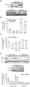
Figure 4. PI3KC2β regulates RhoA and Rac1 activity.
(A) PI3KC2β up-regulates RhoA activity. NIH3T3-V, -C2β-DN and –C2β–WT cells were grown in 10% FCS or serum-starved for 24 h and stimulated with 1 µM LPA for 2 min. Cell lysates were equalized for protein content and subjected to GST-rhotekin pull-down as measure of RhoA activity. GTP-bound and total RhoA were detected with an anti-RhoA antibody. (B) PI3KC2β mediates RhoA-dependent serum response factor (SRF) activation. NIH3T3-V, -C2β-DN and -C2β-WT cells were transiently transfected with the SRF luciferase reporter plasmid p3DA-Luc and pRL-TK control construct. Cells were maintained in 0.5% FCS for 24 h prior to stimulation with 15% FCS for 7 h. Luciferase activity was normalized to pRL-TK control. Figure shows mean ± SD of three independent experiments. (C) PI3KC2β up-regulates Rac1 activity. NIH3T3-V, -C2β-DN and -C2β-WT cells were grown in 10% FCS. Cell lysates were equalized for protein content and subjected to GST-PAK CRIB pull-down as measure of Rac-1 activity. GTP-bound and total Rac1 were detected with anti-Rac1 antibody. Densitometry was performed and signal intensities were normalized against total Rac1. Data are mean with SD from two independent experiments. *, p<0.05. (D) PI3KC2β mediates Rac1-dependent platelet derived growth factor (PDGF) activation. NIH3T3-V, -C2β-DN and -C2β-WT cells were serum-starved for 24 h and stimulated with 1 nM PDGF for 2 min. Cell lysates were equalized for protein content and subjected to GST-PAK CRIB pull-down as measure of Rac-1 activity. GTP-bound and total Rac1 were detected with anti-Rac1 antibody. Densitometry was performed and signal intensities were normalized against total Rac1. Data are mean with SD from two independent experiments. *, p<0.05. A representative western blot is shown. Activation of JNK was assessed in the cell lines treated with PDGF by immunoblotting with phospho-specific SAPK/JNK antibody. WT1, WT2 and DN1-3 indicate individual clones.
Figure 5
Figure 5. The assembly of the Dbl - PI3KC2β complex is not modulated by PI3K activity or cell stimulation with EGF or PDGF.
(A) HEK293 cells were transfected with vectors encoding Dbl in combination with Myc-PI3KC2β WT or DN, or empty vector. Immunoprecipitates prepared with anti-Dbl or anti-Myc tag antibodies were analysed by western blot with the antibodies indicated. (B) Lysates from NIH3T3-V, -C2β-WT or -C2β-DN cells were immunoprecipitated with anti-Dbl antibodies and analysed by western blot. (C) Serum-starved NIH3T3-C2β-WT cells were stimulated with EGF (20 ng/ml) or PDGF (20 ng/ml) for the indicated lengths of time. Immunoprecipitates prepared with anti-Glu (EE) tag antibodies were analysed by western blot with the antibodies indicated. (D) HEK293 cells were transfected with vectors encoding Dbl in combination with Myc-PI3KC2β WT or DN, or empty vector. Immunoprecipitates prepared with anti-Dbl antibodies were analysed for GEF activity towards recombinant RhoA.
Figure 6
Figure 6. PI3KC2β protects NIH3T3 cells against anoikis.
(A) NIH3T3-V, -C2β-DN and -C2β-WT cells were plated in presence of serum on ultra-low attachment matrix. After 16 h Caspase 3/7 activity was measured as readout for detachment-induced apoptosis (anoikis). Data are mean ± SD of two independent experiments. WT1, −2 and DN1, −2 indicate individual clones. (B) NIH3T3 cells transiently transfected with the wild-type Myc-PI3KC2β, constitutively active form of GFP-fused RhoA protein (CA-RhoA-GFP) or myristylated (Akt-myr) were plated 48 h post-transfection on ultra-low attachment matrix. 16 h later Caspase 3/7 activation was analysed as readout for anoikis. Data are mean ± SD of three independent experiments. Expression level of the transfected proteins and associated signalling was analysed 48 h post-transfection by immunoblotting with indicated antibodies. (C) NIH3T3-C2β-WT cells were transiently transfected with dominant-negative RhoA-GFP (DN-Rho-GFP) or control vector (V). 48 h post-transfection cells were plated on ultra-low attachment matrix and caspase 3/7 activation was measured. Data are mean ± SD of three independent experiments. 48 h post-transfection expression level of the transfected proteins was analysed by western blot with indicated antibodies.
Figure 7
Figure 7. PI3KC2β increases MAPK and Akt signalling downstream of EGFR and PDGFR.
(A) and (B) NIH3T3-V, -C2β-DN and -C2β-WT cells were serum-deprived overnight and stimulated for 10 min. with EGF (A) or PDGF (B) as indicated. Cell lysates were analysed by immunobloting and MAPK and Akt pathway activation was assessed with indicated phospho-specific antibodies. (C) and (D) Lysates of NIH3T3-V, -C2β-DN and -C2β-WT cells grown in 10% FCS were analysed for activated signalling molecules implicated in the cell cycle control and caveolae formation such as p53, Bcl2, PTEN and caveolin 1, −2. WT1, −2 and DN1, −2 indicate individual clones.

Similar articles

Cited by

References

    1. Katso R, Okkenhaug K, Ahmadi K, White S, Timms J, et al. (2001) Cellular function of phosphoinositide 3-kinases: implications for development, homeostasis, and cancer. Annu Rev Cell Dev Biol 17: 615–675. - PubMed
    1. Wymann MP, Bjorklof K, Calvez R, Finan P, Thomast M, et al. (2003) Phosphoinositide 3-kinase gamma: a key modulator in inflammation and allergy. Biochem Soc Trans 31: 275–280. - PubMed
    1. Sasaki T, Suzuki A, Sasaki J, Penninger JM (2002) Phosphoinositide 3-kinases in immunity: lessons from knockout mice. J Biochem 131: 495–501. - PubMed
    1. DiNitto JP, Cronin TC, Lambright DG (2003) Membrane recognition and targeting by lipid-binding domains. Sci STKE 2003: re16. - PubMed
    1. Engelman JA, Luo J, Cantley LC (2006) The evolution of phosphatidylinositol 3-kinases as regulators of growth and metabolism. Nat Rev Genet 7: 606–619. - PubMed

Publication types

MeSH terms