. 2012;7(9):e45036.
doi: 10.1371/journal.pone.0045036. Epub 2012 Sep 11.

Progression of cartilage degradation, bone resorption and pain in rat temporomandibular joint osteoarthritis induced by injection of iodoacetate

Affiliations

Progression of cartilage degradation, bone resorption and pain in rat temporomandibular joint osteoarthritis induced by injection of iodoacetate

Xue-Dong Wang et al. PLoS One. 2012.

Abstract

Background: Osteoarthritis (OA) is an important subtype of temporomandibular disorders. A simple and reproducible animal model that mimics the histopathologic changes, both in the cartilage and subchondral bone, and clinical symptoms of temporomandibular joint osteoarthritis (TMJOA) would help in our understanding of its process and underlying mechanism.

Objective: To explore whether injection of monosodium iodoacetate (MIA) into the upper compartment of rat TMJ could induce OA-like lesions.

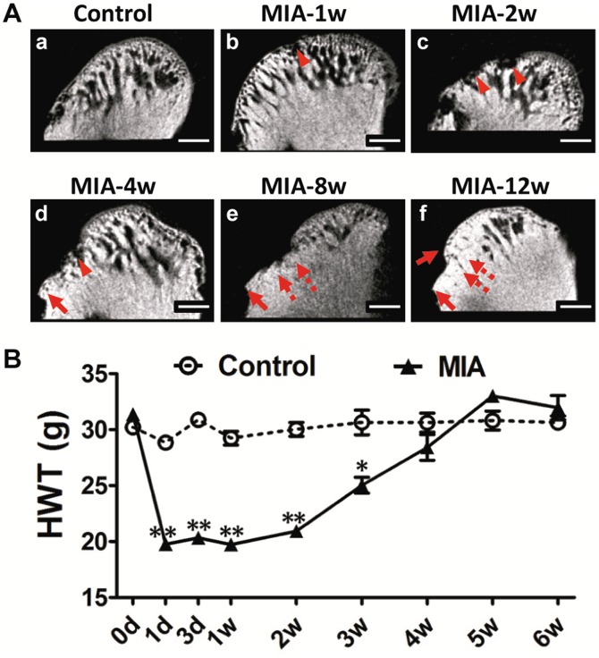
Methods: Female rats were injected with varied doses of MIA into the upper compartment and observed for up to 12 weeks. Histologic, radiographic, behavioral, and molecular changes in the TMJ were evaluated by light and electron microscopy, MicroCT scanning, head withdrawal threshold test, real-time PCR, immunohistochemistry, and TUNEL assay.

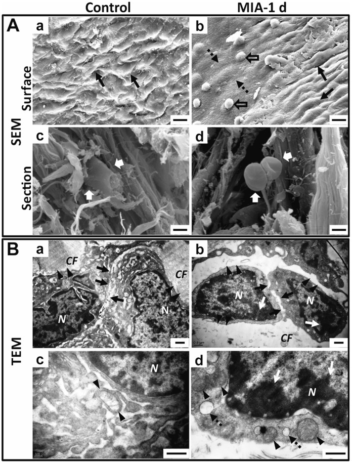
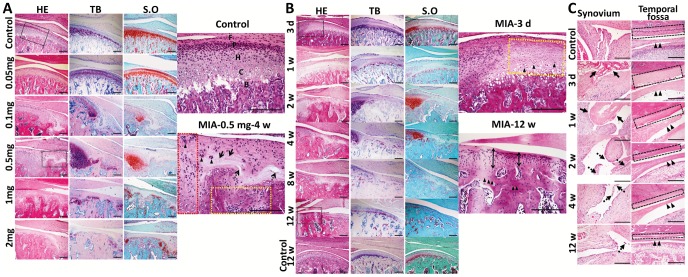
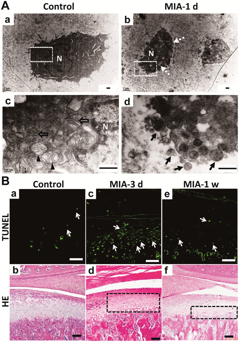
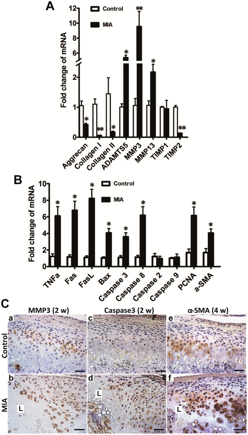
Results: The intermediate zone of the disc loosened by 1 day post-MIA injection and thinned thereafter. Injection of an MIA dose of 0.5 mg or higher induced typical OA-like lesions in the TMJ within 4 weeks. Condylar destruction presented in a time-dependent manner, including chondrocyte apoptosis in the early stages, subsequent cartilage matrix disorganization and subchondral bone erosion, fibrosis, subchondral bone sclerosis, and osteophyte formation in the late stages. Nociceptive responses increased in the early stages, corresponding to severe synovitis. Furthermore, chondrocyte apoptosis and an imbalance between anabolism and catabolism of cartilage and subchondral bone might account for the condylar destruction.

Conclusions: Multi-level data demonstrated a reliable and convenient rat model of TMJOA could be induced by MIA injection into the upper compartment. The model might facilitate TMJOA related researches.


Conflict of interest statement

Competing Interests: The authors have declared that no competing interests exist.

Figures

Figure 1
Figure 1. Outline of experimental design and confirmation of injection site into upper compartment of rat TMJ.
A: Outline of experimental design. B: Photograph of dye (fast green solution) injection into the upper compartment of the left TMJ. (a). Needle insertion was 5 mm anterior to the external auditory canal. (b). Dissection showed that the needle was right under the root of the zygoma (dotted arrow), anterior of the external auditory canal (arrow), stopped at the temporal fossa, and was located in the upper compartment (black circle). (c). 50 µL dye was injected. (d). Opening the capsule revealed that the dye was restricted to the upper compartment of the TMJ (disc and condyle: hollow arrow).
Figure 2
Figure 2. Ultrastructural changes in disc 1 day after MIA injection into upper compartment of TMJ. A.
SEM view of the disc. (a). The furrowed surface (arrow) of the control joint. (b). The surface 1 day after MIA treatment, showing regional flattening in the intermediate zone (dotted arrow) with residual gelatin condensed to a mass (hollow arrow), surrounded by an area with a rough and uneven surface (arrow). (c). Section view of the control disc showed the disc cells studded in the collagen fibrils (white arrow). (d). Section view of the disc of the MIA-treated group showed that disc cells were crimpled and detached from the collagen fibrils (white arrow). B. TEM view of the intermediate zone of the disc. (a). Cells in the control disc were closely attached to the collagen fibrils (arrowhead) and cell junctions were tight (arrow). (b). The cells in the MIA-treated disc were shrunken with condensed chromatin (white arrow), detached from the disrupted ECM (arrowhead), and had loose cell junctions (arrow). (c). Mitochondria were regularly tubular-shaped (arrow) around the nuclei in the control disc. (d). Chromatin compaction (white arrow), swollen mitochondria (arrowhead), and vacuolar degeneration (dotted arrow) were observed in the disc cells 1 day after MIA treatment. (CF: collagen fibers; N: nucleus; Bar = 50 µm in A-a, b; Bar = 20 µm in A-c, d; Bar = 0.5 µm in B).
Figure 3
Figure 3. Dose- and time-dependent histopathologic changes in TMJ tissues.
TMJ was sectioned in saggital for HE, TB and S.O staining. A. Dose course (0.05 mg, 0.1 mg, 0.5 mg, 1 mg, 2mg per joint) of MIA 4 weeks post-injection. Black frames are magnified. Condylar cartilage in the controls stained purple blue by TB and red by S.O; (F: fibrous layer; P: proliferative layer; H: hypertrophy layer; C: calcified layer; B: subchondral bone.). Typical OA-like lesion induced by 0.5 mg MIA, including regional loss of chondrocytes (arrow), chondrocyte cluster formation (arrowhead), horizontal cleft (dotted arrow), peripheral chondrocyte proliferation (red frame), and subchondral bone erosion with adjacent bone marrow full of fibroblast-like cells (yellow frame). Lesions staining by TB and S.O was uneven. (Bar = 200 µm) B. Time-dependent changes in the condyle following MIA injection (0.5 mg/joint; 3 days to 12 weeks). Black frames were magnified. After three days, chondrocytes in the anterior and central areas of the cartilage were lightly stained with nuclear condensation (arrowhead). At 12 weeks, thin cartilage (double arrow) and sclerotic subchondral bone (arrowhead) replaced the lesion. (Bar = 200 µm) C. Time-dependent changes in the synovium, disc, and temporal fossa following MIA injection. Time-dependent changes in synovitis (fibrin-like exudates: arrow; proliferative villi of the synovium: dotted arrow), hypo-cellular change and thinning of the disc (arrowhead), and destruction of temporal fossa cartilage (black frame) following MIA induction are shown. (Bar = 300 µm).
Figure 4
Figure 4. Time-dependent radiographic changes in condylar subchondral bone and nociceptive responses following MIA injection (0.5 mg/joint).
A. Representative images of the condyle by MicroCT scanning with a sagittal section view demonstrated. (a). Control condyle showed intact subchondral bone with a smooth, continuous surface. (b). Regional loss of surface bone (arrowhead) occurred in the frontal bevel of the condyle by 1 week. (c). Multiple erosions of subchondral bone (arrowhead) were observed by 2 weeks. (d). Erosion in the subchondral bone grew deeper and was much more extensive with obvious defects (arrowhead) and osteophyte formation (arrow) by 4 weeks. (e and f). Sclerotic changes (dotted arrow) and osteophytes (arrow) were evident by 8 weeks and 12 weeks. (Bar = 300 µm) B. Changes in animal nociceptive response after MIA injection into TMJ. The HWT was significantly decreased in the first 3 weeks after MIA injection, but gradually recovered to control levels from 4 weeks post-injection. All data were presented as mean ± SEM. (n = 3; **P<0.01; *P<0.05).
Figure 5
Figure 5. Apoptosis of chondrocytes in condyle after MIA treatment.
A. TEM view of condylar chondrocytes. (a). The chondrocytes in the control group were polygonal. (b). Chondrocytes treated by MIA were shrunken with vacuolar degeneration after 1 day (dotted arrow). (c). Magnified photograph of the white frame in (a). Abundant mitochondria (arrowhead) and endoplasmic reticulum (hollow arrow) were observed around the nuclei (N) in the control chondrocyte. (d). Magnified photograph of the white frame in (b). Apoptotic bodies (black arrow) were observed in the chondrocyte following MIA induction. (Bar = 0.5 µm) B. Comparison of TUNEL assay and HE staining results. (a) There were few apoptotic chondrocytes (arrow) in the control group and the corresponding HE staining shown in (b). (c). Diffuse apoptotic chondrocytes were observed in the region corresponding to the lightly stained area with nuclear condensation of HE staining (black frame in d) at 3 days post-MIA injection. The TUNEL positive chondrocytes almost disappeared (e) due to the extreme loss of chondrocytes as shown by HE staining (black frame in f) at 1 week. (Bar = 80 µm).
Figure 6
Figure 6. Changes in gene and protein expression in condyle following MIA injection were evaluated by real-time PCR and IHC, respectively.
A. Two weeks after MIA injection, anabolism-associated aggrecan and collagen I and II were downregulated compared with the control group. Catabolism-associated MMP3, MMP13, and ADAMTS5 were upregulated and TIMP2, but not TIMP1, was correspondingly downregulated. B. Two weeks after MIA (0.5 mg) injection, apoptosis-associated genes of the death receptor family, such as, TNFα, Fas, FasL, caspase8, caspase3, and BAX, but not caspase2 and caspase9, were significantly elevated in the MIA injection group; PCNA and α-SMA, representing proliferation and fibrous restoration, respectively, were upregulated (mean ± SEM; n = 6; **P<0.01; *P<0.05). C. There were very few chondrocytes left in the lesion labeled as L 2 weeks after MIA (0.5 mg) injection. MMP3 was mainly expressed in the hypertrophic layer in the control cartilage (a). Diffuse staining of MMP3 was observed in the chondrocytes adjacent to the lesion (L) at 2 weeks (b). Caspase3 was rarely expressed in the control cartilage (c). Enhanced staining of caspase3 was observed in the proliferative and hypertrophic layers adjacent to the lesion (L) at 2 weeks (d). Expression of α-SMA was mainly in the hypertrophic chondrocytes in the control group (e). Stronger staining of α-SMA was observed adjacent to the lesion (L) at 4 weeks (f). (Bar = 40 µm).

References

    1. Dworkin SF, LeResche L (1992) Research diagnostic criteria for temporomandibular disorders: review, criteria, examinations and specifications, critique. J Craniomandib Disord 6: 301–355. - PubMed
    1. Zarb GA, Carlsson GE (1999) Temporomandibular disorders: osteoarthritis. J Orofac Pain 13: 295–306. - PubMed
    1. Israel HA, Diamond B, Saed-Nejad F, Ratcliffe A (1998) Osteoarthritis and synovitis as major pathoses of the temporomandibular joint: comparison of clinical diagnosis with arthroscopic morphology. J Oral Maxillofac Surg 56: 1023–1027; discussion 1028. - PubMed
    1. Stegenga B, de Bont LG, Boering G (1989) Osteoarthrosis as the cause of craniomandibular pain and dysfunction: a unifying concept. J Oral Maxillofac Surg 47: 249–256. - PubMed
    1. Karsdal MA, Leeming DJ, Dam EB, Henriksen K, Alexandersen P, et al. (2008) Should subchondral bone turnover be targeted when treating osteoarthritis? Osteoarthritis Cartilage 16: 638–646. - PubMed

Publication types

MeSH terms