. 2012;7(9):e45069.
doi: 10.1371/journal.pone.0045069. Epub 2012 Sep 12.

Astrocyte senescence as a component of Alzheimer's disease

Affiliations

Astrocyte senescence as a component of Alzheimer's disease

Rekha Bhat et al. PLoS One. 2012.

Abstract

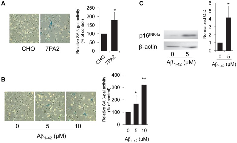
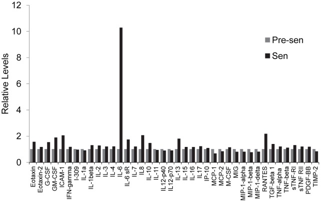
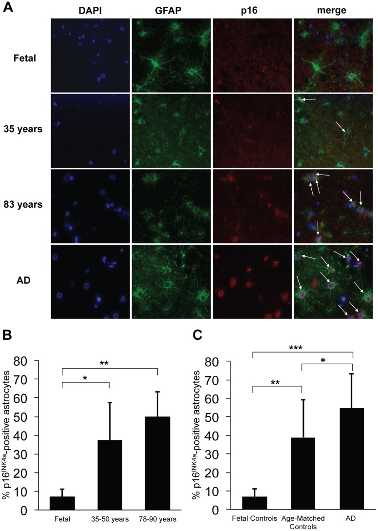
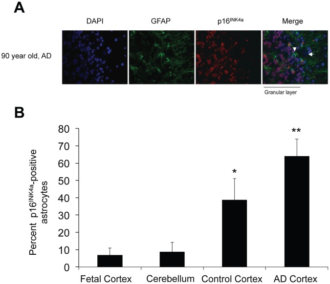
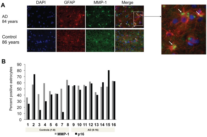
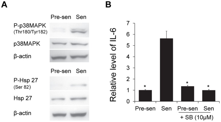
Aging is the main risk factor for Alzheimer's disease (AD); however, the aspects of the aging process that predispose the brain to the development of AD are largely unknown. Astrocytes perform a myriad of functions in the central nervous system to maintain homeostasis and support neuronal function. In vitro, human astrocytes are highly sensitive to oxidative stress and trigger a senescence program when faced with multiple types of stress. In order to determine whether senescent astrocytes appear in vivo, brain tissue from aged individuals and patients with AD was examined for the presence of senescent astrocytes using p16(INK4a) and matrix metalloproteinase-1 (MMP-1) expression as markers of senescence. Compared with fetal tissue samples (n = 4), a significant increase in p16(INK4a)-positive astrocytes was observed in subjects aged 35 to 50 years (n = 6; P = 0.02) and 78 to 90 years (n = 11; P<10(-6)). In addition, the frontal cortex of AD patients (n = 15) harbored a significantly greater burden of p16(INK4a)-positive astrocytes compared with non-AD adult control subjects of similar ages (n = 25; P = 0.02) and fetal controls (n = 4; P<10(-7)). Consistent with the senescent nature of the p16(INK4a)-positive astrocytes, increased metalloproteinase MMP-1 correlated with p16(INK4a). In vitro, beta-amyloid 1-42 (Aβ(1-42)) triggered senescence, driving the expression of p16(INK4a) and senescence-associated beta-galactosidase. In addition, we found that senescent astrocytes produce a number of inflammatory cytokines including interleukin-6 (IL-6), which seems to be regulated by p38MAPK. We propose that an accumulation of p16(INK4a)-positive senescent astrocytes may link increased age and increased risk for sporadic AD.


Conflict of interest statement

Competing Interests: The authors have declared that no competing interests exist.

Figures

Figure 1
Figure 1. Astrocytes trigger senescence in response to Aβ1–42.
(A) Representative images of astrocytes treated with conditioned media from 7PA2 Aβ-secreting Chinese Hamster Ovary (CHO) cells or CHO control cells as described in Figure S2. Graph shows relative amount of SA β-gal-positive cells from 3 independent experiments, *P = 0.039. (B) Representative images of astrocytes treated with oligomerized synthetic Aβ1–42 for 2 hours and assayed for SA β-gal activity after 3 days. Graph shows relative levels of SAβ-gal–positive cells from 3 independent experiments, *P = 0.017, Aβ1–42 (5 µM and **P = 0.009, Aβ1–42 (10 µM) vs. control. (C) Immunoblot for p16INK4a expression in astrocytes treated with 5 µM Aβ1–42 for 24 hours. Lysate was collected 4 days after treatment initiation. β-actin serves as a loading control. Graph depicts normalized optical density of the ratio of p16INK4a:β-actin, *P = 0.02. For all experiments, error bars represent SD and Student’s t-test was used to determine significance.
Figure 2
Figure 2. Senescent astrocytes secrete pro-inflammatory mediators that may constitute a SASP.
Pre-senescent and senescent astrocytes were incubated in serum-free MCDB105 media. Conditioned media was collected after 48 hours and analyzed by antibody array (RayBiotech, Inc.; Norcross, GA) for pro-inflammatory factors as described in Materials and Methods.
Figure 3
Figure 3. Increased frequency of senescent astrocytes during brain aging and AD.
p16INK4a-positive astrocytes were identified in formalin-fixed, paraffin-embedded frontal cortex sections by double immunofluorescence with p16INK4a/GFAP. (A) Double immunofluorescence with p16INK4a/GFAP showing increased p16INK4a-positive astrocytes with increased age and AD (representative images). Blue: DAPI; green: GFAP; red: p16INK4a. Arrows indicate p16INK4a-positive astrocytes. (B) Bar diagram shows increased mean p16 INK4a-positive astrocytes in frontal cortices from non-AD adult subjects (35–50 years, n = 6; 78–90 years, n = 11) as compared to fetal autopsy tissue (n = 4). *P = 0.02, fetal vs. 35–50 year olds; **P<10−6, fetal vs. 78–90 year olds; Student’s t-test. (C) Bar diagram shows increased mean p16 INK4a-positive astrocytes in frontal cortices from AD adult subjects (n = 15) compared to non-AD adult control subjects (n = 25) of similar ages; and fetal controls (n = 4). * P = 0.02, AD vs. adult controls; ***P<10−7, AD vs. fetal controls, and **P<10−6, adult controls vs. fetal controls. Error bars represent SD and Student’s t-test was used to determine significance. At least 200 cells were counted per slide.
Figure 4
Figure 4. Expression level of p16INK4a is not elevated in adult cerebellar astrocytes.
(A) Representative images of cerebellum from an AD patient showing intensely p16INK4a positive granular layer. The Bergmann glia (arrowhead) and Purkinje neurons (arrow) are negative. Blue: DAPI; Green: GFAP; Red: p16INK4a. (B) Bar diagram shows increased mean percentage of p16INK4a-positive astrocytes in formalin-fixed, paraffin-embedded cortical sections from AD subjects (n = 3) and control subjects (n = 3) compared to cerebellum from both AD and aged-matched controls (n = 6). The mean percentage of p16INK4a-positive astrocytes in cerebellum is comparable to fetal cortical levels. *P = 0.04, Student’s t test, cerebellum vs. control cortices; **P = 0.004, Student’s t test, cerebellum vs. AD cortices. Error bars represent SD At least 200 cells per slide were counted for the cortical sections, and at least 100 cells per slide were counted for the cerebellar sections.
Figure 5
Figure 5. MMP-1 expression parallels p16INK4a expression in human astrocytes in vivo.
(A) Double immunofluorescence with MMP-1/GFAP in AD and a similar-aged control (representative images). Blue: DAPI; red: GFAP; green: MMP-1. Arrows indicate MMP-1–positive astrocytes. Inset shows magnified image of MMP-1-positive astrocytes (yellow: cytoplasmic). (B) Bar diagram shows positive correlation between MMP-1 and p16INK4a expression in human frontal cortex, independent of diagnosis, P = 0.02, Spearman’s correlation coefficient = 0.574.
Figure 6
Figure 6. p38MAPK pathway is activated and modulates IL-6 secretion in astrocytes.
(A) Western blots depict the levels of total and phosphorylated p38MAPK and heat shock protein 27 (Hsp27) in pre-senescent and H2O2-induced senescent astrocytes with β-actin as loading control. (B) Pre-senescent and senescent astrocytes were treated with 10 µM SB-203580 or DMSO as a control for 48 hours prior to incubation in serum-free MCDB105 media. Conditioned media was collected after 24 hours and IL-6 was analyzed by ELISA (R&D Systems, Inc.; Minneapolis, MN) and normalized to cell number. Graph depicts the relative level of IL-6 (n = 3), *P<0.01 vs. senescent, Student’s t test.

References

    1. Thies W, Bleiler L (2011) 2011 Alzheimer’s disease facts and figures. Alzheimers Dement 7: 208–244. - PubMed
    1. Herbig U, Ferreira M, Condel L, Carey D, Sedivy JM (2006) Cellular senescence in aging primates. Science 311: 1257. - PubMed
    1. Jeyapalan JC, Sedivy JM (2008) Cellular senescence and organismal aging. Mech Ageing Dev 129: 467–474. - PMC - PubMed
    1. Michaloglou C, Vredeveld LC, Soengas MS, Denoyelle C, Kuilman T, et al. (2005) BRAFE600-associated senescence-like cell cycle arrest of human naevi. Nature 436: 720–724. - PubMed
    1. Jeyapalan JC, Ferreira M, Sedivy JM, Herbig U (2007) Accumulation of senescent cells in mitotic tissue of aging primates. Mech Ageing Dev 128: 36–44. - PMC - PubMed

Publication types

MeSH terms