. 2012 Sep 17:13:175.
doi: 10.1186/1471-2474-13-175.

A method to screen and evaluate tissue adhesives for joint repair applications

Affiliations

A method to screen and evaluate tissue adhesives for joint repair applications

Tilo Dehne et al. BMC Musculoskelet Disord. .

Abstract

Background: Tissue adhesives are useful means for various medical procedures. Since varying requirements cause that a single adhesive cannot meet all needs, bond strength testing remains one of the key applications used to screen for new products and study the influence of experimental variables. This study was conducted to develop an easy to use method to screen and evaluate tissue adhesives for tissue engineering applications.

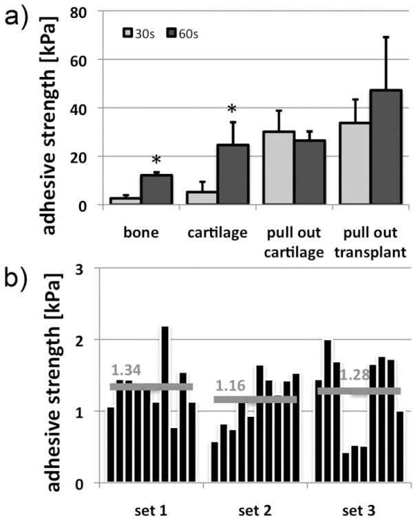
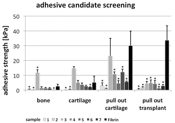
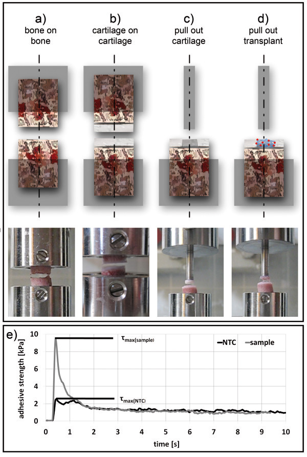
Method: Tissue grips were designed to facilitate the reproducible production of substrate tissue and adhesive strength measurements in universal testing machines. Porcine femoral condyles were used to generate osteochondral test tissue cylinders (substrates) of different shapes. Viability of substrates was tested using PI/FDA staining. Self-bonding properties were determined to examine reusability of substrates (n = 3). Serial measurements (n = 5) in different operation modes (OM) were performed to analyze the bonding strength of tissue adhesives in bone (OM-1) and cartilage tissue either in isolation (OM-2) or under specific requirements in joint repair such as filling cartilage defects with clinical applied fibrin/PLGA-cell-transplants (OM-3) or tissues (OM-4). The efficiency of the method was determined on the basis of adhesive properties of fibrin glue for different assembly times (30 s, 60 s). Seven randomly generated collagen formulations were analyzed to examine the potential of method to identify new tissue adhesives.

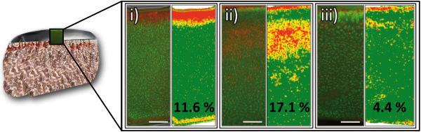
Results: Viability analysis of test tissue cylinders revealed vital cells (>80%) in cartilage components even 48 h post preparation. Reuse (n = 10) of test substrate did not significantly change adhesive characteristics. Adhesive strength of fibrin varied in different test settings (OM-1: 7.1 kPa, OM-2: 2.6 kPa, OM-3: 32.7 kPa, OM-4: 30.1 kPa) and was increasing with assembly time on average (2.4-fold). The screening of the different collagen formulations revealed a substance with significant higher adhesive strength on cartilage (14.8 kPa) and bone tissue (11.8 kPa) compared to fibrin and also considerable adhesive properties when filling defects with cartilage tissue (23.2 kPa).

Conclusion: The method confirmed adhesive properties of fibrin and demonstrated the dependence of adhesive properties and applied settings. Furthermore the method was suitable to screen for potential adhesives and to identify a promising candidate for cartilage and bone applications. The method can offer simple, replicable and efficient evaluation of adhesive properties in ex vivo specimens and may be a useful supplement to existing methods in clinical relevant settings.


Figures

Figure 1
Figure 1
Viability of substrate tissue. (left) Sections of PI/FDA-stained cartilage components from (i) leveled and (ii) hole-punched substrate cylinders, and (iii) cartilage discs were analyzed by fluorescence microscopy. Viable cells appear green and apoptotic cells red. (right) Histomorphometric analysis was performed to determine the percentage of apoptotic cells as measure for the viability of the tissue. For each image areas of different red tones were determined and presented in new colors. High content of apoptotic cells was indicated in red, moderate in orange, low in yellow and areas with high content of viable cells were shown in green (see Methods). Only a small proportion of cells was found apoptotic (<18%), hence 48 h post preparation cartilage components of the substrates are in a viable condition for adhesive strength measurements. Bar = 200 μm.
Figure 2
Figure 2
Establishing the test system. (a) Different modes of operation were applied to determine adhesive characteristics of fibrin. Varying bonding strengths were detected in the specific tests with highest values in fixing transplants and lowest in bonding bone tissue. T-test: 30 s vs. 60 s, * p < 0.05. (b) The reusability of tissue cylinders was investigated for cartilage. Three different biological replicates (set 1–3) of cartilage cylinders were investigated. After each measurement with fibrin, the adhesive was removed and a new control measurement with PBS was performed. This proceeding was repeated 10 times. Technical variations for each set range from 0.4 and 2.4 kPa, but do not increase with frequency of use suggesting that the cylinders can be reused. Differences according to biological variation are marginal (mean values range from 1.16 – 1.34 kPa, grey bar) demonstrating good reproducibility of the preparation method.
Figure 3
Figure 3
Screening for potential adhesives. Screening was performed in different operation modes (see Figure 3). Sample 3 demonstrated higher bonding strength than fibrin when used on cartilage and bone tissue. In settings filling “artificial defects” with cartilage tissue or TE transplants, fibrin showed highest bonding strength. These results demonstrate high bonding properties of fibrin in all settings. Sample 3 can be identified as potential tissue adhesive that is not suitable for fixing fibrin-based TE transplants. T-test vs. fibrin, * p < 0.05.
Figure 4
Figure 4
Tissue grips. Pictures and technical drawings of grips used for (a) substrate tissue cylinder preparations and (b) adhesive strength measurements.
Figure 5
Figure 5
Preparation of substrate tissues. Schematic graphs and pictures illustrating the preparation of substrate tissue cylinders used for the determination of bonding strength. Osteochondral discs of 1–1.5 cm thickness (b) were obtained from the lateral and medial condyle of pig femoral bones (a) using a rotary reciprocating saw. (c) The removed discs were hole-punched to generate a cylindrical tissue specimen with a diameter of 10 mm. A bench vise facilitated a controlled agitation of the punch orthogonal to the sectional plane. (d) The raw tissue cylinder was placed in a customized grip that is fitting into the jig of a standard microtome device (see also Figure 4a). (e) Using the blade of the microtome the surface of the tissue cylinder was leveled to obtain regular bone or cartilage cylinder ends. Thus, starting from a raw cylinder (d), cartilage and bone substrate cylinders with plane surfaces can be generated (h). Furthermore, with the help of a smaller punch (ø 6 mm) another hole was created (f) providing an artificial cartilage defect to be filled with cartilage tissues or transplants (g, i).
Figure 6
Figure 6
Modes of operation. For measuring adhesive strength in bone (a) or cartilage tissue (b), two cylinders were placed opposite to each other. For tissues and transplants a cylindrical indenter was placed opposing the inserted tissue (c) or transplant (d). The tip was coated with cyanoacrylate glue to facilitate a strong bonding between steel tip and inserted tissue or transplant after contact. After preparing the surfaces with the test adhesive, opposite sites were joined under defined initial load (e.g. 0.5 N). Once bonding time was reached opposite sites were separated with constant speed until rupture. This pulled the inserted tissue or transplants out of the “artificial defect”. (e) The progress of adhesive strength was recorded over time. The maximum bonding strength (τmax) was determined and the mean value of the base line signal was subtracted. The bonding strength was calculated from difference between “no template control” (NTC) and sample value.

Similar articles

Cited by

References

    1. Reece TB, Maxey TS, Kron IL. A prospectus on tissue adhesives. Am J Surg. 2001;182(2 Suppl):40S–44S. - PubMed
    1. Berthelsen B, Lofgren J, Svendsen P. Embolization of cerebral arteriovenous malformations with bucrylate. Experience in a first series of 29 patients. Acta Radiol. 1990;31(1):13–21. - PubMed
    1. Sawamura Y, Asaoka K, Terasaka S, Tada M, Uchida T. Evaluation of application techniques of fibrin sealant to prevent cerebrospinal fluid leakage: a new device for the application of aerosolized fibrin glue. Neurosurgery. 1999;44(2):332–337. doi: 10.1097/00006123-199902000-00048. - DOI - PubMed
    1. Donkerwolcke M, Burny F, Muster D. Tissues and bone adhesives–historical aspects. Biomaterials. 1998;19(16):1461–1466. doi: 10.1016/S0142-9612(98)00059-3. - DOI - PubMed
    1. Spotnitz WD. Fibrin sealant: past, present, and future: a brief review. World J Surg. 2010;34(4):632–634. doi: 10.1007/s00268-009-0252-7. - DOI - PubMed

Publication types

MeSH terms