. 2012 Dec;13(12):1653-66.
doi: 10.1111/tra.12009. Epub 2012 Oct 11.

A common clathrin-mediated machinery co-ordinates cell-cell adhesion and bacterial internalization

Affiliations

A common clathrin-mediated machinery co-ordinates cell-cell adhesion and bacterial internalization

Matteo Bonazzi et al. Traffic. 2012 Dec.

Abstract

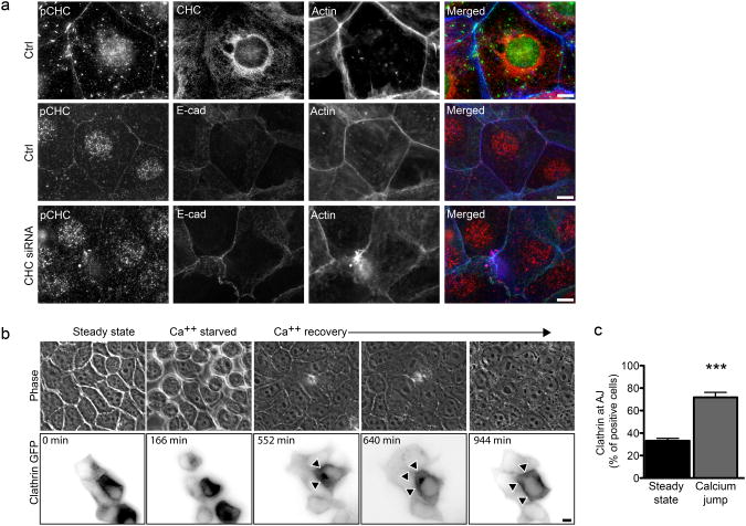
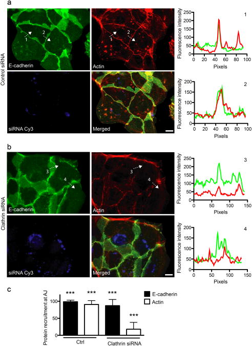
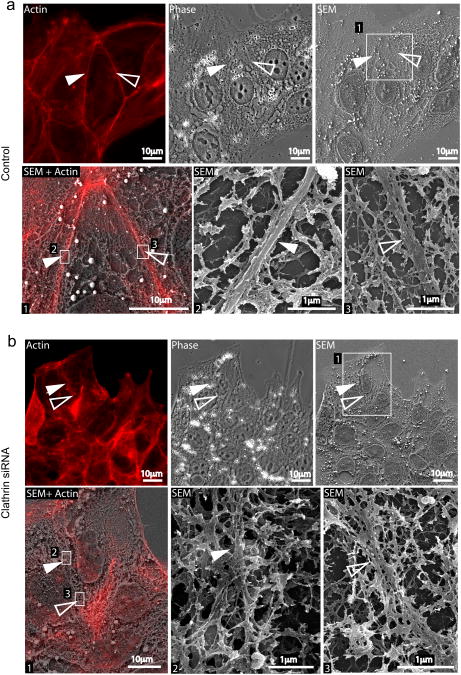
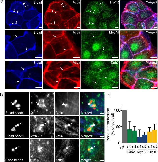
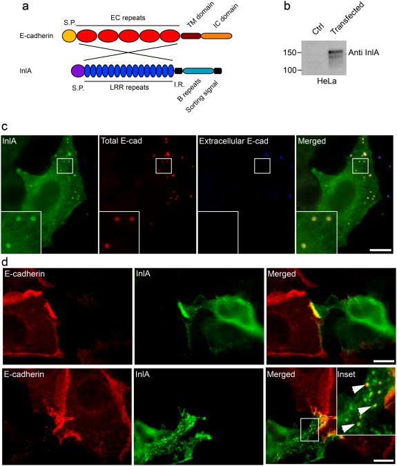
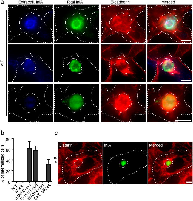
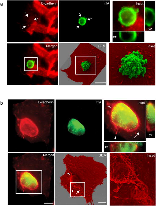
Invasive bacterial pathogens often target cellular proteins involved in adhesion as a first event during infection. For example, Listeria monocytogenes uses the bacterial protein InlA to interact with E-cadherin, hijack the host adherens junction (AJ) machinery and invade non-phagocytic cells by a clathrin-dependent mechanism. Here, we investigate a potential role for clathrin in cell-cell adhesion. We observed that the initial steps of AJ formation trigger the phosphorylation of clathrin, and its transient localization at forming cell-cell contacts. Furthermore, we show that clathrin serves as a hub for the recruitment of proteins that are necessary for the actin rearrangements that accompany the maturation of AJs. Using an InlA/E-cadherin chimera, we show that adherent cells expressing the chimera form AJs with cells expressing E-cadherin. We demonstrate that non-adherent cells expressing the InlA chimera, as bacteria, can be internalized by E-cadherin-expressing adherent cells. Together these results reveal that a common clathrin-mediated machinery may regulate internalization and cell adhesion and that the relative mobility of one of the interacting partners plays an important role in the commitment to either one of these processes.


Figures

Figure 1
Figure 1
AJ formation induces CHC phosphorylation and the recruitment of clathrin at cell-cell contacts a. Jeg3 cells were left untreated (top), incubated with non-targeting (middle) or CHC-targeting (bottom) siRNA sequences, seeded and allowed to form AJs for 12 to 16 hours. Cells were then fixed and labeled for immunofluorescence as indicated. b. Representative images (acquired live) of MDCK cells transfected with CLC-GFP and subjected to the calcium jump assay to follow clathrin recruitment at cell-cell contacts (arrowheads) during the formation of adherens junctions. c. MDCK cells at steady state or 12h after the calcium jump were fixed and labeled for clathrin, E-cadherin and actin. The frequency of clathrin recruitment at cell-cell junctions was assayed by fluorescence microscopy. In c values are means (± standard deviation) of three independent experiments. Asterisks represent p values (***= p ≤ 0.001, Student t-test). Scale bars 10 μm.
Figure 2
Figure 2
Clathrin is required for actin recruitment at cell-cell contacts. Jeg3 cells were transfected with Cy3-tagged control (a) or clathrin-targeted siRNAs (b). 48h after transfection cells were trypsinized and seeded on glass coverslips to allow the formation of new adherens junctions. 12 h after seeding, cells were fixed and labeled for E-cadherin (green) and actin (red). The fluorescence intensity profiles along arbitrary lines drawn perpendicular to cell-cell contacts (dashed arrows 1, 2, 3 and 4) was measured for E-cadherin (green) and actin (red) (Right charts, numbers correspond to the dashed profiles on the left panels). c. Quantification of control and clathrin knocked-down cells presenting E-cadherin and actin at cell-cell junctions. Values are means (± standard deviation) of three independent experiments where approximately 100 cells were analyzed for each condition. Asterisks represent p values (***= p ≤ 0.001, Student t-test). Scale bars 10 μm.
Figure 3
Figure 3
CLSEM analysis of actin at cell-cell contacts in control and clathrin depleted cells. Jeg3 cells transfected with Cy3-tagged control (a) or anti-clathrin siRNAs (b) were permeabilized and labeled for actin (red). Cells were first imaged with an epifluorescence microscope (Actin and phase panels in a and b) and then processed for scanning electron microscopy (SEM). The ultrastructure of the cytoskeleton in control and clathrin knocked down cells was compared. In both a and b, panels 2 and 3 represent higher magnifications of the boxed areas in panel 1. Full and empty arrowheads point at actin structures at cell-cell contacts.
Figure 4
Figure 4
The clathrin/actin interaction machinery is recruited at sites of adherens junctions formation. a. Jeg3 cells were seeded and allowed to form AJs for 12 hours. Cells were then fixed and labeled for E-cadherin (blue), actin (red) and Hip1R, Myosin VI, Dab2 (green). Arrows indicate sites where Hip1R Myosin VI and Dab2 accumulate with E-cadherin and actin at adherens junctions. Scale bars 10 μm. b Jeg3 cells were incubated with E-cadherin-coated latex beads for 15 min, fixed and processed for immunofluorescence. Arrows point at sites of colocalization. c. Jeg3 cells were transfected with two siRNA sequences targeted to either Dab2, Myosin VI or Hip1R or with scrambled control siRNA sequences and incubated 1 h with E-cadherin-coated latex beads. Cells were then fixed and differentially labeled to discriminate intracellular and extracellular beads and the efficiency of beads internalization was quantified by fluorescence microscopy. Values are means (± standard deviation) of three independent experiments where approximately 100 beads were counted for each condition. Asterisks represent p values. Scale 10 μm (d).
Figure 5
Figure 5
InlA/E-cadherin interactions generate hybrid cell-cell junctions. a. The InlA/E-cadherin chimeric protein was generated by substituting the EC repeats of E-cadherin with the LRR repeats of the Listeria monocytogenes surface protein InlA. b. HeLa cells were transfected with a control plasmid or with a plasmid containing the chimeric DNA and the expression of the chimera was tested by Western blots with an InlA-specific antibody. c. HeLa cells transfected with the InlA-E-cadherin chimera were incubated with E-cadherin-coated beads for 1 h. Beads were labeled with an anti-E-cadherin antibody before (blue) and after permeabilization (red) to distinguish extracellular and intracellular beads and cells were labeled with an anti-InlA antibody (green) to detect transfected cells. d. HeLa cells transfected with the InlA/E-cadherin chimera were co-cultured with Jeg3 cells expressing endogenous E-cadherin. After an overnight incubation cells were fixed and labeled with an antibody against the extracellular domain of E-cadherin, that specifically recognizes endogenous E-cadherin (red), and with an InlA-specific antibody (green). When cell-cell contacts were more interdigitated we could observe the presence of endocytic vesicles positive for both InlA (green) and E-cadherin (red). Scale bars 10 μm.
Figure 6
Figure 6
Cell adhesion mediates cell-in-cell events. a. Maximum intensity projections (MIP) of image stacks acquired along the z-axis of HeLa cells transfected with the InlA/E-cadherin chimera (long-dashed outline), trypsinized and incubated for 1 h on a confluent layer of Jeg3 cells (short-dashed outline). HeLa cells were labeled for InlA before permeabilization (blue) to detect extracellular cells and after permeabilization (green) to detect total HeLa cells. Jeg3 cells were labeled for E-cadherin (red). Images represent different stages of cell-cell internalization. b. HeLa cells were either left untransfected (N.T.), transfected with an empty vector (mock), with the InlA/E-cadherin chimera (InlA/hE-cad) or with GFP-tagged E-cadherin (E-cad/E-cad). Cells were trypsinized and incubated for 1h on adherent Jeg3 cells. As a control, HeLa cells transfected with the InlA/E-cadherin chimera were trypsinized and incubated for 1h on ELB1 cells expressing mouse E-cadherin (InlA/mEcad). To test the role of clathrin in cell-in-cell events HeLa cells transfected with the InlA/E-cadherin chimera were incubated for 1h with adherent Jeg3 cells where clathrin heavy chain (CHC) had been previously knocked down by siRNA. In all cases the frequency of cell-in-cell events was quantified by fluorescence microscopy were HeLa cells were differentially labeled before and after permeabilization. Values are means (± standard deviation) of three independent experiments where approximately 100 cells were analyzed for each condition. c. Maximum intensity projections (MIP) of image stacks acquired along the z-axis of HeLa cells transfected with the InlA/E-cadherin chimera (long-dashed outline), trypsinized and incubated for 1 h on a confluent layer of Jeg3 cells (short-dashed outline). Cells were labeled for InlA (green) and clathrin (red). Scale bars 10 μm.
Figure 7
Figure 7
CLSEM analysis of cell-in-cell events Early (a) and late (b) stages of cell-in-cell events. HeLa cells transfected with the InlA/E-cadherin chimera were trypsinized and incubated for 1 h on a confluent layer of Jeg3 cells. HeLa cells were labeled for InlA (green) and Jeg3 cells were labeled for E-cadherin (red). Cells were imaged along the z-axis to visualize E-cadherin recruitment around transfected HeLa cells (xz and yz orthogonal views of insets). Using CLSEM the same cells were scanned at the electron microscope to better visualize cell-cell internalization (SEM and corresponding insets. Jeg3 cells are pseudo-colored in red, transfected HeLa cells are pseudo-colored in green). Arrows indicate examples of correlated structures visualized by immunofluorescence and scanning electron microscopy. Scale bars 10 μm.

Similar articles

Cited by

References

    1. Cavallaro U, Christofori G. Cell adhesion and signalling by cadherins and Ig-CAMs in cancer. Nat Rev Cancer. 2004;4:118–132. - PubMed
    1. Bonazzi M, Cossart P. Impenetrable barriers or entry portals? The role of cell-cell adhesion during infection. JCB. 2011;195:349–358. - PMC - PubMed
    1. Knodler LA, Celli J, Finlay BB. Pathogenic trickery: deception of host cell processes. Nat Rev Mol Cell Biol. 2001;2:578–588. - PubMed
    1. Pizarro-Cerdá J, Cossart P. Bacterial adhesion and entry into host cells. Cell. 2006;124:715–727. - PubMed
    1. Bonazzi M, Veiga E, Pizarro-Cerdá J, Cossart P. Successive post-translational modifications of E-cadherin are required for InlA-mediated internalization of Listeria monocytogenes. Cell Microbiol. 2008;10:2208–2222. - PubMed

Publication types

LinkOut - more resources