. 2012 Sep 17:12:148.
doi: 10.1186/1471-2431-12-148.

Circulating hematopoietic stem cell count is a valuable predictor of prematurity complications in preterm newborns

Affiliations

Circulating hematopoietic stem cell count is a valuable predictor of prematurity complications in preterm newborns

Maciej Kotowski et al. BMC Pediatr. .

Abstract

Background: The frequency of preterm labour has risen over the last few years. Hence, there is growing interest in the identification of markers that may facilitate prediction and prevention of premature birth complications. Here, we studied the association of the number of circulating stem cell populations with the incidence of complications typical of prematurity.

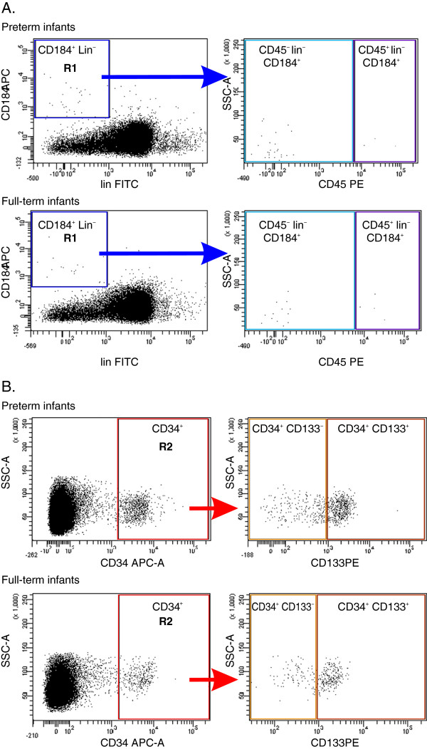
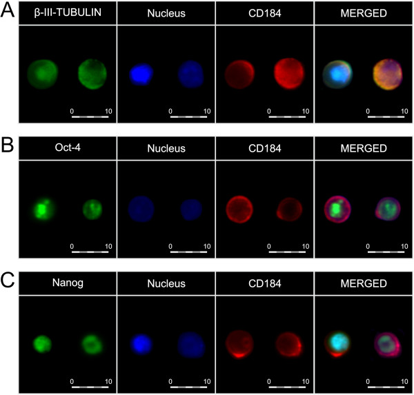
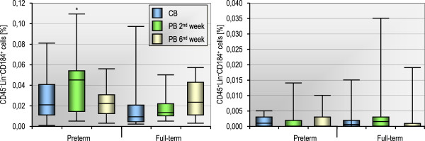
Methods: The study groups consisted of 90 preterm (23-36 weeks of gestational age) and 52 full-term (37-41 weeks) infants. Non-hematopoietic stem cells (non-HSCs; CD45-lin-CD184+), enriched in very small embryonic-like stem cells (VSELs), expressing pluripotent (Oct-4, Nanog), early neural (β-III-tubulin), and oligodendrocyte lineage (Olig-1) genes as well as hematopoietic stem cells (HSCs; CD45+lin-CD184+), and circulating stem/progenitor cells (CSPCs; CD133+CD34+; CD133-CD34+) in association with characteristics of prematurity and preterm morbidity were analyzed in cord blood (CB) and peripheral blood (PB) until the sixth week after delivery. Phenotype analysis was performed using flow cytometry methods. Clonogenic assays suitable for detection of human hematopoietic progenitor cells were also applied. The quantitative parameters were compared between groups by the Mann-Whitney test and between time points by the Friedman test. Fisher's exact test was used for qualitative variables.

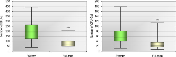
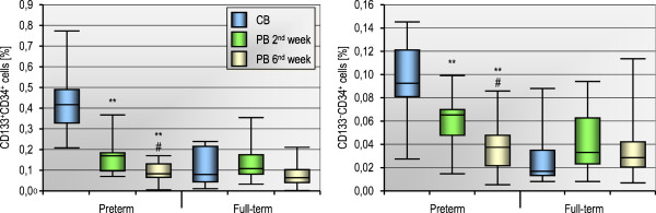
Results: We found that the number of CB non-HSCs/VSELs is inversely associated with the birth weight of preterm infants. More notably, a high number of CB HSCs is strongly associated with a lower risk of prematurity complications including intraventricular hemorrhage, respiratory distress syndrome, infections, and anemia. The number of HSCs remains stable for the first six weeks of postnatal life. Besides, the number of CSPCs in CB is significantly higher in preterm infants than in full-term neonates (p < 0.0001) and extensively decreases in preterm babies during next six weeks after birth. Finally, the growth of burst-forming unit of erythrocytes (BFU-E) and colony-forming units of granulocyte-macrophage (CFU-GM) obtained from CB of premature neonates is higher than those obtained from CB of full-term infants and strongly correlates with the number of CB-derived CSPCs.

Conclusion: We conclude that CB HSCs are markedly associated with the development of premature birth complications. Thus, HSCs ought to be considered as the potential target for further research as they may be relevant for predicting and controlling the morbidity of premature infants. Moreover, the observed levels of non-HSCs/VSELs circulating in CB are inversely associated with the birth weight of preterm infants, suggesting non-HSCs/VSELs might be involved in the maturation of fetal organism.


Figures

Figure 1
Figure 1
A representative study of gating strategy for analyzing/sorting of non-HSCs/VSELs, HSCs, and CSPCs circulating in newborn PB by FACS. Each colored gate illustrates a population of cells which expresses selected surface proteins. Panel A: Cytofluorimetric analysis of non-HSCs and HSC: CD184+lin- cells were gated from a total fraction of immunofluorescence-stained nucleated cells from the PB of preterm and full term infants, then those cells from region R1 were further analyzed for CD45 expression; Panel B: Cytofluorimetric analysis of CSPCs (CD133+CD34+ and CD133-CD34+): CD34+ cells were gated from a total fraction of immunofluorescence-stained nucleated cells from the PB of preterm and full term infants, then those cells from region R2 were further analyzed for CD133 expression.
Figure 2
Figure 2
Immunocytofluorescence of CB-derived non-HSCs. Individual images depict the expression of pluripotent/early neuronal markers, CD184, as well as nuclei in non-HSCs. Merged images show coexpression of β-III-tubulin (panel A), Oct-4 (panel B), Nanog (panel C), and CD184, together with nuclei. Pseudocolors are assigned to each staining as follows: anti-β-III-tubulin, -Oct-4, and -Nanog – green, anti-CD184 – red, nuclei - blue. The cells were captured with × 40 objective magnification. The expression of each antigen was examined in cells in six independent experiments. Representative data are shown.
Figure 3
Figure 3
The number of CD45-lin-CD184+ (non-HSCs) and CD45+lin-CD184+ (HSCs) blood stem cell populations counted in CB at birth as well as in PB two and six weeks after birth in 13 preterm and 18 full-term infants. *p < 0.05 for comparison between preterm and full-term infants (Mann–Whitney test). No statistically significant changes were observed within each group (p > 0.15, Friedman test).
Figure 4
Figure 4
Clonogenic growth efficiency of CD34-positive cells obtained from the CB of 44 preterm or 24 full-term infants. Formation of burst-forming unit-erythroid (BFU-E) or colony-forming unit-granulocyte-monocyte (CFU-GM) was assessed in clonogenic in vitro assays. Colony counts are expressed in absolute values per 2 × 104 plated cells and represent the median [min–max] from all the performed assays in each group. ***p < 0.001 for comparison between preterm and full-term infants (Mann–Whitney test).
Figure 5
Figure 5
The number of CD133+CD34+ and CD133-CD34+ blood cells counted in CB at birth as well as in PB two and six weeks after delivery in 13 preterm and 18 full-term infants. **p < 0.01 for comparison between PB in 2nd and 6th week vs CB. #p < 0.05 for PB in 6th week vs PB in 2nd week (Mann–Whitney test).

Similar articles

Cited by

References

    1. Norman JE, Morris C, Chalmers J. The effect of changing patterns of obstetric care in Scotland (1980–2004) on rates of preterm birth and its neonatal consequences: perinatal database study. PLoS Med. 2009;6:e1000153. doi: 10.1371/journal.pmed.1000153. - DOI - PMC - PubMed
    1. Di Tommaso M, Tranquilli A. A checklist to identify the origin of cerebral palsy. J Matern Fetal Neonatal Med. 2004;15:281–286. doi: 10.1080/14767050410001712121. - DOI - PubMed
    1. Goldenberg RL, Culhane JF, Iams JD, Romero R. Epidemiology and causes of preterm birth. Lancet. 2008;371:75–84. doi: 10.1016/S0140-6736(08)60074-4. - DOI - PMC - PubMed
    1. Machalińska A, Baumert B, Kuprjanowicz L, Wiszniewska B, Karczewicz D, Machaliński B. Potential application of adult stem cells in retinal repair–challenge for regenerative medicine. Curr Eye Res. 2009;34:748–760. doi: 10.1080/02713680903050592. - DOI - PubMed
    1. Paczkowska E, Kucia M, Koziarska D, Halasa M, Safranow K, Masiuk M, Karbicka A, Nowik M, Nowacki P, Ratajczak MZ, Machalinski B. Clinical evidence that very small embryonic-like (VSEL) stem cells are mobilized into peripheral blood in patients after stroke. Stroke. 2009;40:1237–1244. doi: 10.1161/STROKEAHA.108.535062. - DOI - PubMed

Publication types

MeSH terms