. 2012 Sep 17:9:206.
doi: 10.1186/1743-422X-9-206.

Sf-PHB2, a new transcription factor, drives WSSV Ie1 gene expression via a 12-bp DNA element

Affiliations

Sf-PHB2, a new transcription factor, drives WSSV Ie1 gene expression via a 12-bp DNA element

Guoda Ma et al. Virol J. .

Abstract

Background: The WSSV immediate early gene ie1 is highly expressed throughout viral infection cycle and may play a central role in initiating viral replication during infection.

Results: Here, a detailed characterization of the ie1 promoter was performed using deletion and mutation analyses to elucidate the role of the individual promoter motifs. Three results were obtained: 1) the ie1 promoter is a classical eukaryotic promoter that contains the initiator element (Inr) and TATA box responsible for the basal promoter activity; 2) mutation or truncation of a predicted Sp1 site decreased the level of promoter activity by about 3-fold, indicating that the Sp1 site is an important cis-element of the promoter; and 3) truncation of a 12-bp sequence that resides at -78/-67 of the ie1 promoter decreased the level of promoter activity by about 14-fold, indicating that the 12-bp motif is a critical upstream element of the ie1 promoter for binding of a strong transcription factor to drive the ie1 gene expression in the cells. Further, the 12-bp DNA binding protein was purified from the nuclear proteins of Sf9 cells using DNA affinity chromatography, and was identified as a homologue of the prohibitin2 protein (named as Sf-PHB2) using mass spectrometry. Furthermore, the DNA binding activity of Sf-PHB2 was verified using a super shift analysis.

Conclusion: These results support that the Sf-PHB2 is a novel transcription factor that drives WSSV ie1 gene expression by binding to the 12-bp DNA element.


Figures
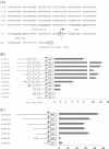
Figure 1
Figure 1
Functional analysis of the WSSV ie1 promoter through deletion and mutation. (A) The 388 bp sequence of the WSSV ie1 promoter is shown. The numbers are relative to the transcription start site (+1). The specific protein binding sites such as Sp1 and the TATA box are underlined. The transcription start site is shown in italics and bold. (B) The schematic depicts the features of the WSSV ie1 promoter that is positioned between the putative translational start site and its 388-bp upstream sequence. A series of truncated promoter fragments were constructed in front of the luciferase gene in a promoter-less vector phRG-B. For the 5' end and the 3' ends, each deletion and mutation of the promoter is depicted and named as shown in the figure. The putative Sp1-binding site and TATA box were mutated [p(-55/+53Δ)and p(-35/+53Δ)] using PCR with a mutated primer. (C) Functional mapping of the cis-elements within the 80-bp sequence (from -135 to -55 bp). The region spanning 80 bp of the promoter was progressively deleted from its 5'-end.
Figure 2
Figure 2
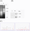
Purification of the 12-p DNA-binding protein using a tandem DNA-affinity columns. (A) Concatamerization of specific DNA oligonucleotides using self-priming PCR. PCR products with the concatamers of the 12-bp oligonucleotide were analyzed using agarose electrophoresis. 1, self-primer PCR products; M, DNA ladder. (B) SDS-PAGE analysis of the purified 12-bp DNA-binding protein. 1, the purified protein; M, protein markers. (C) Identification of the captured proteins using mass spectrometry, two representative peptide mass fingerprint profiles VPWFQYPIIYDIR (A) and FNASQLITQR (B).
Figure 3
Figure 3
Evolutional analysis of the prohibitin 2 gene family. (A) Multiple sequence alignment of the deduced amino acid sequence of the Sf-PHB2 gene with 9 ancient PHB2 family members. These sequences are available from the GenBank under the accessions: Dm = Drosophila melanogaster (NP_725832.2), Sf = Spodoptera frugiperda (HQ337624), Pm = Penaeus monodon (ACD13589.1), Hu = Homo sapiens (NP_009204.1), Cq = Culex quinquefasciatus (EDS26618.1), Nv = Nematostella vectensis (XP_001634411.1), Ga = Gallus gallus (NP_001074354.1), Bm = Bombyx mori L (NP_001040326.1), Ec = Equus caballus (XP_001497915.1), Xe = Xenopus (NP_001016551.1). (B) Sequence homology of the prohibitin2. The matrix of the percentage similarities of the amino acids for the 10 members of the PHB2 family. (C) Phylogenetic analysis of the PHB2 protein family members.
Figure 4
Figure 4
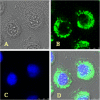
Intracellular localizations of the Sf-PHB2 in the Sf9 cells. (A) Normal Sf9 cells. (B) The Sf-PHB2 was visualized using mouse antibody against Sf-PHB2 and FITC-conjugated goat anti-mouse IgG antibody. (C) The nuclei were visualized by counterstaining with DAPI. (D) Merged FITC and DAPI signals. Original magnification: 400 x. bar = 1 μm.
Figure 5
Figure 5
Sf-PHB2 specifically binds to the 12-bp sequence of the WSSV ie1 promoter. The biotin-labeled probe was incubated in the absence (lane 1) or presence (lanes 2-8) of the nuclear extracts from the Sf9 cells. For the competition experiments, an equal amount of unlabelled EBNA oligos (lane 6) or 2-, 5- and 10-fold molar excesses of the unlabelled probe (lanes 2, 3 and 4) were added to the binding reaction. Lanes 7 and 8, the anti-PHB2 antibody-mediated supershift experiment. The arrow indicates the supershift.
Figure 6
Figure 6
Comparison of the early and late promoter subsequences between the WSSV ie1 gene and the insect baculovirus genes. The numbers indicate the number of nucleotides between the regulatory elements shown. ATG is the translation start of the coding sequence. The underlined sequences are the late promoter sequences. The boxed regions are the early promoter sequences, and the arrow is an early mRNA start site.

Similar articles

Cited by

References

    1. Chou HY, Huang CY, Wang CH, Chiang HC, Lo CF. Pathogenicity of a baculovirus infection causing white spot syndrome in cultured penaeid shrimp in Taiwan. Dis Aquat Org. 1995;23:165–173.
    1. Mayo MA. A summary of taxonomic changes recently approved by ICTV. Arch Virol. 2002;147:1655–1663. doi: 10.1007/s007050200039. - DOI - PubMed
    1. van Hulten MC, Witteveldt J, Peters S, Kloosterboer N, Tarchini R, Fiers M, Sandbrink H, Lankhorst RK, Vlak JM. The white spot syndrome virus DNA genome sequence. Virology. 2001;286:7–22. doi: 10.1006/viro.2001.1002. - DOI - PubMed
    1. Chen LL, Wang HC, Huang CJ, Peng SE, Chen YG, Lin SJ, Chen WY, Dai CF, Yu HT, Wang CH, Lo CF, Kou GH. Transcriptional analysis of the DNA polymerase gene of shrimp white spot syndrome virus. Virology. 2002;301:136–147. doi: 10.1006/viro.2002.1536. - DOI - PubMed
    1. Yang F, He J, Lin X, Li Q, Pan D, Zhang X, Xu X. Complete genome sequence of the shrimp white spot bacilliform virus. J Virol. 2001;75:11811–11820. doi: 10.1128/JVI.75.23.11811-11820.2001. - DOI - PMC - PubMed

Publication types

MeSH terms

LinkOut - more resources