. 2012 Nov;51(5):902-12.
doi: 10.1016/j.bone.2012.08.113. Epub 2012 Aug 16.

The collection of NFATc1-dependent transcripts in the osteoclast includes numerous genes non-essential to physiologic bone resorption

Affiliations

The collection of NFATc1-dependent transcripts in the osteoclast includes numerous genes non-essential to physiologic bone resorption

Julia F Charles et al. Bone. 2012 Nov.

Abstract

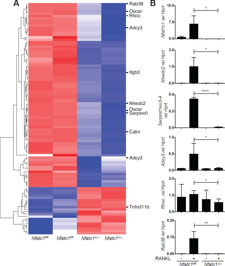
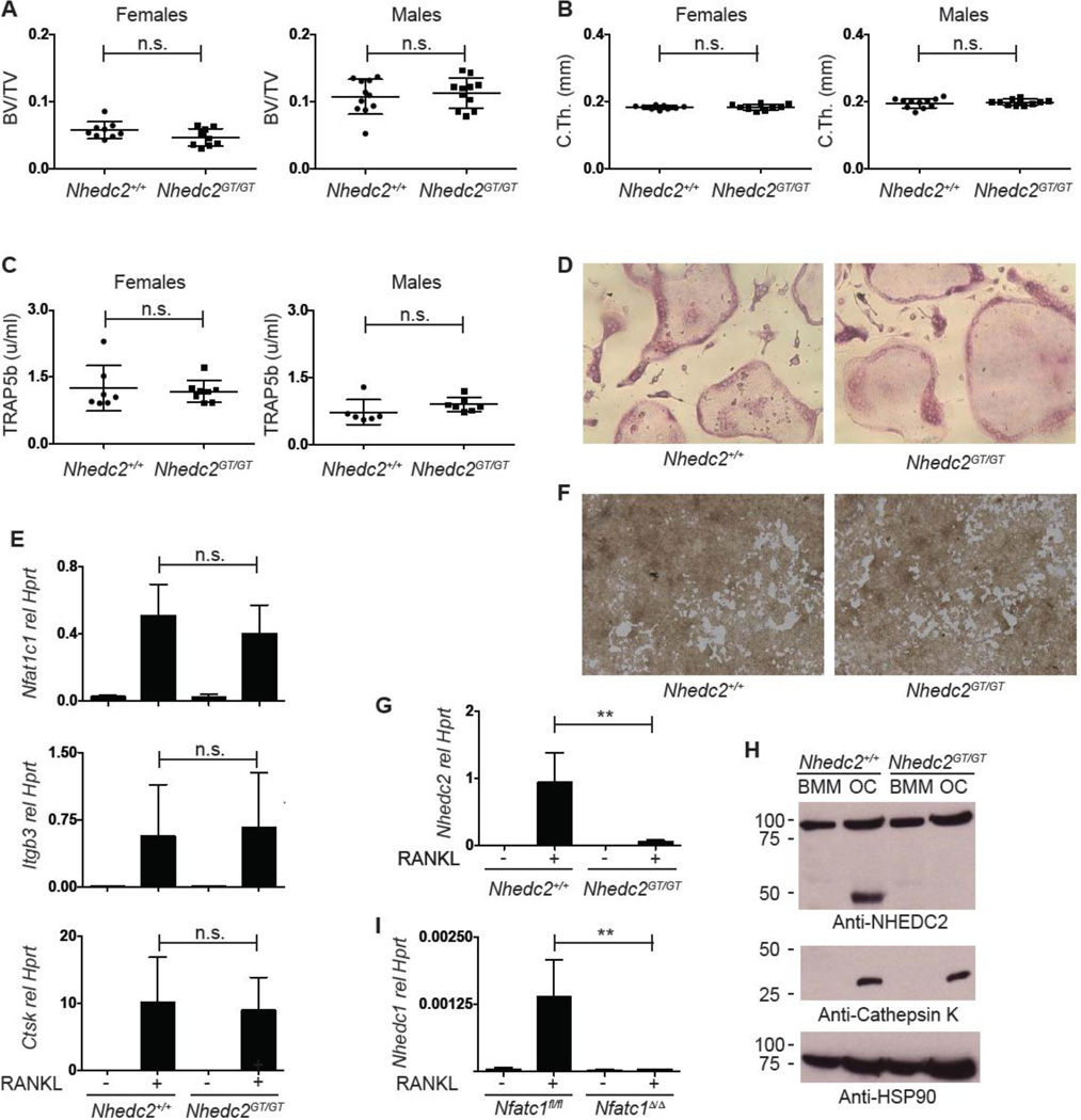
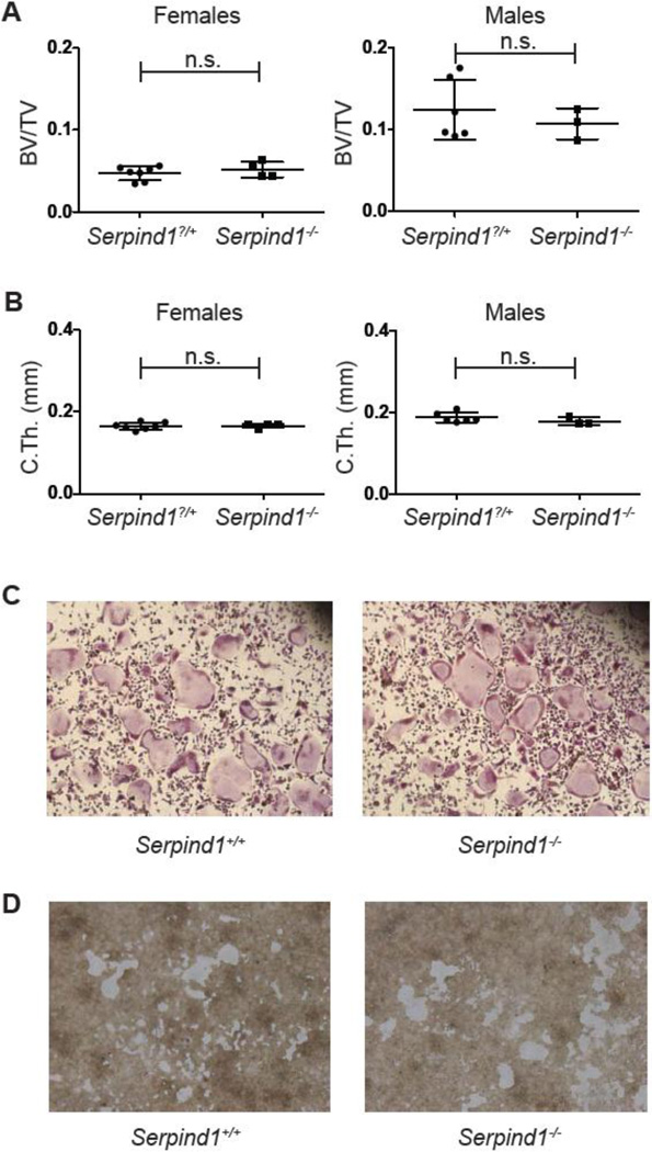
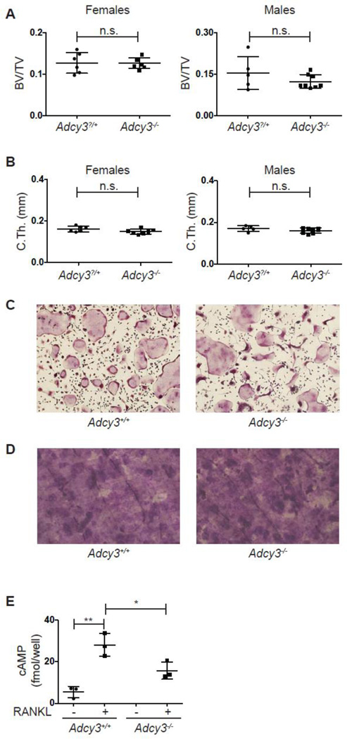
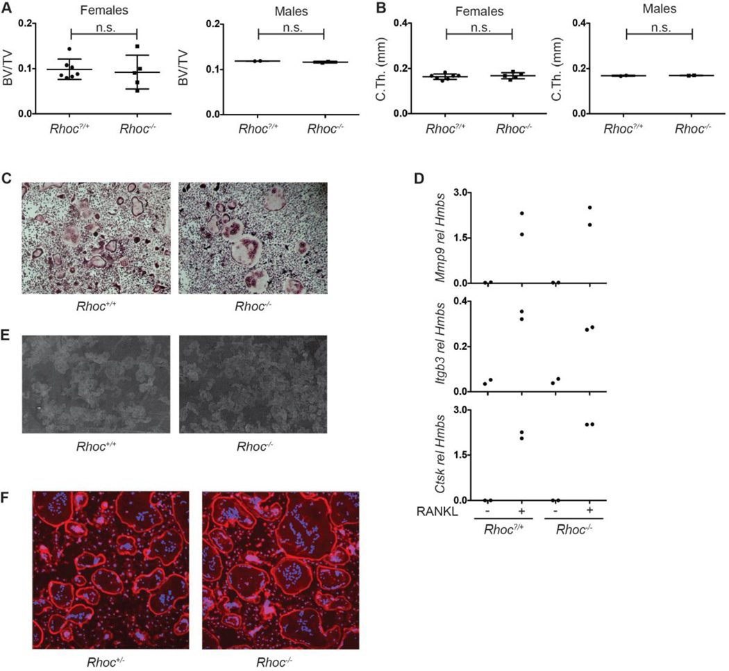
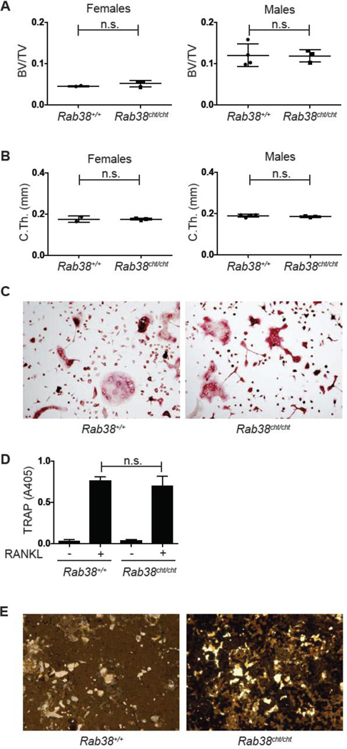
Osteoclasts are specialized secretory cells of the myeloid lineage important for normal skeletal homeostasis as well as pathologic conditions of bone including osteoporosis, inflammatory arthritis and cancer metastasis. Differentiation of these multinucleated giant cells from precursors is controlled by the cytokine RANKL, which through its receptor RANK initiates a signaling cascade culminating in the activation of transcriptional regulators which induce the expression of the bone degradation machinery. The transcription factor nuclear factor of activated T-cells c1 (NFATc1) is the master regulator of this process and in its absence osteoclast differentiation is aborted both in vitro and in vivo. Differential mRNA expression analysis by microarray is used to identify genes of potential physiologic relevance across nearly all biologic systems. We compared the gene expression profile of murine wild-type and NFATc1-deficient osteoclast precursors stimulated with RANKL and identified that the majority of the known genes important for osteoclastic bone resorption require NFATc1 for induction. Here, five novel RANKL-induced, NFATc1-dependent transcripts in the osteoclast are described: Nhedc2, Rhoc, Serpind1, Adcy3 and Rab38. Despite reasonable hypotheses for the importance of these molecules in the bone resorption pathway and their dramatic induction during differentiation, the analysis of mice with mutations in these genes failed to reveal a function in osteoclast biology. Compared to littermate controls, none of these mutants demonstrated a skeletal phenotype in vivo or alterations in osteoclast differentiation or function in vitro. These data highlight the need for rigorous validation studies to complement expression profiling results before functional importance can be assigned to highly regulated genes in any biologic process.


Figures

Fig. 1
Fig. 1. Identification of potential osteoclast regulators by expression profiling
(A) Microarray analysis of Nfatc1fl/fl and Nfatc1Δ/Δ OCPs stimulated for 3 d with MCSF and RANKL. Shown is a heat map depicting genes up or down regulated 6-fold. NFATc1-dependent transcripts selected for further study, as well as representative known regulators of osteoclast function are highlighted. (B) Real-time PCR analysis for the expression of the indicated genes in cultures of Nfatc1fl/fl and Nfatc1Δ/Δ OCPs stimulated with MCSF alone or with MCSF and RANKL to stimulate osteoclast differentiation. (*, p<0.05; **, p<0.01; ****, p<0.0001).
Fig. 2
Fig. 2. Analysis of Nhedc2GT/GT mice and osteoclasts
(A, B) Micro-CT quantification of femoral (A) metaphyseal trabecular BV/TV and (B) midshaft cortical thickness of 12–14 week old female and male Nhedc2+/+ (n=9, female; n=11, male) and Nhedc2GT/GT (n=9, female; n=11, male) mice. (C) Serum TRAP5b levels in 10-week old female and male Nhedc2+/+ (n=7, female; n=6, male) and Nhedc2GT/GT (n=8, female; n=7, male) mice. (D) TRAP stain of Nhedc2+/+ and Nhedc2GT/GT osteoclast cultures. (E) Real time PCR analysis for osteoclast marker genes in samples from Nhedc2+/+ (n=5) and Nhedc2GT/GT (n=4) OCPs stimulated with MCSF alone or with MCSF and RANKL to induce osteoclast differentiation. (F) resorption of calcium phosphate coated tissue culture wells incubated with Nhedc2+/+ and Nhedc2GT/GT osteoclasts. (G) Real time PCR analysis for Nhedc2 expression in Nhedc2+/+ (n=5) and Nhedc2GT/GT (n=4) OCPs stimulated with MCSF alone or with MCSF and RANKL to induce osteoclast differentiation. (H) Western blot for NHEDC2 protein on samples from Nhedc2+/+ and Nhedc2GT/GT OCPs stimulated with MCSF alone or with MCSF and RANKL to induce osteoclast differentiation. Immunoblots for Cathepsin K and HSP90 are shown as positive controls for osteoclast differentiation and protein loading, respectively. (I) Real time PCR analysis for Nhedc1 expression in Nfatc1fl/fl and Nfatc1Δ/Δ OCPs stimulated with MCSF alone or with MCSF and RANKL to induce osteoclast differentiation. (n.s., not significant; **, p<0.01.)
Fig. 3
Fig. 3. Analysis of Serpind1−/− mice and osteoclasts
(A, B) Micro-CT quantification of femoral (A) metaphyseal trabecular BV/TV and (B) midshaft cortical thickness of 12 week old female and male Serpind1?/+ (female, n=7; male, n=6) and Serpind1−/− (female, n=4; male, n=3) mice. (C) TRAP stain of Serpind1+/+ and Serpind1−/− osteoclast cultures. (D) resorption of calcium phosphate coated tissue culture wells incubated with Serpind1+/+ and Serpind1−/− osteoclasts. (n.s., not significant; **, p<0.01.)
Fig. 4
Fig. 4. Analysis of Adcy3−/− mice and osteoclasts
(A, B) Micro-CT quantification of femoral (A) metaphyseal trabecular BV/TV and (B) midshaft cortical thickness of 8-week old female and male Adcy3?/+ (female, n=6; male, n=5) and Adcy3−/− (female, n=7; male, n=8) mice. (C) TRAP stain of Adcy3+/+ and Adcy3−/− osteoclast cultures. (D) Toluidine blue stained bovine cortical bone slices cultured with Adcy3+/+ and Adcy3−/− osteoclasts. (E) cAMP levels in cultures of in Adcy3+/+ and Adcy3−/− OCPs stimulated with MCSF alone or MCSF and RANKL to induce osteoclast differentiation (n=3 per group). (n.s., not significant; *, p<0.05; **, p<0.01.)
Fig. 5
Fig. 5. Analysis of Rhoc−/− mice and osteoclasts
(A, B) Micro-CT quantification of femoral (A) metaphyseal trabecular BV/TV and (B) midshaft cortical thickness of 8–10 week old female and male Rhoc?/+ (female, n=7; male, n=2) and Rhoc−/− (female, n=5; male, n=2) mice. (C) TRAP stain of Rhoc+/+ and Rhoc−/− osteoclast cultures. (D) Real time PCR analysis for osteoclast marker genes in mRNA samples from Rhoc?/+ and Rhoc−/− OCPs stimulated with MCSF alone or with MCSF and RANKL to induce osteoclast differentiation (n=2 per group). Lack of differences in gene expression between Rhoc+/+ and Rhoc−/− osteoclasts is representative of 3 independent experiments. (E) Lectin-TRITC stained dentin slices cultured with Rhoc+/+ and Rhoc−/− osteoclasts. (F) Phalloidin stain of Rhoc+/+ and Rhoc−/− osteoclast cultures. (n.s., not significant). (n.s., not significant)
Fig. 6
Fig. 6. Analysis of Rab38cht/cht mice and osteoclasts
(A, B) Micro-CT quantification of femoral (A) metaphyseal trabecular BV/TV and (B) midshaft cortical thickness of 16 week old female and male Rab38+/+ (female, n=2; male, n=4) and Rab38cht/cht (female, n=3; male, n=3) mice. (C) TRAP stain of Rab38+/+ and Rab38cht/cht osteoclast cultures. (D) TRAP assay on the culture supernatants of Rab38+/+ and Rab38cht/cht (n=6 per genotype) OCPs stimulated with MCSF along or MCSF and RANKL to induce osteoclast differentiation. (E) Von kossa stain of calcium phosphate coated tissue culture wells incubated with Rab38+/+ and Rab38cht/cht osteoclasts. (n.s., not significant)

Similar articles

Cited by

References

    1. Sims NA, Gooi JH. Bone remodeling: Multiple cellular interactions required for coupling of bone formation and resorption. Semin Cell Dev Biol. 2008;19:444–451. - PubMed
    1. Miller PD. Denosumab: anti-RANKL antibody. Curr Osteoporos Rep. 2009;7:18–22. - PubMed
    1. Kendler DL, Roux C, Benhamou CL, Brown JP, Lillestol M, Siddhanti S, Man HS, San Martin J, Bone HG. Effects of denosumab on bone mineral density and bone turnover in postmenopausal women transitioning from alendronate therapy. J Bone Miner Res. 2010;25:72–81. - PubMed
    1. Rizzoli R, Reginster JY, Boonen S, Breart G, Diez-Perez A, Felsenberg D, Kaufman JM, Kanis JA, Cooper C. Adverse reactions and drug-drug interactions in the management of women with postmenopausal osteoporosis. Calcif Tissue Int. 89:91–104. - PMC - PubMed
    1. Woo SB, Hellstein JW, Kalmar JR. Narrative [corrected] review: bisphosphonates and osteonecrosis of the jaws. Ann Intern Med. 2006;144:753–761. - PubMed

Publication types

MeSH terms