. 2012 Dec 13:226:29-39.
doi: 10.1016/j.neuroscience.2012.08.045. Epub 2012 Sep 15.

Reversal of inhibition of putative dopaminergic neurons of the ventral tegmental area: interaction of GABA(B) and D2 receptors

Affiliations

Reversal of inhibition of putative dopaminergic neurons of the ventral tegmental area: interaction of GABA(B) and D2 receptors

S Nimitvilai et al. Neuroscience. .

Abstract

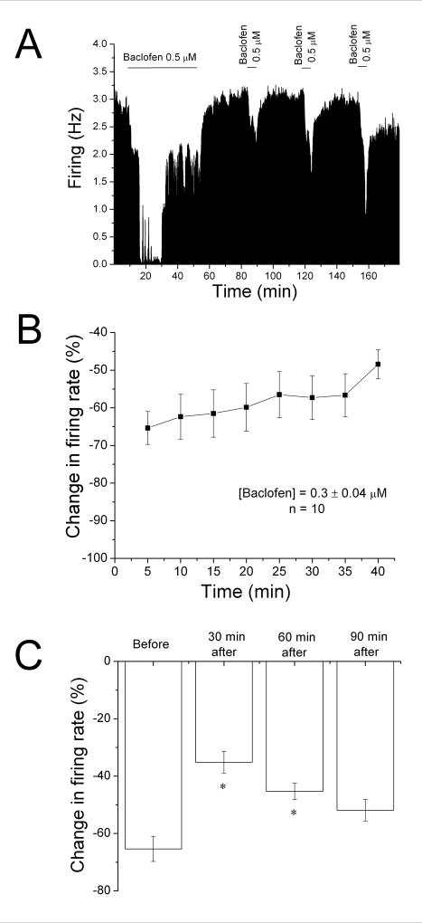
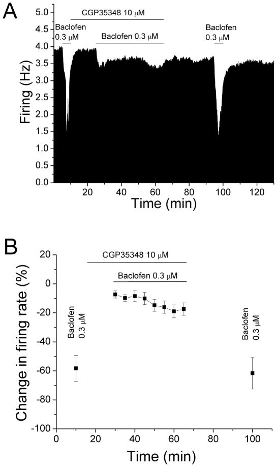
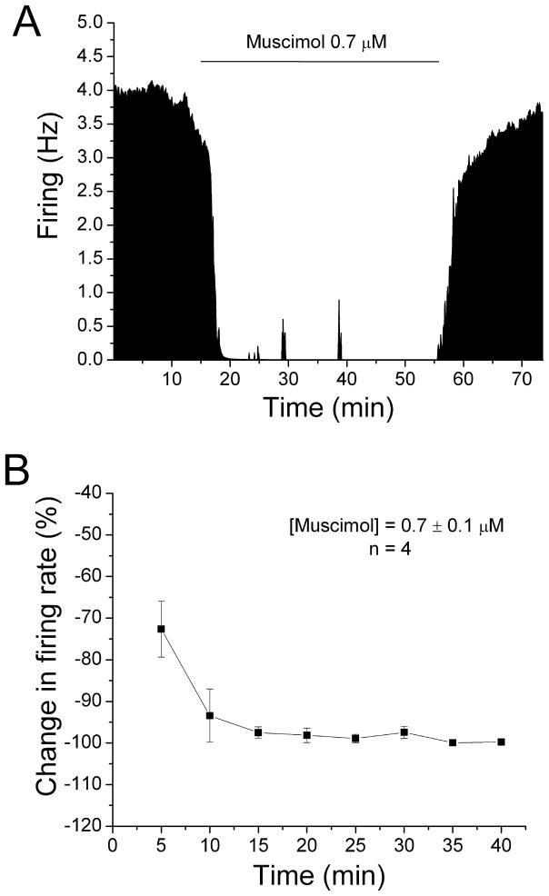
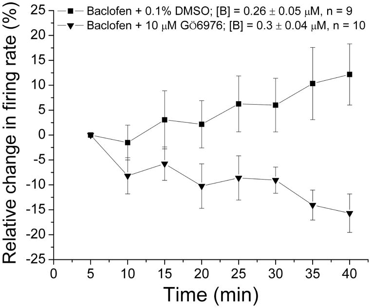
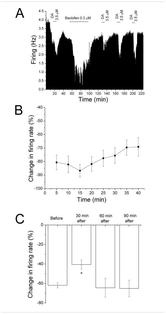
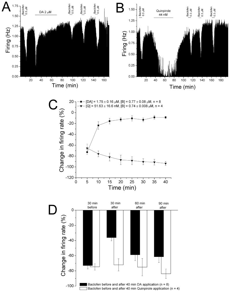
Neurons of the ventral tegmental area (VTA) are critical in the rewarding and reinforcing properties of drugs of abuse. Desensitization of VTA neurons to moderate extracellular concentrations of dopamine (DA) is dependent on protein kinase C (PKC) and intracellular calcium levels. This desensitization is called DA inhibition reversal, as it requires concurrent activation of D2 and D1-like receptors; activation of D2 receptors alone does not result in desensitization. Activation of other G-protein-linked receptors can substitute for D1 activation. Like D2 receptors, GABA(B) receptors in the VTA are coupled to G-protein-linked potassium channels. In the present study, we examined interactions between a GABA(B) agonist, baclofen, and dopamine agonists, dopamine and quinpirole, to determine whether there was some interaction in the processes of desensitization of GABA(B) and D2 responses. Long-duration administration of baclofen alone produced reversal of the baclofen-induced inhibition indicative of desensitization, and this desensitization persisted for at least 60 min after baclofen washout. Desensitization to baclofen was dependent on PKC. Dopamine inhibition was reduced for 30 min after baclofen-induced desensitization and conversely, the magnitude of baclofen inhibition was reduced for 30 min by long-duration application of dopamine, but not quinpirole. These results indicate that D2 and GABA(B) receptors share some PKC-dependent mechanisms of receptor desensitization.


Conflict of interest statement

Disclosures: None of the authors have any conflict of interest associated with the content of this report.

Figures

Figure 1
Figure 1. Reversal of baclofen inhibition and persistence of desensitization
A. Mean ratemeter graph of the effects of baclofen (0.5 μM) on the firing rate of a single DA VTA neuron. Vertical bars indicate the firing rate over 5 sec intervals. Horizontal bars indicate the duration of baclofen application (concentrations indicated above bar). Baclofen initially produced a decrease in firing rate of 84% at 5 min, and a maximum inhibition of 86%. By the end of the 40 min baclofen administration, the inhibition of firing rate subsided so that the firing rate was 50%. Then, Baclofen was washed out for 30 min. Following this washout period, baclofen was administered for 5 min at 30 min intervals, producing inhibitions of 27% at 30 min, 49% at 60 min, and 69% at 90 min after the long baclofen administration period. B. The mean firing rate of 10 DA VTA neurons during the 40 min baclofen administration in experiments similar to the one depicted in Figure 1A. Baclofen concentration was adjusted so that greater than 50% inhibition was produced; the mean concentration of baclofen that was administered was 0.3 ± 0.04 μM. There was a statistically significant reduction in baclofen-induced inhibition (One-way repeated measures ANOVA, F6,73 = 2.91, p < 0.05). C. The effect of 5 min application of baclofen during and after the 40-min application period. The inhibitory effect of baclofen was significantly reduced at 30 and 60 min after the 40 min baclofen administration period compared to the effect of baclofen at 5 min during that initial baclofen exposure (One-way ANOVA, F3,29 = 12.01, p < 0.05).
Figure 2
Figure 2. Baclofen inhibition reversal blocked by CGP35348
A. Mean ratemeter graph of the effects of baclofen and CGP35348 on the firing rate of a single DA VTA. Vertical bars indicate the firing rate over 5 sec intervals. Horizontal bars indicate the duration of baclofen (0.3 μM) or baclofen+CGP35348 (10 μM) application. Initially, a 5 min baclofen (0.3 μM) administration produced inhibition of 53.0%. During the 40 min application of baclofen and CGP35348, there was a decrease in firing rate of 10% at 5 min that further decreased to 13% by the end of the 40 min period. Baclofen was administered for 5 min after the cessation of baclofen and CGP35348 treatment, producing an inhibition of 60.9% at 30 min. B. The mean firing rate of DA VTA neurons in response to baclofen and CGP35348 in experiments similar to the one depicted in Figure 2A. The mean concentration of baclofen at 0.3 μM from experiments such as that shown in Figure 1 was used. The 5 min application of baclofen initially produced an inhibition in firing rate of 58.3%. In the presence of CGP35348 (10 μM), baclofen produced only 7% decrease in firing rate at 5 min, and this inhibition was further increased to 17% at 40 min. After the cessation of baclofen and CGP35348 treatment, baclofen produced an inhibition in firing rate of 61.6%. There was no significant reduction in baclofen-induced inhibition (paired t-test, p > 0.05).
Figure 3
Figure 3. No reversal of muscimol-induced inhibition
A. Mean ratemeter graph of the effects of muscimol (0.7 μM) on the firing rate of a single DA VTA neuron. Vertical bars indicate the firing rate over 5 sec intervals. Horizontal bars indicate the duration of muscimol application (concentrations indicated above bar). Muscimol initially produced a decrease in firing rate of 63.53% at 5 min, and the inhibition increase further so that by the end of the 40 min muscimol administration, the inhibition of firing rate was 99.96%. B. The mean firing rate of 4 DA VTA neurons during the 40 min muscimol administration in experiments similar to the one depicted in Figure 3A. Muscimol concentration was adjusted so that greater than 50% inhibition was produced at 5 min; the mean concentration of muscimol that was administered was 0.7 ± 0.1 μM. Muscimol produced an inhibition in firing rate of 72.6% at 5 min and the inhibition increased significantly over time (one-way repeated measures ANOVA, F(7,21) = 7.16, p < 0.05).
Figure 4
Figure 4. Baclofen inhibition reversal blocked by Gö6976
The mean firing rate of DA VTA neurons during the 40 min baclofen administration in experiments (similar to Figure 1B). In these experiments, the recording pipette included either DMSO vehicle (0.1%) or 10 μM Gö6976. Baclofen concentration was adjusted so that greater than 50% inhibition was produced. For recordings in which DMSO vehicle (0.1%) was included in the recording pipette, the mean concentration of baclofen that was administered was 0.3 ± 0.04 μM (■, n=9); for recordings in which 10 μM Gö6976 was included in the recording pipette, the mean concentration of baclofen that was administered was 0.26 ± 0.05 μM (▼, n=10). There was a significant difference in the response to baclofen over time for the two groups (Two-way ANOVA, F2,222 = 53.16, p < 0.05).
Figure 5
Figure 5. Desensitization to dopamine inhibition following reversal of baclofen inhibition
A. Mean ratemeter graph of the effects of dopamine (3.5 μM) and baclofen (0.3 μM) on the firing rate of a single DA VTA neuron. Vertical bars indicate the firing rate over 5 sec intervals. Horizontal bars indicate the duration of dopamine or baclofen application (concentrations indicated above bar). Initially, a 5 min dopamine administration produced inhibition of 59%. During the 40 min application, baclofen produced a decrease in firing rate of 73% at 5 min, with a maximum inhibition of 84%, and inhibition of only 57% by the end of the 40 min period. During the 30 min washout period, the baseline stabilized at a new, lower level. Dopamine was administered for 5 min at 30 min intervals after the cessation of baclofen treatment, producing inhibitions of 29% at 30 min, 43% at 60 min, and 45% at 90 min. B. The mean firing rate of DA VTA neurons during the 40 min baclofen administration in experiments similar to the one depicted in Figure 3A. Baclofen concentration was adjusted so that greater than 50% inhibition was produced; the mean concentration of baclofen that was administered was 0.29 ± 0.04 μM. There was a statistically significant reduction in baclofen-induced inhibition over time (One-way repeated measures ANOVA, F7,42 = 4.63, p < 0.05). C. The effect of 5 min application of dopamine during and after the 40-min baclofen application period. The inhibitory effect of dopamine was significantly reduced only at 30 min after the 40 min baclofen administration period compared to the effect of dopamine before the baclofen treatment (One-way ANOVA, F3,29 = 12.01, p < 0.05).
Figure 6
Figure 6. Desensitization to baclofen inhibition following reversal of dopamine inhibition
A. Mean ratemeter graph of the effects of baclofen (0.5 μM) and dopamine (2 μM) on the firing rate of a single DA VTA neuron. Vertical bars indicate the firing rate over 5 sec intervals. Horizontal bars indicate the duration of baclofen or dopamine application (concentrations indicated above bar). Initially, a 5 min baclofen administration produced inhibition of 68%. During the 40 min application, dopamine produced a decrease in firing rate of 62% at 5 min (which was the maximum), and inhibition of only 7% by the end of the 40 min period. Baclofen was administered for 5 min at 30 min intervals after the cessation of dopamine treatment, producing inhibitions of 31% at 30 min, 52% at 60 min, and 51% at 90 min. B. Mean ratemeter graph of the effects of baclofen (0.3 μM) and quinpirole (44nM) on the firing rate of a single DA VTA neuron. Vertical bars indicate the firing rate over 5 sec intervals. Horizontal bars indicate the duration of baclofen or quinpirole application (concentrations indicated above bar). Initially, a 5 min baclofen administration produced inhibition of 67%. During the 40 min application, quinpirole produced a decrease in firing rate of 63% at 5 min, and a maximum inhibition of 95%; inhibition at the end of the 40 min period was 87%. Baclofen was administered for 5 min at 30 min intervals after the cessation of quinpirole treatment, producing inhibitions of 55% at 30 min, 43% at 60 min, and 69% at 90 min. C. The mean firing rate of DA VTA neurons during the 40 min administration of dopamine (■, n=8) or quinpirole (▼, n=4) in experiments similar to the ones depicted in Figures 4A and 4B. Concentrations were adjusted so that greater than 50% inhibition was produced; the mean concentration of dopamine that was administered was 1.75 ± 0.16 μM; mean quinpirole concentration was 51.6 ± 16.6 nM. D. The effect of 5 min application of baclofen during and after the 40-min period of application of dopamine (Filled bars) or quinpirole (open bars). The inhibitory effect of baclofen was significantly reduced only at 30 min after the 40 min dopamine administration period compared to prior to the dopamine treatment (One-way ANOVA, F3,26 = 7.08, p < 0.05); there was no difference in baclofen-induced inhibition before and after the quinpirole treatment (One-way ANOVA, F3,9 = 0.67, p > 0.05).

Similar articles

Cited by

References

    1. Ackerman JM, Johansen PA, Clark D, White FJ. Electrophysiological effects of putative autoreceptor-selective dopamine agonists on A10 dopamine neurons. J Pharmacol Exp Ther. 1993;265:963–970. - PubMed
    1. Arora D, Hearing M, Haluk DM, Mirkovic K, Fajardo-Serrano A, Wessendorf MW, Watanabe M, Lujan R, Wickman K. Acute cocaine exposure weakens GABA(B) receptor-dependent G-protein-gated inwardly rectifying K+ signaling in dopamine neurons of the ventral tegmental area. J Neurosci. 2011;31:12251–12257. - PMC - PubMed
    1. Bartlett SE, Enquist J, Hopf FW, Lee JH, Gladher F, Kharazia V, Waldhoer M, Mailliard WS, Armstrong R, Bonci A, Whistler JL. Dopamine responsiveness is regulated by targeted sorting of D2 receptors. Proc Natl Acad Sci U S A. 2005;102:11521–11526. - PMC - PubMed
    1. Bofill-Cardona E, Kudlacek O, Yang Q, Ahorn H, Freissmuth M, Nanoff C. Binding of calmodulin to the D2-dopamine receptor reduces receptor signaling by arresting the G protein activation switch. J Biol Chem. 2000;275:32672–32680. - PubMed
    1. Bonci A, Malenka RC. Properties and plasticity of excitatory synapses on dopaminergic and GABAergic cells in the ventral tegmental area. J Neurosci. 1999;19:3723–3730. - PMC - PubMed

Publication types

MeSH terms