. 2012 Dec 13:226:130-44.
doi: 10.1016/j.neuroscience.2012.09.008. Epub 2012 Sep 15.

Spontaneous activity in the developing gerbil auditory cortex in vivo involves GABAergic transmission

Affiliations

Spontaneous activity in the developing gerbil auditory cortex in vivo involves GABAergic transmission

V C Kotak et al. Neuroscience. .

Abstract

A salient feature of the developing brain is that spontaneous oscillations (SOs) and waves may influence the emergence of synaptic connections. While GABA produces depolarization and may support SOs in the neurons of developing rodents, it elicits hyperpolarization and diminishes SOs in developing gerbil auditory cortex (ACx). Therefore, we asked whether SOs exist in developing gerbil ACx in vivo and if GABAergic involvement can be manipulated. In vivo extracellular recordings in P3-5 ACx revealed SOs with longer burst durations and shorter inter-event intervals compared to ACx SOs in slices. ACx was then validated by gross anatomical features and lesions created at the in vivo recording site that corresponded with the electrophysiological coordinates of thalamorecipient ACx in slices. Further, NeuroVue Red, a lipophilic dye loaded at the in vivo recording sites, stained anatomically identifiable fiber tracks between the ACx and the auditory thalamus, medial geniculate body (MG). Separately, to chronically perturb GABAergic role in SOs, P2-5 pups were administered daily with GABA(A) receptor blocker, bicuculline (BIC). We then recorded from P14-17 ACx neurons in slices generated after hearing onset. ACx neurons from BIC-administered pups exhibited spontaneous action potentials in contrast to subthreshold synaptic potentials in neurons from sham-injected animals. Finally, to elucidate whether the gap junction blocker mefloquine (MFQ) previously shown to dampen ACx SOs in slices affected GABAergic transmission, MFQ was acutely applied in P3-5 slices while spontaneous inhibitory postsynaptic currents (sIPSCs) were recorded. Whereas MFQ increased the amplitude and frequency of sIPSCs in ACx neurons, the broad-spectrum gap junction blocker carbenoxolone decreased sIPSC amplitudes only. Together, we show that P2-5 gerbil ACx can endogenously generate SOs in vivo. Persistence of activity in ACx in P14-17 slices from pups administered with BIC at P2-5 implies that inhibitory GABAergic activity linked with gap-junction participates in the maturation of ACx.


Figures

Fig. 1
Fig. 1
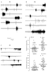
SOs exist in the P3-5 developing gerbil cortex. (A-C) Six consecutive sweeps (five in C) show recurring neuronal bursts recorded by a tethered wire electrode. Sporadic tonic spikes are also observed interspersed among the bursts. (D) Scatter plots of SO characteristics recorded in the ACx slice preparation (N=6) vs. those recorded in P2-5 pups using tethered wire preparations (N=7). Horizontal bars are means. Note that the in vivo intra-burst firing frequency is higher (upper panel) and the inter-burst interval is significantly longer (lower panel). In this and all subsequent figures, asterisks indicate that the differences are statistically significant. Details on the levels of significance for each figure panel marked by asterisks appear in the results section.
Fig. 2
Fig. 2
Validation of in vivo recording site. Following in vivo recordings, 500 μm thalamocortical slices were generated from fixed or fresh brains to validate recording sites (white arrows). Postnatal ages are indicated on top right of each panel. (A) The lesion site ~ 3.4 mm from the rostral end, corresponding to the region in fresh slices that respond to the stimulation of the auditory thalamus, MG, and thus ACx. This slice was derived from the animal recordings shown in Fig. 1. For anatomical landmarks, see B. (B) The lesion site indicates the lesion is ~ 1.4 mm from the rostral end, and therefore possibly somatosensory. MG: medial geniculate body, ACx: auditory cortex, H: hippocampus, Peri: perirhinal cortex. (C) The lesion site is 3.3 mm from the rostral end indicates a more temporal and deeper location and correspond to coordinates of thalamorecipient ACx as follows. (D). The lesion site 5.5 mm indicates a caudal location for the recording site, corresponding to the perirhinal cortex. This lesion had opened up appearing as a wedge during vibratome sectioning, and thus appears deeply cut. (E, F) Photomicrographs of thalamocortical slices from which coordinates of thalamorecipient ACx were measured. When the auditory thalamus (MG) was stimulated extracellularly (bipolar stimulating electrode immediately rostral to the MG at the emerging afferent fibers, blue arrows in the recordings indicate stimulus artifact, 5 mA/500 μS), brief field potentials are recorded, validating that the site of recordings was thalamorecipient ACx. Such responses are shown at the bottom panels of E and F where MG-evoked field responses are shown (blue traces). Note the several milliseconds time delay between the stimulus artifact and the onset of inward response. Recording sites were chosen 1 mm apart to illustrate the approximate caudorostral dimension of ACx. The gray traces represent failed synaptic transmission when subthreshold stimulation at lower intensities (100- 200 μA/100-200 μS duration, blue arrows indicate stimulus artifacts) did not elicit any detectable response. G and H are images of brain slices from 2 pups in which in which < 3 min in vivo recordings were first performed (Fig. 1B and C). Pups were then anesthetized and brain slices made immediately and taken to the recording chamber. When the auditory thalamus (MG) was stimulated field responses could be recorded in areas (asterisks) adjacent to the lesion (< 500 μm) created during the in vivo recordings (white arrows), confirming the in vivo recordings were from thalamorecipient auditory cortex (ACx). The bottom 2 traces show thalamically-evoked responses. Blue arrows indicate stimulus artifacts.
Fig. 3
Fig. 3
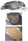
Identification of auditory cortex (ACx) in postnatal pups. Top panel. View of an adult gerbil brain showing key regions and vasculature landmarks around the ACx (Büdinger et al., 2000, 2006). The 2 major vasculature landmarks, the mid-cerebral artery (MCA) and inferior cerebral vein (ICV) are labeled respectively by white and black-arrowheads while the arrows point at their major branches. The outlined ACx is shown to contain primary auditory cortex (A1) as a gray shaded oblong while the adjacent smaller anterior and posterior association auditory cortices are similarly shaded. Scale bar = 2 mm; d: dorsal, r: rostral. Middle panel. Top view of a P14 gerbil brain that was transcardially perfused with ink, showing similar vascular landmarks as above (2A). The white arrow indicates the NeuroVue Red filter. Bottom panel. A P4 gerbil brain without any ink perfusion, showing the NeuroVue loaded filter (arrow). The rulers in the middle and bottom panels display millimeter scales.
Fig. 4
Fig. 4
Anatomical connections between cortex and MG. The lipophilic dye NeuroVue Red was loaded at the putative ACx area in intact brains and they were left for a month in a sucrose paraformaldehyde fixative. Fig 4A-D are fluorescent micrographs of 50 μm coronal slices passing through the thalomorecipient auditory cortex from several brains that show track-traced dye that had traveled to the MG several millimeters medial (MG, arrow heads). Arrows in each panel indicate the lesion mark that was created in the intact brains during insertion of the filter paper with the dye. Asterisks indicate en passant thalamocortical fibers. The first 3 images are from different P3 pups, while panel 4D micrograph is from a P4 pup. E is a de-saturated, enhanced fluorescent micrograph of a DAPI stained coronal section to identify some major landmarks around the ACx and MG. DG, dentate gyrus (labeled on contra side), 1, 2, and 3 are respectively CA1, CA2 and CA3 of the hippocampus, ACx, auditory cortex. Scale bar = 1.2 mm. The MG is seen as a bulbous structure between the ipsilateral dentate gyrus and CA3 (MG, arrowhead); its location corresponds with the NeuroVue-filled MG (A-D). F, G, and H are fluorescent micrographs from another P3 brain section respectively showing DAPI stained, NeuroVue stain and a merged image of the two. Arrow in H indicates the site of dye loading. Scale bar = 1 mm in F also for G and H.
Fig. 5
Fig. 5
GABA application blocks SOs. (A) Acute bath application of GABA (30 μl, ~1 mM effective concentration, arrowhead) dampens of SOs (top trace) leading to single action potentials followed by prolonged hyperpolarization in a P3 ACx neuron in brain slice. B. In the middle panel, a similar damping effect of GABA is shown for a P2 neuron. Action potentials within an SO are slowed and this is followed by prolonged hyperpolarization. C. In another slice (P3) a much lesser volume of GABA (10μm, effective concentration ~400 μM) leads to disruption of an SO (see second burst, arrow); action potentials are stunted, followed by hyperpolarization and a rebound SO. In a P5 neuron, 20 μl application of GABA (effective concentration ~ 700 μM) leads to similar block of spikes in an SO (arrow) followed by hyperpolarization. After recovery, SOs are irregular with some IPSPs. D. Application of 30 μl GABA (effective concentration ~ 1 mM) led to SO blockade (arrowhead) and prolonged hyperpolarization, in this case, ~ 10 mV. The cell resumed SOs about 4 minutes thereafter.
Fig. 6
Fig. 6
GABAergic transmission may influence the maturation of spontaneous activity in vivo. (A) A representative cell-attached recordings from a P16 ACx neuron previously (P2-5) administered with BIC displays some SO-like and tonic discharge. (B) Bar graphs representing mean intra-burst discharge rate (left panel) and tonic firing (right panel) from all BIC treated and age-matched control neurons. Asterisks indicate that the means are significantly different. The numbers above or within bars graphs represent N values.
Fig. 7
Fig. 7
Gap junctions affect spontaneous inhibitory currents in vitro: Facilitation of inhibitory function by MFQ. (A) The top panel shows two representative sweeps of sIPSCs recorded at VH=−60 mV in the presence of DNQX and AP-5 for 30 seconds each in a P4 neuron. (B) Note that 15′ after bath application of MFQ (25 μM), the sIPSC amplitude and frequency have increased. (C) Bar graphs summarizing the mean amplitude and frequency of sIPSCs all recorded neurons. Note that the amplitude of mean sIPSCs (left panel) and frequency (filled bar graphs) are greater after the application of MFQ. The numbers within bars graphs represent N values.
Fig. 8
Fig. 8
Gap junctions affect spontaneous inhibitory currents in vitro: Disinhibition by CBN may disrupt SOs. (A) A representative cell-attached recording from a control P5 ACx neuron shows 2 inward deflecting SO bursts and a few spikes between inter-burst interval (top trace). Bath application of CBN (200 μM) disrupts SO leading to greater discharge. (B) Bar graph representation of total discharge rate before and 15 minutes after the application of 200 μM CBN from all recorded L2/3 ACx neurons (mean Hz ± SEM).

Similar articles

Cited by

References

    1. Adelsberger H, Garaschuk O, Konnerth A. Cortical calcium waves in resting newborn mice. Nat Neurosci. 2005;8:988–990. - PubMed
    1. Baltz T, de Lima AD, Voigt T. Contribution of GABAergic interneurons to the development of spontaneous activity patterns in cultured neocortical networks. Front Cell Neurosci. 2010;4:1–17. - PMC - PubMed
    1. Ben-Ari Y. Excitatory actions of GABA during development: the nature of the nurture. Nat Rev Neursoci. 2002;3:728–739. - PubMed
    1. Ben-Ari Y, Gaiarsa JL, Tyzio R, Khazipov R. GABA: a pioneer transmitter that excites immature neurons and generates primitive oscillations. Physiol Rev. 2007;87:1215–1284. - PubMed
    1. Büdinger Heil P., Scheich H. Functional organization of auditory cortex in the Mongolian gerbil (Meriones unguiculatus). Anatomical subdivisions and corticocortical connections. Eur J Neurosci. 2000;12:2425–2451. - PubMed

Publication types

MeSH terms