Review
. 2013 Jun;70(12):2099-121.
doi: 10.1007/s00018-012-1156-5. Epub 2012 Sep 18.

Multiple roles for the actin cytoskeleton during regulated exocytosis

Affiliations
Review

Multiple roles for the actin cytoskeleton during regulated exocytosis

Natalie Porat-Shliom et al. Cell Mol Life Sci. 2013 Jun.

Abstract

Regulated exocytosis is the main mechanism utilized by specialized secretory cells to deliver molecules to the cell surface by virtue of membranous containers (i.e., secretory vesicles). The process involves a series of highly coordinated and sequential steps, which include the biogenesis of the vesicles, their delivery to the cell periphery, their fusion with the plasma membrane, and the release of their content into the extracellular space. Each of these steps is regulated by the actin cytoskeleton. In this review, we summarize the current knowledge regarding the involvement of actin and its associated molecules during each of the exocytic steps in vertebrates, and suggest that the overall role of the actin cytoskeleton during regulated exocytosis is linked to the architecture and the physiology of the secretory cells under examination. Specifically, in neurons, neuroendocrine, endocrine, and hematopoietic cells, which contain small secretory vesicles that undergo rapid exocytosis (on the order of milliseconds), the actin cytoskeleton plays a role in pre-fusion events, where it acts primarily as a functional barrier and facilitates docking. In exocrine and other secretory cells, which contain large secretory vesicles that undergo slow exocytosis (seconds to minutes), the actin cytoskeleton plays a role in post-fusion events, where it regulates the dynamics of the fusion pore, facilitates the integration of the vesicles into the plasma membrane, provides structural support, and promotes the expulsion of large cargo molecules.


Figures
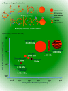
Fig. 1
Fig. 1
Cargo sorting into secretory vesicles and their morphological diversity. a Cargo sorting at the TGN. Molecules destined for secretion are sorted into regulated secretory vesicles at the TGN by two main mechanisms. According to the “sorting by entry” model, cargo molecules bind to specific receptors and are sorted into the nascent secretory vesicles, which detach from the TGN and are transported towards the plasma membrane. According to the “sorting by retention” model, cargo molecules freely enter newly formed vesicles that are released into the cytoplasm. These “immature” vesicles undergo an extensive “maturation” step, which is mediated by the removal of specific molecules and membranes via clathrin-coated vesicles. b Correlation between the sizes of the secretory vesicles and the duration of exocytosis. The sizes of the secretory vesicles ranges from 50 nm up to 2–3 μm in diameter and roughly correlate with the molecular weights of the cargo molecules (from 0.1 kDa up to >200 kDa). The duration of exocytosis, as measured from the opening of the fusion pore to the moment in which the secretory vesicle is completely integrated into the plasma membrane, inversely correlates with vesicles size. Small synaptic vesicles complete exocytosis in a time scale ranging from a microsecond to a few milliseconds. Dense core vesicles, lytic and insulin granules complete exocytosis within a few seconds, whereas large secretory granules in exocrine glands last for several seconds up to a minute, and Weibel–Palade bodies, lamellar bodies, and cortical granules last for several minutes
Fig. 2
Fig. 2
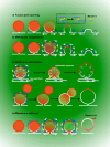
Steps in regulated exocytosis. Newly formed secretory vesicles are transported toward the cell periphery using microtubules and their associated motors kinesin and dynein (a). At the cell periphery, the secretory vesicles associate with the actin cytoskeleton by recruiting and activating myosin motors. Vesicles at the plasma membrane may dock using multiple mechanisms (b). The initial tether may be initiated by the exocyst complex, whereas the docking may be achieved by the interactions between the SNARE complex and Munc18. Both processes are coordinated by small GTPases, such as Rab3a, Rab8, and RalA (b). In order to become fusion-competent, docked secretory vesicles have to undergo priming that is mediated by proteins such as Mun13 and CAPS, which displaces Mun18 from the SNARE complex and facilitates the binding of complexin (c). Upon increase of Ca++ levels of, synaptotagmin, binds to the phospholipid at the plasma membrane, displaces complexin enabling the SNARE complex to promote the fusion of the bilayers (d). After fusion, vesicles can undergo full fusion, kiss and run/cavicapture or compound exocytosis
Fig. 3
Fig. 3
The actin cytoskeleton in pre-fusion steps. Before fusion, the actin cytoskeleton may provide a scaffold for anchoring secretory vesicles in close proximity to the plasma membrane (a), act as a physical barrier (b), or provide a track to transport the vesicles to the fusion site (c). These models are not mutually exclusive (d). A possible scenario envisions that under resting conditions, secretory vesicles may bind to the cortical actin cytoskeleton via multiple molecules (which include SynapsinI, myosin Va, and MARCKS) and be “functionally” entrapped by actin filaments. Upon stimulation, the small GTPase-mediated disassembly and reassembly of the actin cytoskeleton would transport the vesicles into a position suitable for tethering. Localized actin disassembly permits the vesicles to access the plasma membrane and requires the Ca++-mediated activation of actin severing proteins and the activation of GAPs for specific GTPases. Actin reassembly enables the activation of myosin motors and requires the GEF-mediated activation of selected GTPases and their effectors (d). Myosins and the actin cytoskeleton interact with the exocyst, facilitating tethering, and with the munc18/SNARE complex, regulating docking and priming (e)
Fig. 4
Fig. 4
The actin cytoskeleton in post-fusion steps. The contractile activity of the actomyosin complex is required to expand the fusion pore by exerting a force parallel to the plasma membrane (a) and/or to pull the membranes of the secretory vesicles toward the plasma membrane by exerting a force tangential to the surface of the vesicles (b). Both processes favor the expulsion of cargo molecules (a, b). In exocrine glands, the actomyosin complex may also serve as a scaffold to stabilize the secretory vesicles that are fused with the plasma membrane (c). This would prevent the uncontrolled expansion of the vesicles that is caused by either an increase in hydrostatic pressures, as shown to occur in exocrine glands in live animals, or fusion with adjacent secretory vesicles (c). The main myosin motor used for these functions is the non-muscle myosin II. The actomyosin complex may also control the retrieval of the vesicular membranes in order to maintain plasma membrane homeostasis. This process requires myosin II, Ie, and Ic (d)
Fig. 5
Fig. 5
Role of the actin cytoskeleton and the morphology of the secretory apparatus. The actin cytoskeleton performs two main functions in regulated exocytosis: (1) the tight control and coordination of the delivery of the secretory vesicles to their fusion sites at the plasma membrane, and (2) the regulation of the vesicle membranes dynamics after fusion. The first function is prevalent in cells in which the secretory vesicles have relatively small diameters, exhibit fast rates of fusion, and release small molecular weight cargo (synaptic vesicles, dense core vesicles, insulin, and lytic granules). From a biophysical point of view, the integration of these small vesicles into the plasma membrane is energetically favorable and proceeds very rapidly. In these systems, the actin cytoskeleton may provide an additional layer of regulation to prevent the uncontrolled delivery of secretory vesicles to fusogenic areas of the plasma membrane that may result in severe pathological conditions. The second function is more prevalent in cells in which secretory vesicles have large diameters and the post-fusion process is kinetically slow. Since the integration of large vesicles that fuse with curved membranes, such as canaliculi and ducts, is not energetically favorable and proceeds slowly (seconds to minutes), the recruitment of a contractile actomyosin complex onto the membranes of the secretory vesicles may provide the energy to facilitate this process. The contractile activity may also facilitate the expulsion of very large cargo, the retrieval of membranes by compensatory endocytosis, and to serve as a scaffold to stabilize them and prevent their disruption, which can be caused by either an increase in hydrostatic pressures fusion with adjacent secretory vesicles. For some small vesicles, such as in neuroendocrine cells, the actomyosin complex regulates the fusion pore and the fate of the vesicles after fusion (full fusion vs. kiss and run) without being recruited onto the membrane of the vesicles

Similar articles

Cited by

References

    1. Burgoyne RD, Morgan A. Secretory granule exocytosis. Physiol Rev. 2003;83(2):581–632. - PubMed
    1. De Matteis MA, Luini A. Exiting the Golgi complex. Nat Rev Mol Cell Biol. 2008;9(4):273–284. - PubMed
    1. Stockli J, Fazakerley DJ, James DE. GLUT4 exocytosis. J Cell Sci. 2011;124(Pt 24):4147–4159. - PMC - PubMed
    1. Bossi G, Griffiths GM. CTL secretory lysosomes: biogenesis and secretion of a harmful organelle. Semin Immunol. 2005;17(1):87–94. - PubMed
    1. Sudhof TC. Calcium control of neurotransmitter release. Cold Spring Harb Perspect Biol. 2012;4(1):a011353. - PMC - PubMed

Publication types

LinkOut - more resources