. 2012 Jul;31(6):360-7.
doi: 10.1016/j.matbio.2012.07.003. Epub 2012 Aug 6.

Quantitative microtiter fibronectin fibrillogenesis assay: use in high throughput screening for identification of inhibitor compounds

Affiliations

Quantitative microtiter fibronectin fibrillogenesis assay: use in high throughput screening for identification of inhibitor compounds

Bianca R Tomasini-Johansson et al. Matrix Biol. 2012 Jul.

Abstract

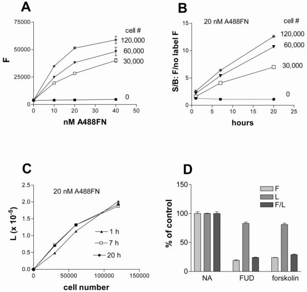
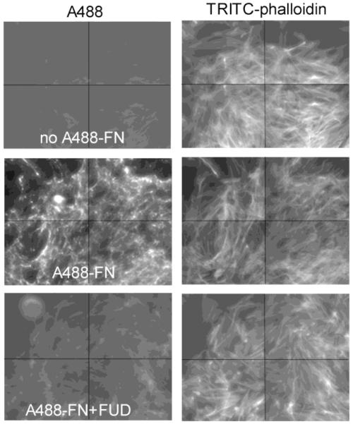
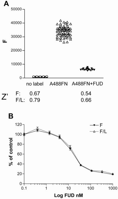
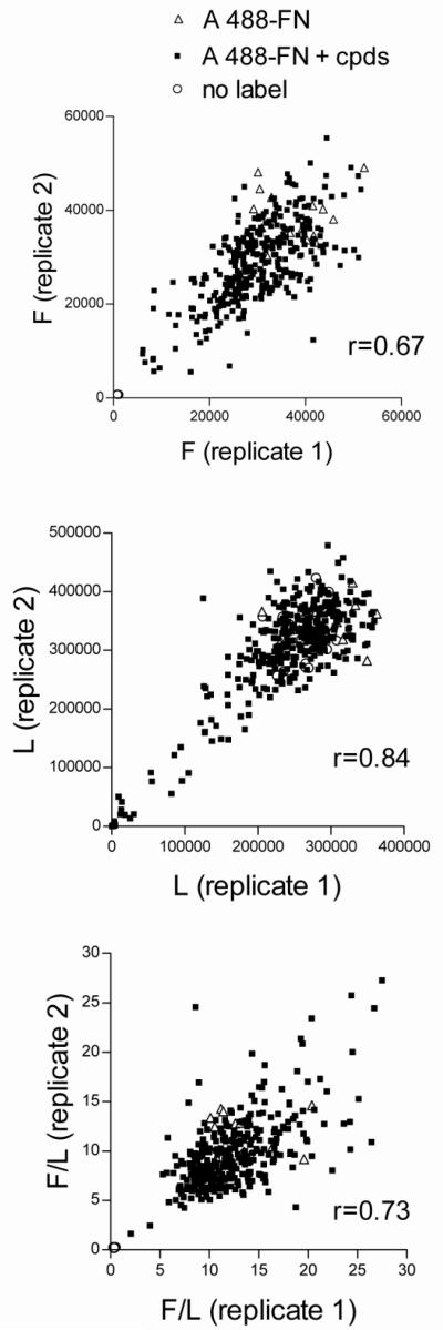
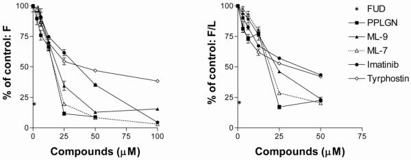
Fibronectin (FN) is a plasma glycoprotein that circulates in the near micromolar concentration range and is deposited along with locally produced FN in the extracellular matrices of many tissues. The control of FN deposition is tightly controlled by cells. Agents that modulate FN assembly may be useful therapeutically in conditions characterized by excessive FN deposition, such as fibrosis, inflammatory diseases, and malignancies. To identify such agents by high throughput screening (HTS), we developed a microtiter assay of FN deposition by human fibroblasts. The assay provides a robust read-out of FN assembly. Alexa 488-FN (A488-FN) was added to cell monolayers, and the total fluorescence intensity of deposited A488-FN was quantified. The fluorescence intensity of deposited A488-FN correlated with the presence of FN fibrils visualized by fluorescence microscopy. The assay Z' values were 0.67 or 0.54, respectively, when using background values of fluorescence either with no added A488-FN or with A488-FN added together with a known inhibitor of FN deposition. The assay was used to screen libraries comprising 4160 known bioactive compounds. Nine compounds were identified as non- or low-cytotoxic inhibitors of FN assembly. Four (ML-9, HA-100, tyrphostin and imatinib mesylate) are kinase inhibitors, a category of compounds known to inhibit FN assembly; two (piperlongumine and cantharidin) are promoters of cancer cell apoptosis; and three (maprotiline, CGS12066B, and aposcopolamine) are modulators of biogenic amine signaling. The latter six compounds have not been recognized heretofore as affecting FN assembly. The assay is straight-forward, adapts to 96- and 384-well formats, and should be useful for routine measurement of FN deposition and HTS. Screening of more diverse chemical libraries and identification of specific and efficient modulators of FN fibrillogenesis may result in therapeutics to control excessive connective tissue deposition.


Figures

Figure 1
Figure 1. Dependence of FN fibrillogenesis on cell number, concentration of A488-FN, time, and presence of known inhibitors
A) The indicated numbers of AH1F cells were plated in 96-well plates in DMEM containing 2% FBS, and following a 1-h incubation A488-FN was added to wells for varying concentrations and incubated for 20-h. Plates were washed and fluorescence was determined. B) The indicated numbers of cells were plated, given 20 nM (9 μg/ml) A488-FN, and then incubated for 1-, 7-, or 20-h at 37°C. Fluorescence (signal, S) was divided by fluorescence from wells without added labeled FN (background, B) to generate signal to background ratios (S/B). C) Luminescence values corresponding to cell number of wells described in panel B) following addition of Cell Titer Glo reagent. D) Cells (60,000 per well) were incubated 20-h with 20 nM Alexa 488-FN in the presence or absence of 500 nM FUD or 100 μM forskolin. Following PBS washes, fluorescence (F) and luminescence (L) were measured and F, L, and F/L of wells containing FUD or forskolin were expressed as percent of values in wells containing no inhibitor (NA, no additions). F values were obtained by subtracting fluorescence from cell monolayers without added label. Error bars = SEM of triplicate wells.
Figure 2
Figure 2. Imaging of FN fibrils and stress fibers
AH1F cells (15,000 per well) were dispensed in transparent 384-well plates with 20 nM Alexa 488-FN in the presence or absence of 500 nM FUD, and monolayers were incubated for 20-h. Microplates were washed twice with PBS containing Ca2+ and Mg2+, fixed, permeabilized and stained with TRITC-phalloidin to identify actin stress fibers. Wells were imaged on an inverted fluorescent microscope using Attovision software. Shown are montages from 4 areas captured per well of fluorescence for Alexa 488 showing FN fibrils inhibited by FUD (compare the middle left and lower left panels), and the corresponding panels to the right for TRITC channels showing stress fibers were unaffected by FUD treatment.
Figure 3
Figure 3. Validation of the assay in a HTS format
AH1F cells in 384-well plates were incubated with nothing, or 20 nM Alexa 488-FN without or with 1 μM FUD followed by 20-h incubation, washing, and quantitation of fluorescence (F) and luminescence (L). (A) Scatter plot of values from wells without or with A488-FN (n=80) or with A488-FN and FUD (n=16). F and F/L data were used for determination of the Z’ values shown under scatter plot. (B) Effect of increasing amounts of FUD on F and F/L as a percentage of positive control (A488-FN without FUD). Error bars = SEM of 16 wells per dose.
Figure 4
Figure 4. Replicate assays of cherry-picked compounds
AH1F cells were dispensed into duplicate 384-well plates in DMEM and incubated for 1 h before addition of 337 compounds chosen from among 4160 compounds as causing enhancement or inhibition of FN assembly. A488-FN, 20 nM, was added followed by 20-h incubation, washing, and quantitation of (A) fluorescence (F), (B) luminescence (L), and (C) normalized fluorescence (F/L). Values from each well in replicate plates are plotted against each other to demonstrate degree of inter-plate variability. Pearson correlation coefficients (r) were, respectively, 0.67, 0.84, and 0.73 for fluorescence, luminescence, and F/L.
Figure 5
Figure 5. Dose-dependence of FN assembly inhibition by four compounds identified from cherry pick testing
AH1F cells were plated into 96-well plates and incubated with increasing doses of piperlongumine (PPLGN), ML-9, imatinib mesylate or tyrphostin. ML-7 and 1 μM FUD were included as known inhibitors of FN assembly. Wells were analyzed for fluorescence (F) and luminescence (L). Points are the averages of 3 assays, with fluorescence (F) and fluorescence/luminescence (F/L) expressed as percentages of values obtained from A488-FN incubated on cells without compounds. Error bars = SD of 3 experiments, each assay point in triplicate.

Similar articles

Cited by

References

    1. Allen-Hoffmann BL, Mosher DF. Matrix assembly sites for exogenous fibronectin are decreased on human fibroblasts after treatment with agents which increase intracellular cAMP. J Biol Chem. 1987;262:14361–14365. - PubMed
    1. Blum G, Gazit A, Levitzki A. Development of new insulin-like growth factor-1 receptor kinase inhibitors using catechol mimics. J Biol Chem. 2003;278:40442–40454. - PubMed
    1. Brunner M, Millon-Fremillon A, Chevalier G, Nakchbandi IA, Mosher D, Block MR, Albiges-Rizo C, Bouvard D. Osteoblast mineralization requires beta1 integrin/ICAP-1-dependent fibronectin deposition. J Cell Biol. 2011;194:307–322. - PMC - PubMed
    1. Chen TC, Hinton DR, Zidovetzki R, Hofman FM. Up-regulation of the cAMP/PKA pathway inhibits proliferation, induces differentiation, and leads to apoptosis in malignant gliomas. Lab Invest. 1998;78:165–174. - PubMed
    1. Chernousov MA, Fogerty FJ, Koteliansky VE, Mosher DF. Role of the I-9 and III-1 modules of fibronectin in formation of an extracellular fibronectin matrix. J Biol Chem. 1991;266:10851–10858. - PubMed

Publication types

LinkOut - more resources