

p53-Independent regulation of p21Waf1/Cip1 expression and senescence by PRMT6
- PMID: 22987071
- PMCID: PMC3479215
- DOI: 10.1093/nar/gks858
p53-Independent regulation of p21Waf1/Cip1 expression and senescence by PRMT6
Abstract
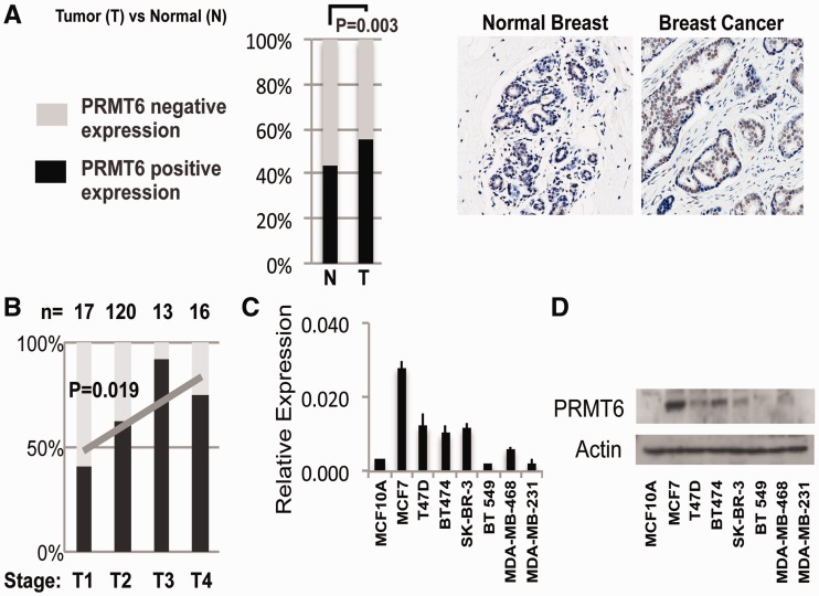
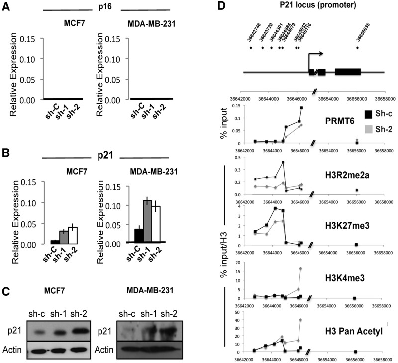
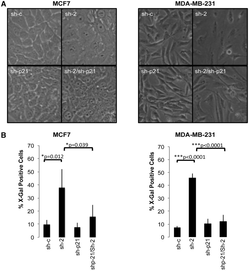
p21 is a potent cyclin-dependent kinase inhibitor that plays a role in promoting G1 cell cycle arrest and cellular senescence. Consistent with this role, p21 is a downstream target of several tumour suppressors and oncogenes, and it is downregulated in the majority of tumours, including breast cancer. Here, we report that protein arginine methyltransferase 6 (PRMT6), a type I PRMT known to act as a transcriptional cofactor, directly represses the p21 promoter. PRMT6 knock-down (KD) results in a p21 derepression in breast cancer cells, which is p53-independent, and leads to cell cycle arrest, cellular senescence and reduced growth in soft agar assays and in severe combined immunodeficiency (SCID) mice for all the cancer lines examined. We finally show that bypassing the p21-mediated arrest rescues PRMT6 KD cells from senescence, and it restores their ability to grow on soft agar. We conclude that PRMT6 acts as an oncogene in breast cancer cells, promoting growth and preventing senescence, making it an attractive target for cancer therapy.
Figures


Similar articles
-
The arginine methyltransferase PRMT6 regulates cell proliferation and senescence through transcriptional repression of tumor suppressor genes.Nucleic Acids Res. 2012 Oct;40(19):9522-33. doi: 10.1093/nar/gks767. Epub 2012 Aug 16. Nucleic Acids Res. 2012. PMID: 22904088 Free PMC article.
-
Cell cycle regulation by the PRMT6 arginine methyltransferase through repression of cyclin-dependent kinase inhibitors.PLoS One. 2012;7(8):e41446. doi: 10.1371/journal.pone.0041446. Epub 2012 Aug 20. PLoS One. 2012. PMID: 22916108 Free PMC article.
-
Depletion of B cell CLL/Lymphoma 11B Gene Expression Represses Glioma Cell Growth.Mol Neurobiol. 2016 Aug;53(6):3528-3539. doi: 10.1007/s12035-015-9231-1. Epub 2015 Jun 23. Mol Neurobiol. 2016. PMID: 26096706
-
p21 in cancer: intricate networks and multiple activities.Nat Rev Cancer. 2009 Jun;9(6):400-14. doi: 10.1038/nrc2657. Nat Rev Cancer. 2009. PMID: 19440234 Free PMC article. Review.
-
Overview of the PRMT6 modulators in cancer treatment: Current progress and emerged opportunity.Eur J Med Chem. 2024 Dec 5;279:116857. doi: 10.1016/j.ejmech.2024.116857. Epub 2024 Sep 12. Eur J Med Chem. 2024. PMID: 39276585 Review.
Cited by
-
Exploring the multifaceted antitumor activity of axitinib in lung carcinoids.Front Endocrinol (Lausanne). 2024 Jul 10;15:1433707. doi: 10.3389/fendo.2024.1433707. eCollection 2024. Front Endocrinol (Lausanne). 2024. PMID: 39050569 Free PMC article.
-
PRMT6 activates cyclin D1 expression in conjunction with the transcription factor LEF1.Oncogenesis. 2021 May 17;10(5):42. doi: 10.1038/s41389-021-00332-z. Oncogenesis. 2021. PMID: 34001852 Free PMC article.
-
PRMT6 serves an oncogenic role in lung adenocarcinoma via regulating p18.Mol Med Rep. 2020 Oct;22(4):3161-3172. doi: 10.3892/mmr.2020.11402. Epub 2020 Aug 3. Mol Med Rep. 2020. PMID: 32945431 Free PMC article.
-
PRMT6 facilitates EZH2 protein stability by inhibiting TRAF6-mediated ubiquitination degradation to promote glioblastoma cell invasion and migration.Cell Death Dis. 2024 Jul 23;15(7):524. doi: 10.1038/s41419-024-06920-2. Cell Death Dis. 2024. PMID: 39043634 Free PMC article.
-
PTEN arginine methylation by PRMT6 suppresses PI3K-AKT signaling and modulates pre-mRNA splicing.Proc Natl Acad Sci U S A. 2019 Apr 2;116(14):6868-6877. doi: 10.1073/pnas.1811028116. Epub 2019 Mar 18. Proc Natl Acad Sci U S A. 2019. PMID: 30886105 Free PMC article.
References
-
- Guccione E, Bassi C, Casadio F, Martinato F, Cesaroni M, Schuchlautz H, Luscher B, Amati B. Methylation of histone H3R2 by PRMT6 and H3K4 by an MLL complex are mutually exclusive. Nature. 2007;449:933–937. - PubMed
-
- Strahl BD, Briggs SD, Brame CJ, Caldwell JA, Koh SS, Ma H, Cook RG, Shabanowitz J, Hunt DF, Stallcup MR, et al. Methylation of histone H4 at arginine 3 occurs in vivo and is mediated by the nuclear receptor coactivator PRMT1. Curr. Biol. 2001;11:996–1000. - PubMed
-
- Santos-Rosa H, Schneider R, Bannister AJ, Sherriff J, Bernstein BE, Emre NC, Schreiber SL, Mellor J, Kouzarides T. Active genes are tri-methylated at K4 of histone H3. Nature. 2002;419:407–411. - PubMed
-
- Barski A, Cuddapah S, Cui K, Roh TY, Schones DE, Wang Z, Wei G, Chepelev I, Zhao K. High-resolution profiling of histone methylations in the human genome. Cell. 2007;129:823–837. - PubMed
MeSH terms
Substances
LinkOut - more resources
Full Text Sources
Other Literature Sources
Medical
Molecular Biology Databases
Research Materials
Miscellaneous
