. 2013 Apr 1;132(7):1624-32.
doi: 10.1002/ijc.27838. Epub 2012 Oct 11.

Single cell molecular recognition of migrating and invading tumor cells using a targeted fluorescent probe to receptor PTPmu

Affiliations

Single cell molecular recognition of migrating and invading tumor cells using a targeted fluorescent probe to receptor PTPmu

Susan M Burden-Gulley et al. Int J Cancer. .

Abstract

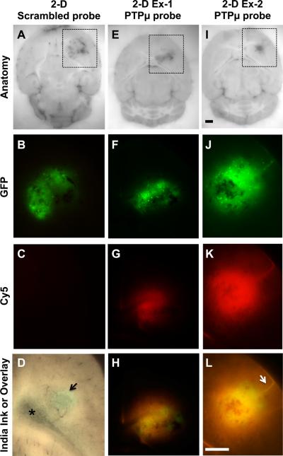
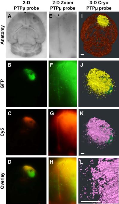
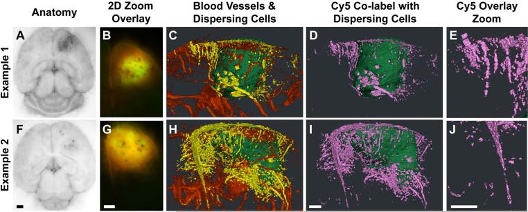
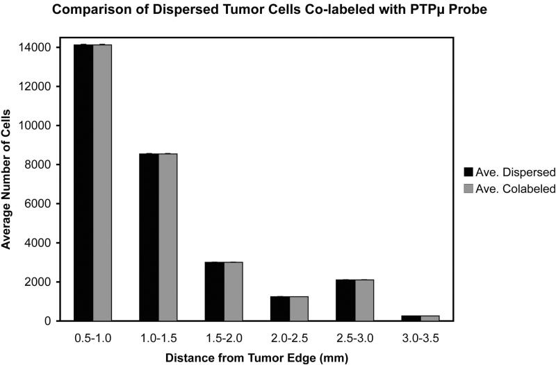
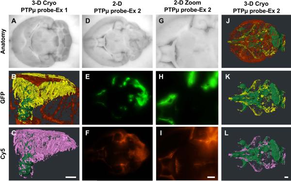
Detection of an extracellular cleaved fragment of a cell-cell adhesion molecule represents a new paradigm in molecular recognition and imaging of tumors. We previously demonstrated that probes that recognize the cleaved extracellular domain of receptor protein tyrosine phosphatase mu (PTPmu) label human glioblastoma brain tumor sections and the main tumor mass of intracranial xenograft gliomas. In this article, we examine whether one of these probes, SBK2, can label dispersed glioma cells that are no longer connected to the main tumor mass. Live mice with highly dispersive glioma tumors were injected intravenously with the fluorescent PTPmu probe to test the ability of the probe to label the dispersive glioma cells in vivo. Analysis was performed using a unique three-dimensional (3D) cryo-imaging technique to reveal highly migratory and invasive glioma cell dispersal within the brain and the extent of colabeling by the PTPmu probe. The PTPmu probe labeled the main tumor site and dispersed cells up to 3.5 mm away. The cryo-images of tumors labeled with the PTPmu probe provide a novel, high-resolution view of molecular tumor recognition, with excellent 3D detail regarding the pathways of tumor cell migration. Our data demonstrate that the PTPmu probe recognizes distant tumor cells even in parts of the brain where the blood-brain barrier is likely intact. The PTPmu probe has potential translational significance for recognizing tumor cells to facilitate molecular imaging, a more complete tumor resection and to serve as a molecular targeting agent to deliver chemotherapeutics to the main tumor mass and distant dispersive tumor cells.


Figures

Figure 1
Figure 1
CNS-1 glioma tumors and dispersing cells are labeled by the PTPμ probe. Unfixed mouse brains containing xenografts of GFP-expressing CNS-1 cells were cryo-imaged following in vivo labeling with scrambled probe (A–C) or PTPμ probe (E–L). Two-dimensional block face images are shown for brightfield (A,E,I), with boxed regions that correspond to the views shown for GFP fluorescence (tumor; B,F,J) and Cy5 fluorescence (scrambled or PTPμ probe; C,G,K) in each column. An overlay of GFP and Cy5 fluorescence demonstrates extensive labeling of the dispersed glioma cells with the PTPμ probe (H,L). In tumors with minimal cell dispersal (F), labeling with the PTPμ probe is localized to the main tumor (G,H). PTPμ probe labels diffusely dispersing cells in a tightly focused pattern when cells migrate as a stream along a defined structure, such as a blood vessel (K,L-see arrow). Brightfield image of a tumor from a brain that was perfused with India Ink (D) to illustrate leakiness of the vasculature. Asterisk in (D) indicates lateral ventricle. Scale bar in (I) represents 1 mm for panels (A, E, I) and the scale bar in (L) represents 1 mm for panels (B–D, F–H, J–L). Burden-Gulley, et al.
Figure 2
Figure 2
The PTPμ probe specifically labels CNS-1 glioma cells that have dispersed from the main tumor. Unfixed mouse brains containing xenografts of GFP-expressing CNS-1 cells were cryo-imaged following in vivo labeling with PTPμ probe (A–H). Two-dimensional block face images are shown for brightfield (A,E), GFP fluorescence (tumor; B,F) and Cy5 fluorescence (PTPμ probe; C,G). An overlay of GFP and Cy5 fluorescence demonstrates extensive labeling of the dispersed glioma cells with the PTPμ probe (D,H). A high magnification image demonstrates that the PTPμ probe labels a stream of dispersing tumor cells (F) at the midline (asterisk) (G,H). Three-dimensional reconstruction of the tumor labeled with PTPμ probe is illustrated as main tumor (green), and dispersing cells (yellow) in a magnified view (J). Complete vasculature for this brain specimen is also shown (I). Dispersed cells co-labeled with PTPμ probe are pseudo-colored pink (K). (L) A magnified view of the tumor shown in (K) illustrates co-labeled cells along the midline, which correspond with the labeled midline cells in the 2-D overlay image (H). Scale bar in (I) represents 1 mm for panels (A–D,I). Scale bar in (K) represents 500 μm for panels (J–K). Scale bar in (L) represents 500 μm for panels (E–H,L). Burden-Gulley, et al.
Figure 3
Figure 3
3-D views of dispersed cells from CNS-1 intracranial tumors labeled by the PTPμ probe. Unfixed mouse brains containing xenografts of GFP-expressing CNS-1 cells were cryo-imaged and reconstructed in 3 dimensions following in vivo labeling with the PTPμ probe. Two-dimensional block face images are shown for brightfield (A,F), and 2-D block face zoomed overlay of GFP (tumor) and Cy5 fluorescence (PTPμ probe; B,G) for two brain tumors. Three-dimensional reconstructions of the same tumor specimens showing the main tumor mass (pseudo-colored green), dispersed tumor cells (pseudo-colored yellow), and vasculature (pseudo-colored red) illustrate that the dispersing cells often migrate on blood vessels (C,H). The total dispersing cell population is extensively co-labeled with the PTPμ probe (D,I), as shown pseudo-colored pink. Magnified views from (D,I) illustrate that PTPμ co-labeled cells are detected several millimeters from the main tumor (E,J). Scale bar in F represents 1 mm for panels (A,F) and scale bar in G represents 1 mm for panels (B,G). Scale bar in I represents 500 μm for panels (C–D, H–I) and the scale bar in J represents 500 μm for panels (E, J). Burden-Gulley, et al.
Figure 4
Figure 4
Histogram of the average number of GFP positive dispersing cells co-labeled with the PTPμ probe per unit distance from the main tumor, +/− standard error (n=4 tumors analyzed). Burden-Gulley, et al.
Figure 5
Figure 5
Dispersing cells from LN-229 intracranial tumors are specifically labeled by the PTPμ probe. Unfixed mouse brains containing xenografts of GFP-expressing LN-229 cells were cryo-imaged and reconstructed in 3 dimensions following in vivo labeling with the PTPμ probe. Three-dimensional reconstruction of an LN229 tumor (A) shows the main tumor mass (pseudo-colored green), dispersed tumor cells (pseudo-colored yellow) and vasculature (pseudo-colored red) (B). PTPμ probe co-labeling of dispersing cells (pseudo-colored pink) was observed at great distances from the main tumor (C). A second tumor example where the LN229 tumor cells spread through the ventricles of the brain is shown (D–L). Two-dimensional block face images are shown for brightfield (D,G), GFP fluorescence (tumor; E,H) and Cy5 fluorescence (PTPμ probe; F,I). Comparison of GFP (E) and Cy5 (F) fluorescence in zoomed images (H,I) demonstrates extensive overlap in signal in this specimen. Three-dimensional reconstruction of the same tumor specimen is shown (J–L). In addition to the main tumor, the total dispersing cell population is extensively co-labeled with the PTPμ probe as shown pseudo-colored pink (L). Scale bar in (L) represents 500 μm for panels (A, D–F, J–L). Scale bar in (C) represents 500 μm for panels (B,C). Scale bar in (I) represents 500 μm for panels (G–I). Burden-Gulley, et al.

References

    1. Kohler BA, Ward E, McCarthy BJ, Schymura MJ, Ries LA, Eheman C, Jemal A, Anderson RN, Ajani UA, Edwards BK. Annual report to the nation on the status of cancer, 1975–2007, featuring tumors of the brain and other nervous system. J Natl Cancer Inst. 2011;103:714–36. - PMC - PubMed
    1. Stupp R, Mason WP, van den Bent MJ, Weller M, Fisher B, Taphoorn MJ, Belanger K, Brandes AA, Marosi C, Bogdahn U, Curschmann J, Janzer RC, et al. Radiotherapy plus concomitant and adjuvant temozolomide for glioblastoma. N Engl J Med. 2005;352:987–96. - PubMed
    1. Pichlmeier U, Bink A, Schackert G, Stummer W. Resection and survival in glioblastoma multiforme: an RTOG recursive partitioning analysis of ALA study patients. Neuro Oncol. 2008;10:1025–34. - PMC - PubMed
    1. Ewelt C, Goeppert M, Rapp M, Steiger HJ, Stummer W, Sabel M. Glioblastoma multiforme of the elderly: the prognostic effect of resection on survival. J Neurooncol. 2011;103:611–8. - PubMed
    1. Stummer W, Reulen HJ, Meinel T, Pichlmeier U, Schumacher W, Tonn JC, Rohde V, Oppel F, Turowski B, Woiciechowsky C, Franz K, Pietsch T. Extent of resection and survival in glioblastoma multiforme: identification of and adjustment for bias. Neurosurgery. 2008;62:564–76. discussion -76. - PubMed

Publication types