. 2012 Nov 1;11(21):3937-44.
doi: 10.4161/cc.21945. Epub 2012 Sep 17.

Understanding the origins of UV-induced recombination through manipulation of sister chromatid cohesion

Affiliations

Understanding the origins of UV-induced recombination through manipulation of sister chromatid cohesion

Shay Covo et al. Cell Cycle. .

Abstract

Ultraviolet light (UV) can provoke genome instability, partly through its ability to induce homologous recombination (HR). However, the mechanism(s) of UV-induced recombination is poorly understood. Although double-strand breaks (DSBs) have been invoked, there is little evidence for their generation by UV. Alternatively, single-strand DNA lesions that stall replication forks could provoke recombination. Recent findings suggest efficient initiation of UV-induced recombination in G1 through processing of closely spaced single-strand lesions to DSBs. However, other scenarios are possible, since the recombination initiated in G1 can be completed in the following stages of the cell cycle. We developed a system that could address UV-induced recombination events that start and finish in G2 by manipulating the activity of the sister chromatid cohesion complex. Here we show that sister-chromatid cohesion suppresses UV-induced recombination events that are initiated and resolved in G2. By comparing recombination frequencies and survival between UV and ionizing radiation, we conclude that a substantial portion of UV-induced recombination occurs through DSBs. This notion is supported by a direct physical observation of UV-induced DSBs that are dependent on nucleotide excision repair. However, a significant role of nonDSB intermediates in UV-induced recombination cannot be excluded.


Figures

None
Figure 1. Mechanisms of DNA damage induced recombination. (A) Recombination induced by a direct DSB. (B) Recombination induced by a secondary (indirect) DSB. (C) Recombination induced by gaps or nicks. The first stage in homologous recombination is processing of the primary lesion (I). For direct DSBs (A) it is resection. Adjacent lesions on opposite strands (represented as stars) are first converted into a DSB and then resected (B). Nicks or gaps (C) may be expanded without forming a DSB. The second step (II) is strand invasion, DNA synthesis and creation of a joint molecule. The third step (III) is resolution of homologous intermediates (for simplicity only one possibility of resolution is presented).
None
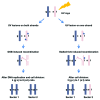
Figure 2. Analysis of UV-induced LOH for cells irradiated at G1. Cells that are heterozygous at a specific locus (“a” allele and “c” allele) are arrested at G1 and irradiated with UV (damage is represented by a star). Sectored colonies are selected after UV exposure. Each sector subcolony represents two sister chromatids. Therefore, the four chromatids are represented in the two sector colonies. Sequencing each sector can reveal which allele, “a” or “c,” was lost. If the number of sequence reads for “a” and “c” alleles is equal then heterozygosity is maintained. If there are no sequence reads for one of them, LOH has occurred. Through analysis of LOH in the two sectors it is possible to establish a 3:1 or 4:0 ratio for the four sister chromatids (see text and ref. 17).
None
Figure 3. Altering cohesin affects damage-induced recombination. (A) Cohesin channels recombination toward sister chromatids and away from homologous chromosomes in G2. This model describes the role of cohesin in preventing homologous recombination (see ref. ; the reproduction of the model was done according to the Creative Commons Attribution License -2.5 of PloS). (B) Tetraploid WT and the hypomorphic “simplex” cohesion mutant with one copy of MCD1 (for details see text and ref. 33) were irradiated with IR or UV in the G1 or G2 phases of the cell cycle. Presented are the frequencies of DNA damage-induced allelic recombination per unit radiation. For the x-axis, the unit dose was J/m2 for UV and krad for IR. Symbols: WT G1, white solid squares; WT G2, white striped squares; MCD1 simplex G1, gray solid squares; MCD1 simplex G2, gray striped squares. (C) Four scenarios that utilize temperature changes to alter cohesin activity in the mcd1–1 temperature-sensitive mutant are presented: 23◦C, light green; 30 C, red. In Scenarios 1, 2 and 4, the logarithmic growth of the culture and the G2 arrest occur at the same temperature. In Scenario 3, the cells are grown to logarithmic phase at 30 C and arrested with nocodazole for 90 min at the same temperature, but then the temperature is shifted to 23 C for 30 min before irradiation while remaining in nocadazole. (D) Recombination induced by 20 krad IR or 40 J/m2 for each Scenario in (C).
None
Figure 4. Significant contribution of DSBs to UV-induced recombination. (A) Survival curve of rad52-null cells arrested in G2 by nocodazole and treated with IR. The survival of rad52-null cells treated with 40 J/m2 is placed on the trend-line. The vertical gray line represents the dose in krad equivalent to 40 J/m2. (B) Dose-dependent IR-induced recombination is presented for two mcd1 mutants; MCD1 simplex (solid line) and mcd1–1 (dashed line). The data for mcd1–1 is taken from Figure 3B, Scenario 2 and for MCD1 simplex from Figure 3A. Recombination induced by 40 J/m2 was calculated for both MCD1 mutants and placed on the trend-line. Vertical gray lines represent the dose in krad equivalent to 40 J/m2. (C) Detection of DSB formation after UVB and ionizing radiation are as previously reported., Briefly, overnight cultures of WT, mcd1–1 and rad14 haploid strains were grown at 30°C, arrested with nocododazole and then exposed to 40J/m2 UVB in ice-cold water. The cultures were then returned to YPDA containing nocodazole to incubate for up to 4 h (30°C). As a control, cells were irradiated with IR (5,10,20,40 krad) using a 137Cs irradiator and harvested immediately after irradiation. Cells were processed for PFGE analysis as described. Linear chromosomes III (details in text) were determined by Southern blotting with a CHA1 probe; the zone corresponding to linear chromosome III or its derivatives is marked by a rectangle. The solid arrow indicates a resected linear DNA molecule and the dashed arrow points to a possible linear dimer of chromosome III (details in text). (D) IR dose-dependent Southern blot signal from a chromosome III specific probe [represented as AU (Arbitrary Units)]. Gray solid and Gray empty circles represent the chromosome III southern signals obtained for WT and mcd1–1 cells irradiated with 40 J/m2 after 4 h, respectively (see lane 11 and 17 of panel 4C).

Similar articles

Cited by

References

    1. Resnick MA. The repair of double-strand breaks in DNA; a model involving recombination. J Theor Biol. 1976;59:97–106. doi: 10.1016/S0022-5193(76)80025-2. - DOI - PubMed
    1. Hicks WM, Kim M, Haber JE. Increased mutagenesis and unique mutation signature associated with mitotic gene conversion. Science. 2010;329:82–5. doi: 10.1126/science.1191125. - DOI - PMC - PubMed
    1. Hicks WM, Yamaguchi M, Haber JE. Real-time analysis of double-strand DNA break repair by homologous recombination. Proc Natl Acad Sci USA. 2011;108:3108–15. doi: 10.1073/pnas.1019660108. - DOI - PMC - PubMed
    1. Westmoreland J, Ma W, Yan Y, Van Hulle K, Malkova A, Resnick MA. RAD50 is required for efficient initiation of resection and recombinational repair at random, gamma-induced double-strand break ends. PLoS Genet. 2009;5:e1000656. doi: 10.1371/journal.pgen.1000656. - DOI - PMC - PubMed
    1. Andersen SL, Sekelsky J. Meiotic versus mitotic recombination: two different routes for double-strand break repair: the different functions of meiotic versus mitotic DSB repair are reflected in different pathway usage and different outcomes. Bioessays. 2010;32:1058–66. doi: 10.1002/bies.201000087. - DOI - PMC - PubMed

Publication types

Substances

LinkOut - more resources