. 2013 Feb;57(2):577-89.
doi: 10.1002/hep.26081.

Toll-like receptor 2 and palmitic acid cooperatively contribute to the development of nonalcoholic steatohepatitis through inflammasome activation in mice

Affiliations

Toll-like receptor 2 and palmitic acid cooperatively contribute to the development of nonalcoholic steatohepatitis through inflammasome activation in mice

Kouichi Miura et al. Hepatology. 2013 Feb.

Abstract

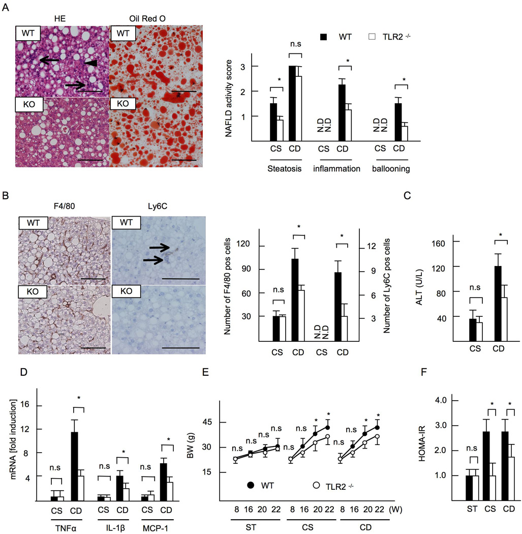
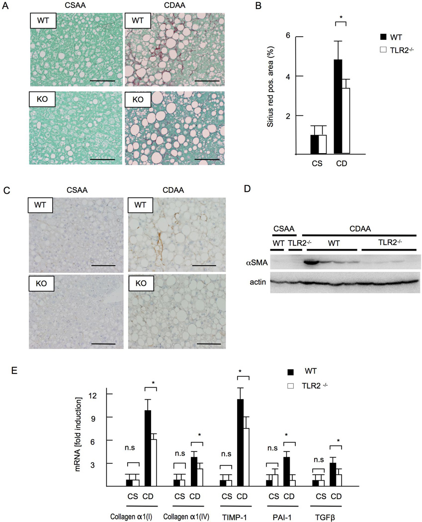
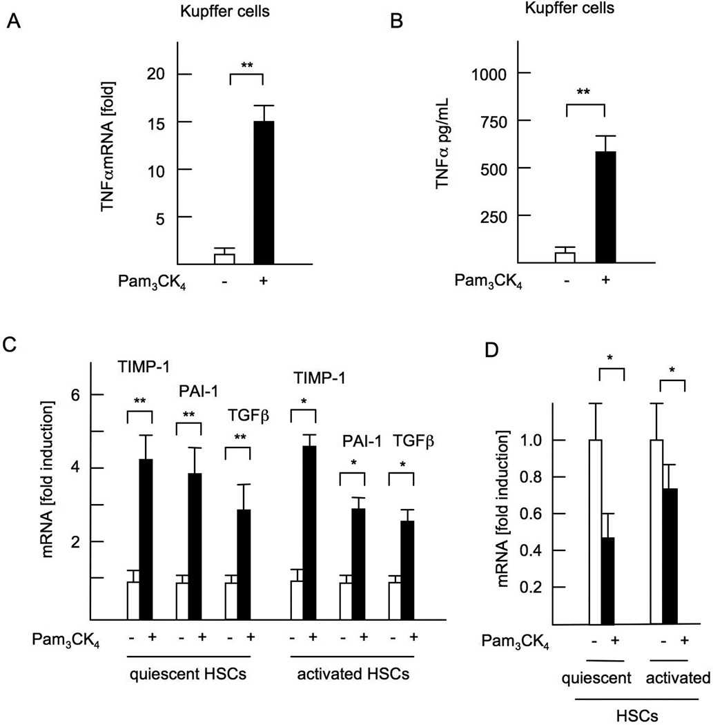
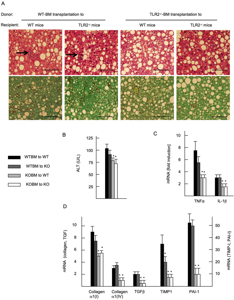
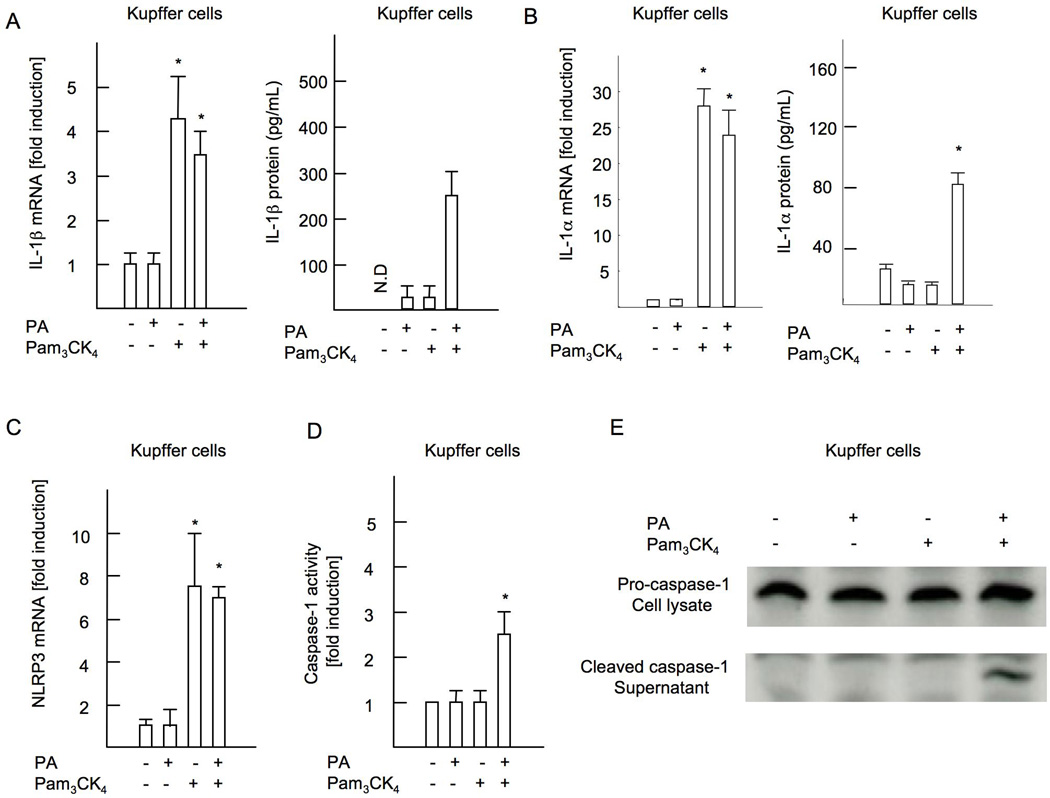
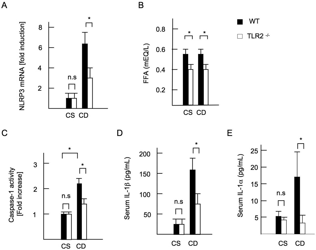
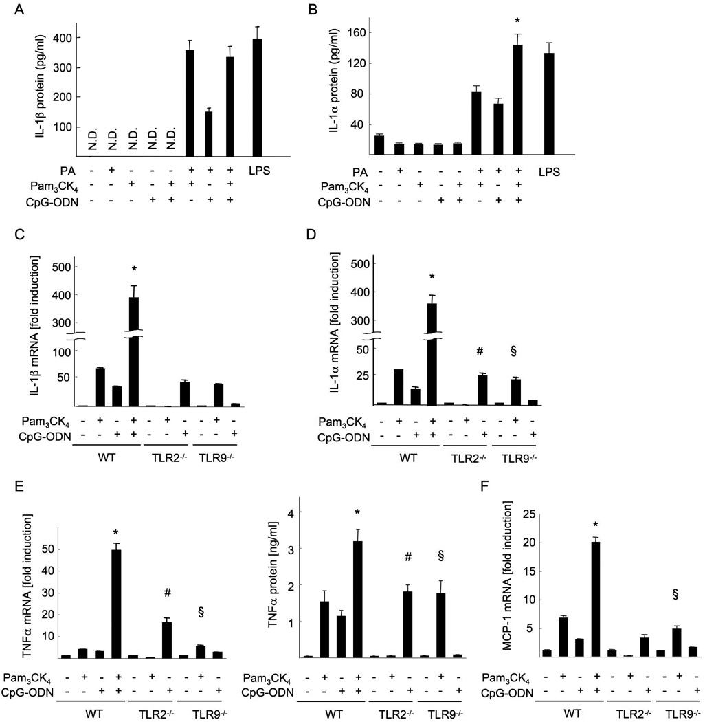
Innate immune signaling associated with Toll-like receptors (TLRs) is a key pathway involved in the progression of nonalcoholic steatohepatitis (NASH). Here we show that both TLR2 and palmitic acid are required for activation of the inflammasome, interleukin (IL)-1α, and IL-1β, resulting in the progression of NASH. Wild-type (WT) and TLR2(-/-) mice were fed a choline-deficient amino acid-defined (CDAA) diet for 22 weeks to induce NASH. Bone marrow-transplanted TLR2 chimeric mice were generated after the recipient mice were lethally irradiated. Kupffer cells and hepatic stellate cells (HSCs) were isolated from WT mice and stimulated with TLR2 ligand and/or palmitic acid. WT mice on the CDAA diet developed profound steatohepatitis and liver fibrosis. In contrast, TLR2(-/-) mice had suppressed progression of NASH. Although both Kupffer cells and HSCs respond to TLR2 ligand, TLR2 bone marrow chimeric mice demonstrated that Kupffer cells were relatively more important than HSCs in TLR2-mediated progression of NASH. In vitro, palmitic acid alone did not increase TLR2 signaling-target genes, including cytokines and inflammasome components in Kupffer cells and HSCs. The TLR2 ligand increased Nod-like receptor protein 3, an inflammasome component, in Kupffer cells but not in HSCs. In the presence of TLR2 ligand, palmitic acid did induce caspase-1 activation and release of IL-1α and IL-1β in Kupffer cells; however, these effects were not observed in HSCs. In vivo, WT mice on the CDAA diet showed increased caspase-1 activation in the liver and elevated serum levels of IL-1α and IL-1β levels, which were suppressed in TLR2(-/-) mice.

Conclusion: TLR2 and palmitic acid cooperatively activate the inflammasome in Kupffer cells and/or macrophages in the development of NASH.


Conflict of interest statement

Disclosures

No conflict of interest

Figures

Figure 1
Figure 1. TLR2−/− mice exhibit less inflammation in CDAA-induced NASH
WT and TLR2−/− mice were fed CSAA diet (CS) or CDAA diet (CD) for 22 weeks. Closed bars indicate WT mice and open bars represent TLR2−/− mice. (A, left) Hematoxylin and eosin (HE) and oil red O staining. Liver sections on CDAA diet are presented. The grades of steatosis were similar levels in WT and TLR2−/− mice whereas inflammatory cell infiltration (arrows) and hepatocyte ballooning (arrow head) were blunted in TLR2−/− mice. Original magnification,×400 for HE and oil red O staining. Bar 100µm. (A, right) NAFLD activity score. (B) Immunohistochemical staining for F4/80 and Ly6C. Liver sections on CDAA diet are presented. Infiltration of F4/80- and Ly6C-positive cells was suppressed in TLR2−/− mice. Original magnifications,×400 for F4/80,×600 for Ly6C. Bar 100 µm. (B, right) The numbers of F4/80-positive and Ly6C-positve cells. (C) Serum ALT levels. (D) mRNA expression of TNFα, IL-1β, and MCP-1. Genes were normalized to 18S RNA as an internal control. (E, F) Data on mice fed standard chow (ST) were included. (E) Body weight. (F) HOMA-IR. ND; not detected. Data represent mean ±SD, *p<0.05. n.s.; not significant.
Figure 2
Figure 2. TLR2−/− mice develop less liver fibrosis
WT and TLR2−/− mice were fed CSAA diet (CS) or CDAA diet (CD) for 22 weeks. Closed bars represent WT mice and open bars indicate TLR2−/− mice. (A) Sirius red staining. Original magnification,×400. Bar 100 µm. (B) Sirius red positive area. Liver fibrosis was attenuated in TLR2−/− mice. (C) Immunohistochemical staining for αSMA. Original magnification,×400. Bar 100 µm. (D) Western blotting for αSMA. (E) mRNA expression of fibrogenic genes. Genes were normalized to 18S RNA as an internal control. Data represent mean ±SD, *p<0.05. n.s.; not significant.
Figure 3
Figure 3. A synthetic TLR2 ligand activates both Kupffer cells and HSCs
WT Kupffer cells and HSCs were isolated and cultured in the presence of 5 µg/ml Pam3CK4. (A) mRNA expression of TNFα in Kupffer cells. (B) TNFα concentrations in the supernatant from WT Kupffer cell-culture. (C) mRNA expression of fibrogenic genes in quiescent HSCs and culture-activated HSCs. (D) Bambi mRNA expression in HSCs. Genes were normalized to 18S RNA as an internal control. Data represent mean ±SD, *p<0.05. **p<0.01. n.s.; not significant.
Figure 4
Figure 4. Hematopoietic cells including Kupffer cells are crucial for the development of liver inflammation and fibrosis in NASH
TLR2 bone marrow (BM) chimeric mice, in which hepatic macrophages were reconstituted with transplanted BM cells, were generated. Mice on CDAA diet are presented. (A) HE staining (upper) and Sirius red staining (lower). WT BM-transplanted mice show inflammatory cell infiltration (arrows), which are blunted in TLR2−/− BM transplanted mice. Liver fibrosis is attenuated in TLR2−/− BM transplanted mice. Original magnification, × 400. Bar 100 µm. (B) Serum ALT levels. (C) mRNA expression of TNFα and IL-1β. (D) mRNA expression of fibrogenic genes. Genes were normalized to 18S RNA as an internal control. Data represent mean ±SD, *p<0.05.
Figure 5
Figure 5. Palmitic acid activates inflammasome in cooperation with TLR2 ligand in Kupffer cells
Kupffer cells were isolated from WT mice and cultured in the presence of 5 µg/ml Pam3CK4 and/or 200 µM palmitic acid. (A) mRNA expression of IL-1β and active IL-1β in the supernatant. (B) mRNA expression of IL-1α and active IL-1α in the supernatant. (C) mRNA expression in NLRP3. Genes were normalized to 18S RNA as an internal control. (D) Caspase-1 activity. (E) Immunobotting for the active form of caspase-1 is shown. N.D; not detected. Data represent mean ±SD, *p<0.05.
Figure 6
Figure 6. Inflammasome activation is abolished in TLR2−/− mice
WT and TLR2−/− mice were fed control CSAA (CS) diet, and CDAA (CD) diet for 22 weeks. Closed bars represent WT mice and open bars indicate TLR2−/− mice. (A) Hepatic mRNA expression of NLRP3. Genes were normalized to 18S RNA as an internal control. (B) FFA concentrations in the portal vein. (C) Caspase-1 activation in the liver. (D) Serum IL-1β concentrations. (E) Serum IL-1α concentrations. N.D; not detected. Data represent mean ±SD, *p<0.05. n.s.; not significant.
Figure 7
Figure 7. TLR2 and TLR9 ligands synergistically produce inflammatory cytokines in Kupffer cells
Kupffer cells were isolated from WT, TLR2−/− and TLR9−/− mice, and cultured in the presence of 5 µg/ml Pam3CK4, 5 µg/ml CpG-ODN and/or 200 µM palmitic acid. (A,B,E) IL-1β (A), IL-1α (B) and TNFα (E, right) proteins in the supernatant. (C-F) mRNA expression of IL-1β (C), IL-1α (D), TNFα (E, left) and MCP-1 (F). Genes were normalized to 18S RNA as an internal control. N.D; not detected. Data represent mean ±SD, *; significant differences to WT KCs treated with Pam3CK4 or CpG-ODN, #; significant differences to WT KCs treated with CpG-DNA, §; no significant difference with WT KCs treated with Pam3CK4.

References

    1. Starley BQ, Calcagno CJ, Harrison SA. Nonalcoholic fatty liver disease and hepatocellular carcinoma: a weighty connection. Hepatology. 2010;51:1820–1832. - PubMed
    1. Park EJ, Lee JH, Yu GY, He G, Ali SR, Holzer RG, Osterreicher CH, et al. Dietary and genetic obesity promote liver inflammation and tumorigenesis by enhancing IL-6 and TNF expression. Cell. 2010;140:197–208. - PMC - PubMed
    1. Maher JJ, Leon P, Ryan JC. Beyond insulin resistance: Innate immunity in nonalcoholic steatohepatitis. Hepatology. 2008;48:670–678. - PMC - PubMed
    1. Miura K, Seki E, Ohnishi H, Brenner DA. Role of toll-like receptors and their downstream molecules in the development of nonalcoholic Fatty liver disease. Gastroenterol Res Pract. 2010;2010:362847. - PMC - PubMed
    1. Kawai T, Akira S. TLR signaling. Semin Immunol. 2007;19:24–32. - PubMed

Publication types