. 2012 Sep 15;26(18):2103-17.
doi: 10.1101/gad.187807.112.

Sonic hedgehog acts cell-autonomously on muscle precursor cells to generate limb muscle diversity

Affiliations

Sonic hedgehog acts cell-autonomously on muscle precursor cells to generate limb muscle diversity

Claire Anderson et al. Genes Dev. .

Abstract

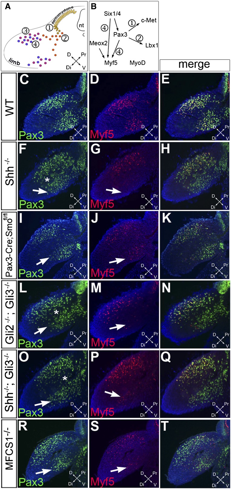
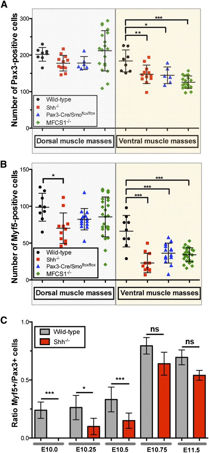
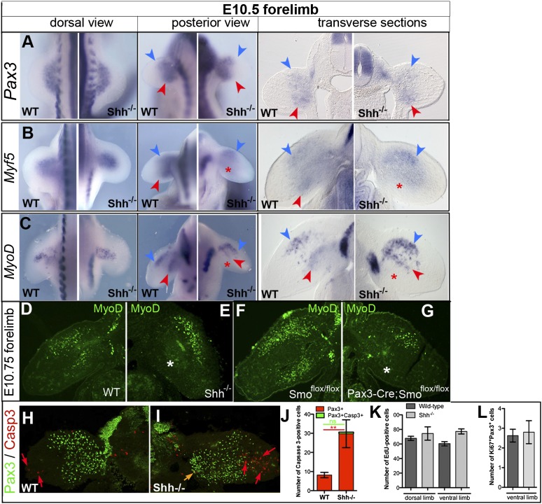
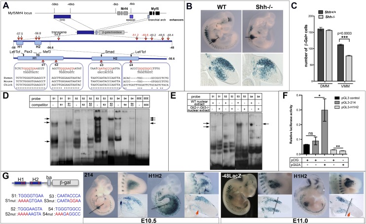
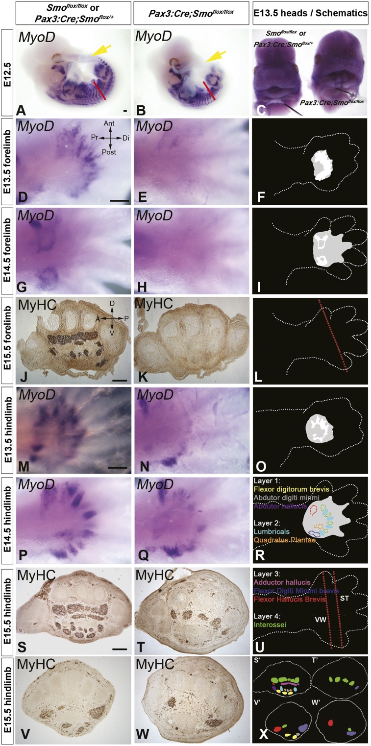
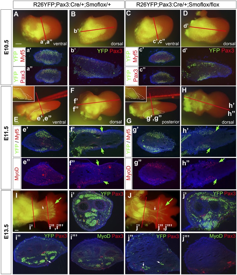
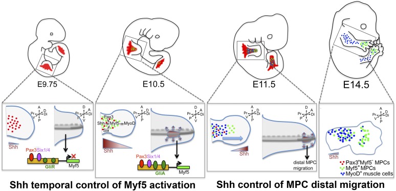
How muscle diversity is generated in the vertebrate body is poorly understood. In the limb, dorsal and ventral muscle masses constitute the first myogenic diversification, as each gives rise to distinct muscles. Myogenesis initiates after muscle precursor cells (MPCs) have migrated from the somites to the limb bud and populated the prospective muscle masses. Here, we show that Sonic hedgehog (Shh) from the zone of polarizing activity (ZPA) drives myogenesis specifically within the ventral muscle mass. Shh directly induces ventral MPCs to initiate Myf5 transcription and myogenesis through essential Gli-binding sites located in the Myf5 limb enhancer. In the absence of Shh signaling, myogenesis is delayed, MPCs fail to migrate distally, and ventral paw muscles fail to form. Thus, Shh production in the limb ZPA is essential for the spatiotemporal control of myogenesis and coordinates muscle and skeletal development by acting directly to regulate the formation of specific ventral muscles.


Figures

Figure 1.
Figure 1.
Shh signaling is required cell-autonomously for the initiation of myogenesis in ventral limb MPCs. (A) Schematic diagram representing the successive steps leading to limb myogenesis with delamination from the ventral dermomyotome (①), migration to the limb bud (②), proliferation and expansion of limb MPCs (③), and activation of the myogenic program with the activation of Myf5 and MyoD (④). (B) Gene network controlling limb myogenesis. (C–T) Immunofluorescence analysis of Pax3 (green) and Myf5 (red) expression in forelimb bud transverse sections from E10.5 wild-type (WT) (C–E), Shh−/− (F–H), Pax3:Cre;Smoflox/flox (I–K), Gli2−/−;Gli3−/− (L–N), Shh−/−;Gli3−/− (O–Q), and MFCS1−/− (R–T) embryos. White arrows indicate the location of ventral limb MPCs. White stars indicate incomplete segregation of Pax3+ MPCs in dorsal and ventral muscle masses. (nt) Neural tube; (D) dorsal; (V) ventral; (Pr) proximal; (Dt) distal. Magnification: 200×.
Figure 2.
Figure 2.
A specific defect in Myf5 activation in the absence of Shh signaling. (A) Quantification of the number of Pax3+ cells per transverse section in E10.5 dorsal (gray shaded area) and ventral (yellow shaded area) forelimbs of mutant mouse embryos. (B) Quantification of the number of Myf5+ cells per transverse section in E10.5 dorsal (gray shaded area) and ventral (yellow shaded area) forelimbs of mutant mouse embryos. (C) Ratio of the number of Myf5+ to Pax3+ cells of wild-type (black) and Shh−/− (red) ventral forelimb muscle masses between E10.0 and E11.5. Statistical analysis is described in Supplemental Table S1.
Figure 3.
Figure 3.
MPC failure to initiate the myogenic program in the absence of Shh is not due to defective proliferation or survival. Expression of Pax3 (A), Myf5 (B), and MyoD (C) was examined by in situ hybridization in E10.5 wild-type (WT) and Shh−/− forelimbs. Blue and red arrowheads indicate the dorsal and ventral muscle masses, respectively. Red asterisks indicate the loss of Myf5 and MyoD expression in the ventral muscle mass. Note the abnormal pattern of ventral MyoD expression in E10.5 Shh−/− forelimbs. Magnification: 200×. (D–G) MyoD distribution (green) in forelimbs from E10.5 wild-type (D), Shh−/− (E), Smoflox/flox (F), and Pax3:Cre;Smoflox/flox (G) mice. White asterisks indicate the loss of MyoD in ventral muscle masses. (H–J) Immunofluorescence analysis of Pax3 (green) and activated Caspase 3 (red) distribution in E10.5 wild-type (H) and Shh−/− (I) forelimbs. Red and orange arrows indicate apoptotic mesenchymal cells and MPCs, respectively. (J) Quantification of the average number of Caspase 3+ cells (red) and Pax3+Caspase 3+ cells (green) per section. One-way ANOVA analysis with Bonferroni post-test showed no significant difference in the number of Pax3+Caspase 3+ cells in wild-type and Shh−/− limbs. (K) Number of EdU+ cells per transverse section in the dorsal and ventral forelimb of E10.5 wild-type and Shh−/− embryos. Unpaired t-test analyses showed no significant differences. (L) Number of Pax3+Ki67+ cells per transverse section of E10.5 wild-type and Shh−/− ventral forelimbs.
Figure 4.
Figure 4.
Gli-binding sites present in the Myf5 limb enhancer are essential for Myf5 activation in the ventral forelimb. (A) Schematic representation of the Myf5/Mrf4 genomic locus indicating the position of the limb (dark blue), epaxial (deep blue) and branchial arch (light blue) enhancers. An enlargement of the 10-kb limb enhancer indicating the position of the 1.4-kb 5′ region demonstrated previously to drive Myf5 expression in the limb is shown. Putative Gli-binding sites are shown in black (with mismatch) and red (no mismatch). An enlargement of the 1.4-kb region is also represented, indicating the position of the putative Gli-binding sites 1–4 in relation to previously identified binding sites. The sequence conservation of sites 1–4 in humans, mice, and chicks is shown. (B) β-Gal staining of E10.5 transgenic embryos expressing LacZ under the control of the 10-kb Myf5 limb enhancer in a wild-type (WT) background (left) and Shh−/− background (right). Transverse sections through the forelimb are shown below. Note the reduced number of β-gal+ cells in the ventral limb. (C) Quantification of the number of β-gal+ cells present in the dorsal muscle mass (DMM) and ventral muscle mass (VMM) of wild-type and Shh−/− transgenic embryos. Unpaired t-test analysis was performed. (D) EMSA analysis using oligonucleotides encompassing sites 1–4 and nuclear extract from E10.5 embryos in the presence or not of cold competitor oligonucleotides (wild type or with mutated Gli-binding sites). “EEE” shows the gel shift with an oligonucleotide encompassing the Gli-–binding site from the Myf5 epaxial enhancer. Arrows point to specific complexes that are not competed by cold oligonucleotides with mutated Gli sites. (E) EMSA analysis as in D in the presence of wild-type or Gli2−/−;Gli3−/− nuclear extract. Arrows indicate complexes that disappear in the presence of Gli2−/−;Gli3−/− nuclear extract. (F) Transactivation assay in C2C12 cells using the 1.4-kb Myf5 enhancer with (H1H2) or without (214) Gli-binding sites 1–4 mutated in a luciferase reporter construct. Relative luciferase activity was determined after cotransfection of a control vector (pCIG) or a construct expressing a constitutively active form of Gli2 (pGli2A). (G) β-Gal staining of E10.5 and E11.0 transgenic embryos expressing LacZ under the control of the 1.4-kb enhancer with mutated Gli sites1–4 (H1H2) compared with control transgenic mice containing either the 10-kb Myf5 enhancer (−48LacZ) or the wild-type 1.4-kb enhancer (214). The left panel depicts the H1H2 construct and the mutations introduced into sites 1–4. Dashed lines indicate the boundary between proximal and distal MPC domains. Red arrows show the total absence of β-gal staining in the distal MPC domain.
Figure 5.
Figure 5.
Sonic hedgehog signaling is required cell-autonomously in limb MPCs for the formation of specific limb muscles. Whole-mount MyoD in situ mRNA hybridization (A–E,G,H,M–Q) or pan-myosin immunodetection on cryosections (J,K,S–W) of sibling (left column) and Pax3:Cre;Smoflox/flox (middle column) mouse embryos. (A,B) Whole mount of MyoD mRNA on Pax3:Cre;Smoflox/flox embryos and sibling E12.5 embryos showing lack of expression in Pax3:Cre;Smoflox/flox tail somites (yellow arrow) and reduction in trunk somites (red line). (C) Heads of E13.5 embryos showing the cleft lip and palette phenotype in Pax3:Cre;Smoflox/flox. (D–W) Ventral (D–H,M–Q; schematized in F,I,O,R) or transverse (J,K,S–W; cut at positions indicated in L,U) views of the autopod showing regions of muscle development. (F,I,O,R) In schematic diagrams, gray areas represent regions of defective muscle, and white areas (shown in F,I) represent the location of MyoD+ cells in siblings. In R and X, colored areas represent the indicated muscles, with those absent in mutants shown as solid (in R). (X) Schematic of muscles lost in the hindlimb.
Figure 6.
Figure 6.
Defects in forelimb autopod myogenesis in mice with Smo-deficient MPCs are first detected at E11.5 in the distal-most migratory MPC pool. (A–D) At E10.5, migration of Pax3 lineage cells, as revealed by Rosa26EYFP labeling, into the forelimb bud of Smo-deficient mice (C,D) is undistinguishable from that of control siblings (A,B). (E–H) At E11.5, a deficit in EYFP+ cells at the distal tip of the migrating ventral cohort of MPCs is observed in Smo-deficient forelimbs (G,H) compared with control forelimbs (E,F). The white dotted lines outline the ventral muscle mass and its distal tip. Note the ventral-specific migration delay of EYFP+ cells in Smo-deficient forelimbs (cf. green arrows in h′,h″ and f′,f′). (I,J) At E13.5, there is a complete absence of Pax3 lineage muscle cells (EYFP+) in the autopod of Smo-deficient mice (green arrow), whereas Pax3 lineage Schwann cell precursors are unaffected (white arrow). The loss of EYFP+ cells in distal muscles correlates with the absence of Pax3+ and MyoD+ cells in the autopod (cf. j″,j″′ and i″,i″′), but not in the zeugopod (i′,j′). Red lines indicate the level of transverse and longitudinal sections. Bar, 20 μm.
Figure 7.
Figure 7.
Model of direct Shh signaling driving muscle diversification in the forelimb through spatiotemporal control of myogenesis and distal MPC migration. In E9.75 wild-type (WT) embryos, Shh is not expressed in the limb, and thus migrating MPCs (red) do not initiate the myogenic program despite the presence of known regulators of Myf5 expression (Pax3 and Six1/4). With Shh expression (brown) initiating in the limb ZPA around E10.0, Gli activator forms (GliA) are generated, which bind to and cooperatively activate the Myf5 limb enhancer together with Pax3 and Six1/4 in ventral limb MPCs (green). Myf5 activation allows for MyoD expression and entry into the myogenic program. At E11.5, both Myf5 and MyoD can now be activated via a Smo-independent mechanism (blue), allowing for a recovery of the myogenic program in Pax3:Cre;Smoflox/flox mice. At E11.5, Shh signaling acts also in a cell-autonomous manner to promote the distal migration of ventral MPCs, thus segregating a limb MPC population to a pool of MPCs destined to form autopod muscles at E14.5 (in green).

References

    1. Ahn S, Joyner AL 2004. Dynamic changes in the response of cells to positive hedgehog signaling during mouse limb patterning. Cell 118: 505–516 - PubMed
    1. Ahn K, Mishina Y, Hanks MC, Behringer RR, Crenshaw EB 3rd 2001. BMPR-IA signaling is required for the formation of the apical ectodermal ridge and dorsal–ventral patterning of the limb. Development 128: 4449–4461 - PubMed
    1. Amthor H, Christ B, Weil M, Patel K 1998. The importance of timing differentiation during limb muscle development. Curr Biol 8: 642–652 - PubMed
    1. Anderson C, Winder SJ, Borycki AG 2007. Dystroglycan protein distribution coincides with basement membranes and muscle differentiation during mouse embryogenesis. Dev Dyn 236: 2627–2635 - PubMed
    1. Anderson C, Thorsteinsdottir S, Borycki AG 2009. Sonic hedgehog-dependent synthesis of laminin α1 controls basement membrane assembly in the myotome. Development 136: 3495–3504 - PMC - PubMed

Publication types

MeSH terms

LinkOut - more resources