. 2013 Apr 15;521(6):1268-88.
doi: 10.1002/cne.23226.

Genetic labeling of steroidogenic factor-1 (SF-1) neurons in mice reveals ventromedial nucleus of the hypothalamus (VMH) circuitry beginning at neurogenesis and development of a separate non-SF-1 neuronal cluster in the ventrolateral VMH

Affiliations

Genetic labeling of steroidogenic factor-1 (SF-1) neurons in mice reveals ventromedial nucleus of the hypothalamus (VMH) circuitry beginning at neurogenesis and development of a separate non-SF-1 neuronal cluster in the ventrolateral VMH

Clement C Cheung et al. J Comp Neurol. .

Abstract

The ventromedial nucleus of the hypothalamus (VMH) influences a wide variety of physiological responses. Here, using two distinct but complementary genetic tracing approaches in mice, we describe the development of VMH efferent projections, as marked by steroidogenic factor-1 (SF-1; NR5A1). SF-1 neurons were visualized by Tau-green fluorescent protein (GFP) expressed from the endogenous Sf-1 locus (Sf-1(TauGFP)) or by crossing the transgenic Sf1:Cre driver to a GFP reporter strain (Z/EG(Sf1:Cre)). Strikingly, VMH projections were visible early, at embryonic (E) 10.5, when few postmitotic SF1 neurons have been born, suggesting that formation of VMH circuitry begins at the onset of neurogenesis. At E14.5, comparison of these two reporter lines revealed that SF1-positive neurons in the ventrolateral VMH (VMH(vl)) persist in Z/EG(Sf1:Cre) embryos but are virtually absent in Sf-1(TauGFP). Therefore, although the entire VMH including the VMH(vl) shares a common lineage, the VMH(vl) further differentiates into a neuronal cluster devoid of SF-1. At birth, extensive VMH projections to broad regions of the brain were observed in both mouse reporter lines, matching well with those previously discovered by injection of axonal anterograde tracers in adult rats. In summary, our genetic tracing studies show that VMH efferent projections are highly conserved in rodents and are established far earlier than previously appreciated. Moreover, our results imply that neurons in the VMH(vl) adopt a distinct fate early in development, which might underlie the unique physiological functions associated with this VMH subregion.


Conflict of interest statement

CONFLICT OF INTEREST STATEMENT

The authors have no conflict of interest and nothing to disclose.

Figures

Figure 1
Figure 1
A: Schematic of the Sf-1TauGFP knock-in targeting vector positioned in the mouse nr5a1 (Sf-1) locus. B: Fluorescence image of the adrenal gland in the Sf-1TauGFP knock-in mouse. C–E: Photomicrographs of nuclear immunostaining of SF-1 (C), cytoplasmic immunostaining of GFP (D), and their colocalization in the VMH at P0 (E). Insets show higher magnifications of the boxed areas and with SF-1 and GFP colocalized in neurons (arrowheads). Scale bars = 0.25 mm.
Figure 2
Figure 2
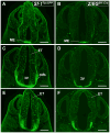
Coronal brain sections from Sf-1TauGFP mouse (A,C) and Z/EGSf1:Cre mouse (B,D) at E10.5. A,B: GFP-labeled neurons are present in the median eminence (ME) of the Sf-1TauGFP mouse. C,D: GFP-labeled fibers in the medial forebrain bundle (mfb) of the Sf-1TauGFP mouse but not the Z/EGSf1:Cre mouse. 3V, third ventricle. Scale bars = 0.5 mm.
Figure 3
Figure 3
Anterior to posterior coronal brain sections from Sf-1TauGFP mouse (A,C,E) and Z/EGSf1:Cre reporter mouse (B,D,F) at E12.5. A,B: GFP-stained neurons in the median eminence (ME). C,D: GFP-labeled neurons in the ME with fibers traveling in the medial forebrain bundle (mfb) in the Sf-1TauGFP mouse but not in the Z/EGSf1:Cre mouse. E,F: GFP-stained neurons in the presumptive VMH and fibers in Sf-1TauGFP mice. Scale bars = 0.75 mm.
Figure 4
Figure 4
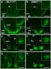
Anterior to posterior coronal brain sections from Sf-1TauGFP mouse (A,C,E,G) and Z/EGSf1:Cre reporter mouse (B,D,F,H) at E 14.5. A,B: Fibers in the ME and extending into the ventral supraoptic commissure (vsoc). C,D: GFP-stained neurons in the VMH and fibers tracts extending dorsally along the vsoc. E,F: Fibers traveling in the mfb angle toward and join the vsoc at the level of the external medullary lamina (eml). E–H: The ventrolateral aspect of the VMH in Sf-1TauGFP (E,G) and in Z/EGSf1:Cre mice (F,H) is indicated by arrows. Insets show expanded view of the VMH and its efferent fibers. Scale bars = 0.5 mm.
Figure 5
Figure 5
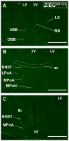
Anterior coronal brain sections from the Z/EGSf1:Cre mouse at E 17.5. A: Fibers are detected in the vertical band of Broca (VBB), the diagonal band of Broca (DBB), and the lateral septal nucleus (LS). Scant fibers are found in the medial septal nucleus (MS). B: Fibers can be seen in the bed nucleus of stria terminalis (BNST) and medial preoptic area (MPoA), sparing the lateral preoptic area (LPoA) and the medial preoptic nucleus (MPoN). C: Fibers are seen in the BNST, by the reticular nucleus of the thalamus (Rt), and in MPoA as well as in the periventricular zone of the third ventricle (3V). Scale bars = 0.5 mm.
Figure 6
Figure 6
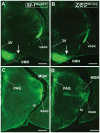
Anterior to posterior coronal brain sections from the Sf-1TauGFP mouse (A,C,E,G,I,K) and the Z/EGSf1:Cre reporter mouse (B,D,F,H,J,L) at E 17.5. A,B: Fibers are seen surrounding, but sparing, the suprachiasmatic nucleus (SCN) in both mouse models. Hypothalamic paraventricular nucleus (PVH) and the anterior hypothalamus (AH) also receive fiber projections. The vsoc is the major fiber tract and travels laterally and dorsally. C,D: Fine fiber tracts are seen projecting toward the HN and the PVH. E,F: Fibers projecting from the dorsolateral region of the VMH join the vsoc at the lateral aspect of the medial lemniscus (ml). Midline fibers travel through the dorsomedial hypothalamic nucleus (DMH) to terminate in the periaqueductal gray (PAG), whereas lateral fibers fanning out from the vsoc travel through the medial geniculate nucleus (MGN). Arrows point to the ventrolateral region of the VMH (VMHvl), where GFP+ neurons are present in the Z/EGSf1:Cre reporter mouse but not the Sf-1TauGFP mouse. G–J: GFP+ neurons in the VMHvl are absent in the Sf-1TauGFP mouse (G,I) but present in the Z/EGSf1:Cre mouse (H,J). Fibers travel through the posterior hypothalamus (PH) and the DMH medially and the MGN laterally. K,L: Fibers can be seen in the PAG and deeper layer of the superior colliculus (SC). For abbreviations see list. Scale bars = 0.5 mm.
Figure 6
Figure 6
Anterior to posterior coronal brain sections from the Sf-1TauGFP mouse (A,C,E,G,I,K) and the Z/EGSf1:Cre reporter mouse (B,D,F,H,J,L) at E 17.5. A,B: Fibers are seen surrounding, but sparing, the suprachiasmatic nucleus (SCN) in both mouse models. Hypothalamic paraventricular nucleus (PVH) and the anterior hypothalamus (AH) also receive fiber projections. The vsoc is the major fiber tract and travels laterally and dorsally. C,D: Fine fiber tracts are seen projecting toward the HN and the PVH. E,F: Fibers projecting from the dorsolateral region of the VMH join the vsoc at the lateral aspect of the medial lemniscus (ml). Midline fibers travel through the dorsomedial hypothalamic nucleus (DMH) to terminate in the periaqueductal gray (PAG), whereas lateral fibers fanning out from the vsoc travel through the medial geniculate nucleus (MGN). Arrows point to the ventrolateral region of the VMH (VMHvl), where GFP+ neurons are present in the Z/EGSf1:Cre reporter mouse but not the Sf-1TauGFP mouse. G–J: GFP+ neurons in the VMHvl are absent in the Sf-1TauGFP mouse (G,I) but present in the Z/EGSf1:Cre mouse (H,J). Fibers travel through the posterior hypothalamus (PH) and the DMH medially and the MGN laterally. K,L: Fibers can be seen in the PAG and deeper layer of the superior colliculus (SC). For abbreviations see list. Scale bars = 0.5 mm.
Figure 7
Figure 7
Comparisons between the Sf-1TauGFP mouse (A,C,E,G,I,K,L) and the Z/EGSf1:Cre mouse (B,D,F,H,J,M,N) at P0. A,B: Comparable projection patterns are seen in the vsoc and fibers traveling in the periventricular system. Cells are noticeably absent in the VMHvl of the Sf-1TauGFP mouse compared with the corresponding region in the Z/EGSf1:Cre mouse (arrow). C,D: Similar fiber projections are seen in the vsoc along the internal capsule (ic), across the medial geniculate nucleus (MGN), and into the PAG. E,F: Fiber projections are seen in the PAG. A solitary GFP+ cell can be seen in the tegmental area of the midbrain of the Z/EGSf1:Cre (arrow in F). G,H: Absence of GFP+ cells in the VMHvl of the Sf-1TauGFP mouse (G). Arrows indicate the presence of GFP+ cells in the presumptive tuberal region of both mouse models. I,J: Colabeling of cytoplasmic GFP+ staining (green) and nuclear SF-1+ staining (red) in the VMH. Absence of SF-1+ staining is noted in the VMHvl of both mouse lines. Boxed areas are shown as expanded views in K–N. K,L: Expanded views of the VMH in Sf-1TauGFP mice from Figure 7I showing colabeling (arrowheads) in the compact region (K) and the absence of any labeling in the VMHvl (L). M,N: Expanded views of the VMH in the Z/EGSf1:Cre mouse from Figure 7J showing colabeling (arrowheads) in the compact region (M) with only GFP+ labeling (arrows) in the VMHvl (N). For abbreviations see list. Scale bars = 0.5 mm.
Figure 7
Figure 7
Comparisons between the Sf-1TauGFP mouse (A,C,E,G,I,K,L) and the Z/EGSf1:Cre mouse (B,D,F,H,J,M,N) at P0. A,B: Comparable projection patterns are seen in the vsoc and fibers traveling in the periventricular system. Cells are noticeably absent in the VMHvl of the Sf-1TauGFP mouse compared with the corresponding region in the Z/EGSf1:Cre mouse (arrow). C,D: Similar fiber projections are seen in the vsoc along the internal capsule (ic), across the medial geniculate nucleus (MGN), and into the PAG. E,F: Fiber projections are seen in the PAG. A solitary GFP+ cell can be seen in the tegmental area of the midbrain of the Z/EGSf1:Cre (arrow in F). G,H: Absence of GFP+ cells in the VMHvl of the Sf-1TauGFP mouse (G). Arrows indicate the presence of GFP+ cells in the presumptive tuberal region of both mouse models. I,J: Colabeling of cytoplasmic GFP+ staining (green) and nuclear SF-1+ staining (red) in the VMH. Absence of SF-1+ staining is noted in the VMHvl of both mouse lines. Boxed areas are shown as expanded views in K–N. K,L: Expanded views of the VMH in Sf-1TauGFP mice from Figure 7I showing colabeling (arrowheads) in the compact region (K) and the absence of any labeling in the VMHvl (L). M,N: Expanded views of the VMH in the Z/EGSf1:Cre mouse from Figure 7J showing colabeling (arrowheads) in the compact region (M) with only GFP+ labeling (arrows) in the VMHvl (N). For abbreviations see list. Scale bars = 0.5 mm.
Figure 8
Figure 8
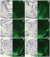
Anterior to posterior coronal brain sections from the Z/EGSf1:Cre reporter mouse at P0. A–R: Projection patterns of GFP-labeled fibers (right) and the corresponding contralateral Nissl-stained sections (left). S–U: High-power photomicrographs showing the corresponding amygdaloid nuclei in the field of GFP-labeled fibers. For abbreviations see list. Scale bars = 0.675 mm in A–R; 0.25 mm in S–U.
Figure 8
Figure 8
Anterior to posterior coronal brain sections from the Z/EGSf1:Cre reporter mouse at P0. A–R: Projection patterns of GFP-labeled fibers (right) and the corresponding contralateral Nissl-stained sections (left). S–U: High-power photomicrographs showing the corresponding amygdaloid nuclei in the field of GFP-labeled fibers. For abbreviations see list. Scale bars = 0.675 mm in A–R; 0.25 mm in S–U.
Figure 9
Figure 9
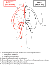
Simplified graphic representation of the fiber projection patterns in the Z/EGSf1:Cre reporter mouse at P0 and the PHA-L-injected rat at the adult stage, modified from Canteras et al. (1994). For abbreviations see list. ENT, entorhinal area.

Similar articles

Cited by

References

    1. Bingham NC, Anderson KK, Reuter AL, Stallings NR, Parker KL. Selective loss of leptin receptors in the ventro-medial hypothalamic nucleus results in increased adiposity and a metabolic syndrome. Endocrinology. 2008;149:2138–2148. - PMC - PubMed
    1. Braz JM, Rico B, Basbaum AI. Transneuronal tracing of diverse CNS circuits by Cre-mediated induction of wheat germ agglutinin in transgenic mice. Proc Natl Acad Sci U S A. 2002;99:15148–15153. - PMC - PubMed
    1. Budefeld T, Tobet SA, Majdic G. Altered position of cell bodies and fibers in the ventromedial region in SF-1 knockout mice. Exp Neurol. 2011;232:176–184. - PMC - PubMed
    1. Bunting M, Bernstein KE, Greer JM, Capecchi MR, Thomas KR. Targeting genes for self-excision in the germ line. Genes Dev. 1999;13:1524–1528. - PMC - PubMed
    1. Cameron NM, Soehngen E, Meaney MJ. Variation in maternal care influences ventromedial hypothalamus activation in the rat. J Neuroendocrinol. 2011;23:393–400. - PubMed

Publication types

Substances