. 2012 Sep 24;209(10):1769-79.
doi: 10.1084/jem.20120905. Epub 2012 Sep 17.

Autoreactive T cells bypass negative selection and respond to self-antigen stimulation during infection

Affiliations

Autoreactive T cells bypass negative selection and respond to self-antigen stimulation during infection

Sarah Enouz et al. J Exp Med. .

Abstract

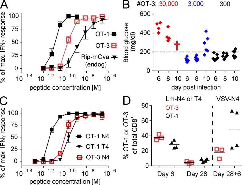
Central and peripheral tolerance prevent autoimmunity by deleting the most aggressive CD8(+) T cells but they spare cells that react weakly to tissue-restricted antigen (TRA). To reveal the functional characteristics of these spared cells, we generated a transgenic mouse expressing the TCR of a TRA-specific T cell that had escaped negative selection. Interestingly, the isolated TCR matches the affinity/avidity threshold for negatively selecting T cells, and when developing transgenic cells are exposed to their TRA in the thymus, only a fraction of them are eliminated but significant numbers enter the periphery. In contrast to high avidity cells, low avidity T cells persist in the antigen-positive periphery with no signs of anergy, unresponsiveness, or prior activation. Upon activation during an infection they cause autoimmunity and form memory cells. Unexpectedly, peptide ligands that are weaker in stimulating the transgenic T cells than the thymic threshold ligand also induce profound activation in the periphery. Thus, the peripheral T cell activation threshold during an infection is below that of negative selection for TRA. These results demonstrate the existence of a level of self-reactivity to TRA to which the thymus confers no protection and illustrate that organ damage can occur without genetic predisposition to autoimmunity.


Figures

Figure 1.
Figure 1.
Low avidity Kb/Ova-specific T cells in the periphery of Rip-mOva mice. Rip-mOva or transgene negative littermate control C57BL/6 mice were infected with Lm-Ova. A fraction of the mice were rechallenged 4 wk later with VSV-Ova. 6 d after the last infection, splenocytes were isolated and either left untreated or were briefly in vitro restimulated with SIINFEKL peptide. In A, CD8+ gated flow cytometry plots of cells intracellularly stained for IFN-γ, and in B, the frequencies of Ova-specific T cells are depicted (n = 3–4 mice per group). Dose–response curves graphing peptide concentration against the fraction of maximum IFN-γ response in splenic CD8+ T cells are shown in C (n = 4 and 5 per group, error bars show SD). The data shown in A–C are representative of three independently performed experiments. (D) Calculated mean half-maximum effective concentrations (EC50) obtained in three independent experiments (each including three to five mice per group) are presented. (E) Maximum blood glucose levels between days 7 and 13 after the secondary infection. The data are pooled from eight independent experiments (total number of mice: 56 Rip-mOva and 26 littermate control, transgene-negative C57BL/6 mice). The horizontal bars in B and D represent the mean value of the individual data points.
Figure 2.
Figure 2.
T cells infiltrate the islets of Rip-mOva mice after sequential Lm-Ova and VSV-Ova infection. C57BL/6 or Rip-mOva mice were infected with Lm-Ova and, 4 wk later, with VSV-Ova. Representative images of anti-CD8–stained pancreas sections obtained at day 8 after the secondary infection are presented. Bars, 50 µm. Results from one of two representative experiments are shown with n = 5 mice per group.
Figure 3.
Figure 3.
OT-3 T cells respond with similar functional avidity to Kb/Ova as OT-1 T cells respond to SIITFEKL. (A) 800 OT-3 T cells were transferred into Rip-mOva mice which were infected with Lm-Ova and, 4 wk later, with VSV-Ova. 104 OT-1 T cell–engrafted control C57BL/6 mice were infected with VSV-Ova. Splenocytes from both groups were restimulated with SIINFEKL peptide and stained for IFN-γ. The corresponding peptide dose–response curves for OT-3, OT-1, and endogenous T cells in Rip-mOva mice are shown (n = 3 or 4, error bars show SD). The data are representative of two independent experiments. (B) Rip-mOva mice, supplemented with titrated numbers of OT-3 T cells, were infected with Lm-Ova, and blood glucose levels were determined 6, 8, and 10 d after infection. Data are representative of three independent experiments (n = 4–6 mice). Because of the diabetes, the mice engrafted with the highest number of OT-3 T cells had to be sacrificed after day 8 (marked with a cross). (C) 2 × 10⁴ naive OT-1 and 105 OT-3 T cells were transferred into CD45.1 congenic mice which were infected with Lm-Ova. Splenocytes were stimulated with titrated doses of SIINFEKL (N4) or SIITFEKL (T4) peptide (OT-1 only) at 7 d after infection and intracellularly stained for IFN-γ. The resulting dose–response curves are shown (n = 4, data are representative of three experiments, SD error bars are shown). (D) 105 OT-1 and OT-3 T cells were transferred into CD45.1 congenic hosts. OT-1 mice were infected with Lm-T4Ova and OT-3 mice with Lm-Ova. OT-1 and OT-3 T cell frequencies at days 6 and 28 after infection and 6 d after a secondary VSV-Ova infection are presented. The horizontal bars represent the mean value of the individual data points. The data are representative of two independent experiments with n = 3–4 mice per group.
Figure 4.
Figure 4.
Survival of a large fraction of OT-3 T cells in the thymus of Rip-mOva mice. (A and B) Lethally irradiated Rip-mOva or C57BL/6 mice (both coexpress CD45.1 and CD45.2) were injected with a 4:1 mix of C57BL/6 (CD45.1) and OT-3 (CD45.2) bone marrow. (A–C) Frequency of OT-3 T cells among total CD8+ T cells in these mice was measured at 6.5 wk (n = 12 per group) or 20 wk (n = 8 and 6 per group) after reconstitution in the peripheral blood (A), at 9 wk in the pancreatic lymph nodes (n = 4 and 5 mice per group; B), and at 5 wk among CD8+ CD24 low single-positive thymocytes (n = 8 and 6 per group; C). Six independent experiments were performed to assess the frequency of OT-3 at 5.5–7 wk in the blood and three experiments at the later time point. The measurements in the pancreatic lymph nodes are representative of two independent experiments. The horizontal bars in A, B, and C represent the mean value of the individual data points.
Figure 5.
Figure 5.
Endogenous Kb/Ova complexes activate OT-3 T cells during an infection. CD45.1 Rip-mOva or C57BL/6 mice were injected with 5 × 10⁵ CellTrace Violet–labeled OT-3 T cells and infected with LCMV Armstrong. 4 d after infection, splenocytes were analyzed for dye dilution and CD62L expression levels. (A) Representative OT-3 gated flow cytometry plots. (B) Data for all mice (n = 3 or 4 per group). The data are representative of three independent experiments. The horizontal bars represent the mean value of the individual data points.
Figure 6.
Figure 6.
pMHC ligands with avidities far below the negative selection threshold induce functional effector and memory T cells. (A) An OT-3 T cell line was briefly stimulated with SIINFEKL (N4) or the APLs SIFNFEKL (F3) or SIVNFEKL (V3). Shown are dose–response curves graphing the peptide concentration against the fraction of IFN-γ–producing T cells. (B–E) 105 CD45.1 congenic OT-3 T cells were transferred into C57BL/6 mice which were then infected with Lm-Ova, Lm-F3, or Lm-V3. Another group of mice was infected with nonrecombinant wild-type Lm (wt). (B) The frequency of OT-3 T cells on day 7 after infection (n = 4 or 5 mice per group). (C) Representative flow cytometry plots of total CD8+ T cells (left) or OT-3 T cells (right) analyzed on day 5.5 after infection for granzyme B, CD44, and CD62L expression. (D) The same cells briefly restimulated with SIINFEKL peptide and stained for TNF and IFN-γ are shown (n = 3 or 4 mice per group in C and D). (E) The frequency of OT-3 T cells in mice infected with Lm-V3 or control Lm-wt at 6 and 28 d and at 4 d after a secondary VSV-Ova infection are displayed (n = 4 or 5 mice per group). (F) Rip-mOva mice received nothing or 2 × 10⁴ OT-3 T cells and infected with Lm-V3. At 5.5 d after infection, peptide-pulsed CFSEbright and control CFSElow splenocytes were injected and analyzed 24 h later for the elimination of the CFSEbright population. The numbers indicate the percentage and SD of CFSEbright cells in Rip-mOva mice supplemented with or without OT-3 T cells (n = 3 per group). (G) Blood glucose levels at 6–8 d after Lm-F3 infection and 105 OT-3 transfer (n = 8 mice). The horizontal bars in B and E represent the mean value of the individual data points. All the experiments shown were performed two times and we observed similar outcomes.
Figure 7.
Figure 7.
Autoreactive OT-3 T cells differentiate into normal effector and memory T cells. (A) Lethally irradiated Rip-mOva or C57BL/6 mice were reconstituted as described in Fig. 4. 6 wk later, expression levels of CD5, CD44, CD25, and PD-1 on OT-3 T cells in C57BL/6 mice (termed foreign-OT-3) and in Rip-mOva mice (self–OT-3 T cells) were determined (representative data for n = 5 mice per group are shown). The black line represents the level of CD5 expression by endogenous T cells. The data are representative of two experiments. (B) Similar chimeric mice were infected at 11 wk after reconstitution with Lm-Ova and, 7 d later, splenocytes were stained with GrzB, anti-CD127, and anti-KLRG1. They were also restimulated with SIINFEKL peptide and then stained intracellularly for IFN-γ and TNF. Shown are OT-3 gated flow cytometry plots representative of n = 3 mice per group. The dashed lines on the left are endogenous CD8+ T cells. (C) Rip-mOva mice received 5 × 10⁴ foreign– or self–OT-3 T cells (described in A), and blood glucose levels and T cell expansion were determined after an Lm-Ova infection (n = 5 mice per group). Values >200 mg/dl were considered diabetic. The experiments shown in B and C were performed two times and showed a similar outcome. (D) H-2Kbm1/Rip-mOva mice were lethally irradiated and grafted with C57BL/6 bone marrow. 8 wk after the reconstitution, 5 × 10⁴ OT-3 Rag2−/− T cells were injected and the mice were infected with Lm-Ova. The frequency of OT-3 T cells was determined 6 and 28 d after the Lm-Ova infection and at day 6 after a VSV-Ova rechallenge. The data are pooled from two independently performed experiments with n = 3 mice per group and experiment. The horizontal bars in C and D represent the mean value of the individual data points.

References

    1. Alam S.M., Travers P.J., Wung J.L., Nasholds W., Redpath S., Jameson S.C., Gascoigne N.R. 1996. T-cell-receptor affinity and thymocyte positive selection. Nature. 381:616–620 10.1038/381616a0 - DOI - PubMed
    1. Anderson M.S., Venanzi E.S., Klein L., Chen Z., Berzins S.P., Turley S.J., von Boehmer H., Bronson R., Dierich A., Benoist C., Mathis D. 2002. Projection of an immunological self shadow within the thymus by the aire protein. Science. 298:1395–1401 10.1126/science.1075958 - DOI - PubMed
    1. Baldwin T.A., Sandau M.M., Jameson S.C., Hogquist K.A. 2005. The timing of TCRα expression critically influences T cell development and selection. J. Exp. Med. 202:111–121 10.1084/jem.20050359 - DOI - PMC - PubMed
    1. Belz G.T., Behrens G.M., Smith C.M., Miller J.F., Jones C., Lejon K., Fathman C.G., Mueller S.N., Shortman K., Carbone F.R., Heath W.R. 2002. The CD8α+ dendritic cell is responsible for inducing peripheral self-tolerance to tissue-associated antigens. J. Exp. Med. 196:1099–1104 10.1084/jem.20020861 - DOI - PMC - PubMed
    1. Bergthaler A., Merkler D., Horvath E., Bestmann L., Pinschewer D.D. 2007. Contributions of the lymphocytic choriomeningitis virus glycoprotein and polymerase to strain-specific differences in murine liver pathogenicity. J. Gen. Virol. 88:592–603 10.1099/vir.0.82428-0 - DOI - PubMed

Publication types

MeSH terms