. 2012 Sep 24;209(10):1841-52, S1-24.
doi: 10.1084/jem.20120219. Epub 2012 Sep 17.

Bcl6 expression specifies the T follicular helper cell program in vivo

Affiliations

Bcl6 expression specifies the T follicular helper cell program in vivo

Xindong Liu et al. J Exp Med. .

Abstract

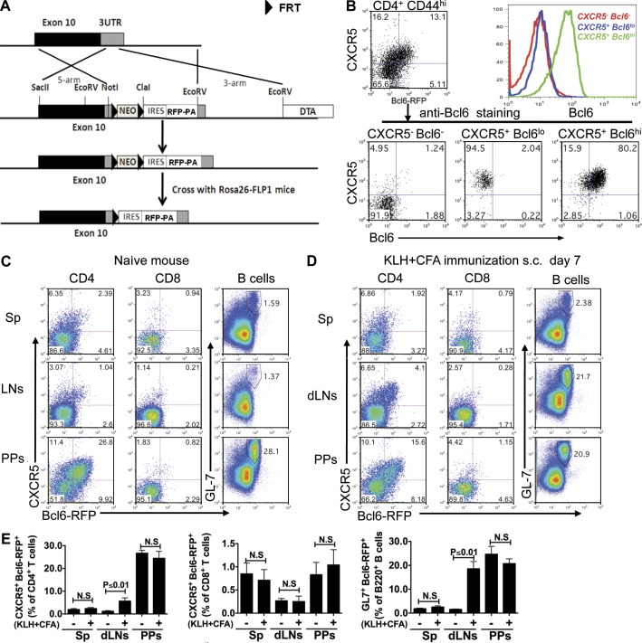
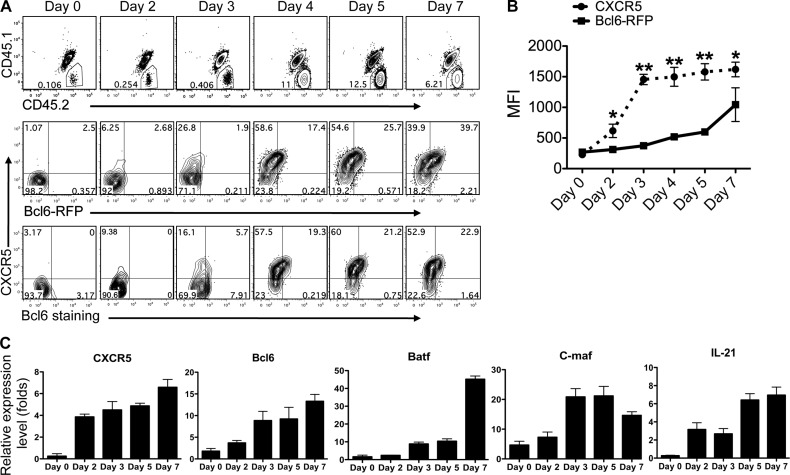
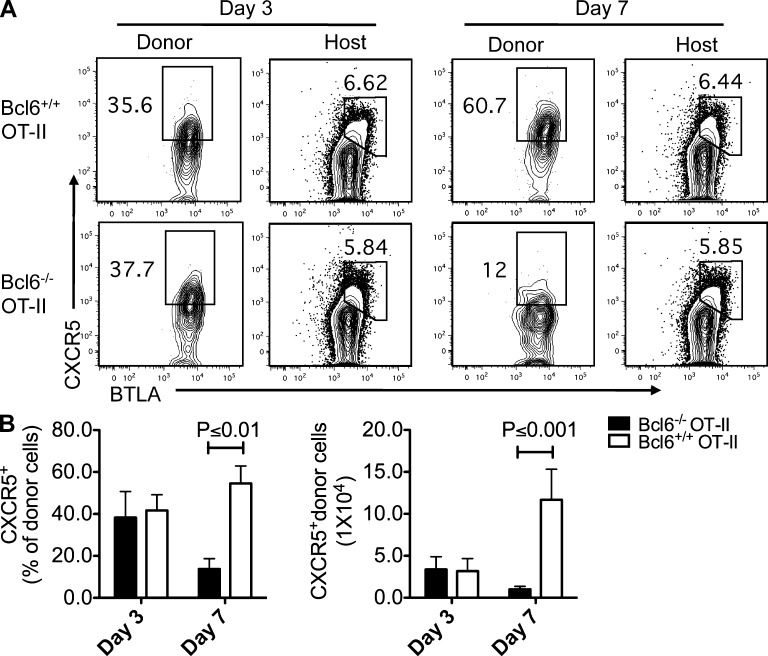
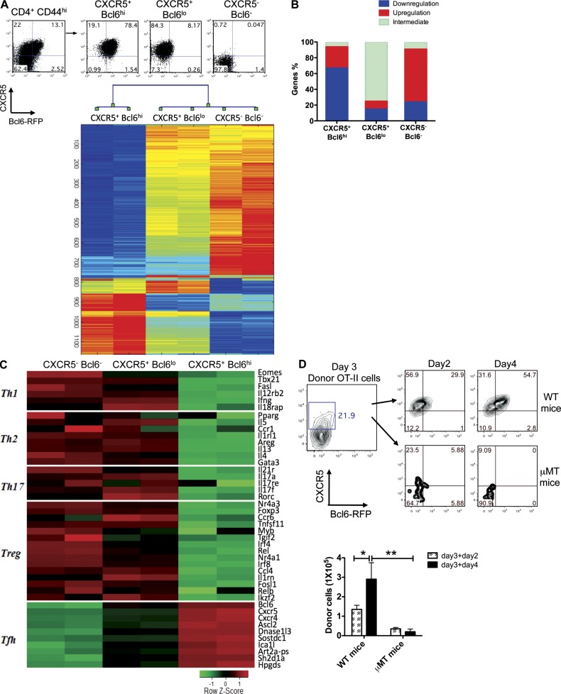
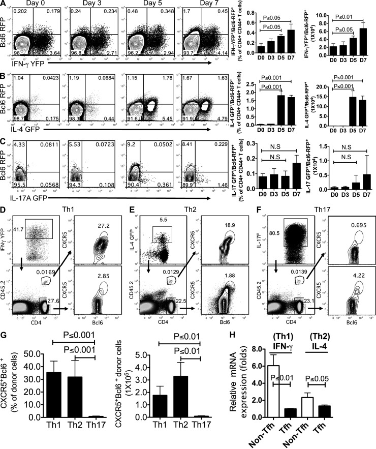
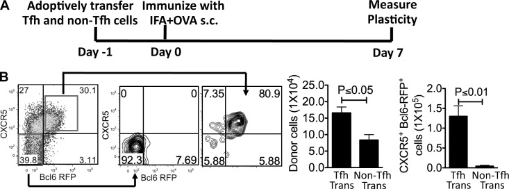
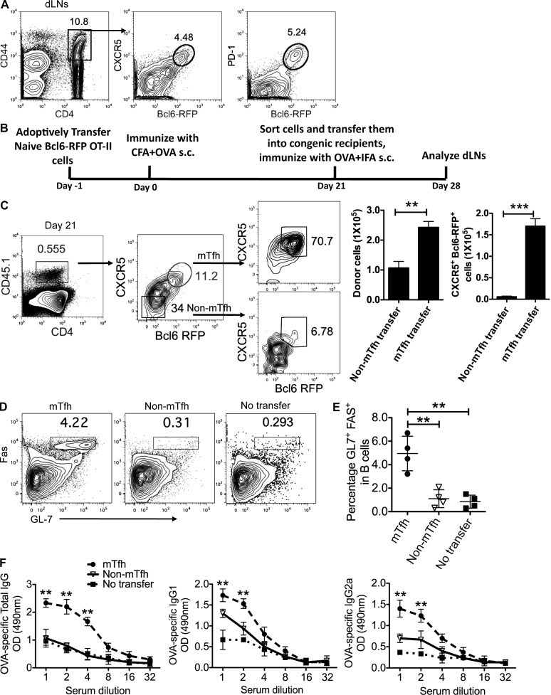
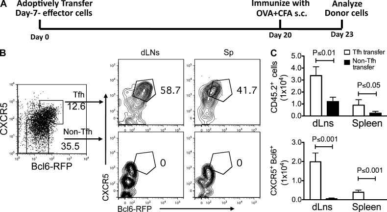
T follicular helper cells (Tfh cells) play a pivotal role in germinal center reactions, which require B cell lymphoma 6 (Bcl6) transcription factor. To analyze their relationships with other effector T cell lineages and their stability in vivo, we developed and analyzed a new Bcl6 reporter mouse alone or together with other lineage reporter systems. Assisted with genome-wide transcriptome analysis, we show substantial plasticity of T cell differentiation in the early phase of immune response. At this stage, CXCR5 appears to be expressed in a Bcl6-independent manner. Once Bcl6 is highly expressed, Tfh cells can persist in vivo and some of them develop into memory cells. Together, our results indicate Bcl6 as a bona fide marker for Tfh polarized program.


Figures

Figure 1.
Figure 1.
Generation and validation of Bcl6-RFP reporter mice. (A) Schematic map for the mouse Bcl6 gene locus, targeting DNA construct, and targeted Bcl6 locus. (B) Bcl6 intracellular staining on sorted cells from Bcl6-RFP reporter mice. Histogram overlay of three populations of T cells: CXCR5Bcl6, CXCR5+Bcl6lo, and CXCR5+Bcl6hi. Data are a representative of two independent experiments. (C and D) In naive (C) and KLH/CFA s.c. immunized (D) reporter mice, CXCR5 and Bcl6-RFP expression was assessed on gated CD4+ T cells and CD8+ T cells from spleens (Sps), dLNs, and Peyer’s patches (PPs) by flow cytometry. Also, analysis of GL7+Bcl6-RFP+ cells is shown in gated B220+ B cells. Data are representative of two independent experiments. (E) Summary of data from C and D. Bar graphs display the number of donor cells as mean ± SD. n = 3 per group. N.S., no statistically significant difference.
Figure 2.
Figure 2.
Ontogeny analysis of Bcl6 expression and Tfh cell development. (A) Naive Bcl6-RFP OT-II CD4+ T cells (CD45.1CD45.2+) were transferred into CD45.1+CD45.2+ congenic mice, which were subsequently immunized s.c. with OVA in CFA. Donor cell expansion (top) and CXCR5 and Bcl6-RFP expression in donor cells (middle) were determined by flow cytometry. Intracellular staining of Bcl6 was also performed (bottom). Data are representative of two independent experiments. (B) Expression of CXCR5 and Bcl6-RFP in donor cells was determined at various time points after immunization and shown in line graph as median fluorescence intensity (MFI) ± SD. *, P < 0.05; **, P < 0.01. (C) Quantitative RT-PCR measurement of Tfh-specific genes in sorted donor cells. The graphs display mean ± SD. (A–C) n = 3 per group.
Figure 3.
Figure 3.
Bcl6 is not required for CXCR5 initiation. (A) Naive bcl6−/− and bcl6+/+ OT-II T cells were transferred into CD45.1+ recipient mice, respectively, and then followed with OVA/CFA immunization s.c., and 3 and 7 d after immunization, donor cells and endogenous host CD4+ T cells from dLNs were characterized by anti-CXCR5 and BTLA staining. Data are representative of two independent experiments. n = 3 per group. (B) Summary of data from A. Bar graphs display the frequency and number of CXCR5+ donor cell as mean ± SD.
Figure 4.
Figure 4.
Global transcriptional changes are associated with Tfh cell development. Bcl6-RFP reporter mice were s.c. immunized with KLH in CFA. 7 d later, mice were sacrificed, and activated CD44+CD4+ T cell from dLNs were sorted into three populations: CXCR5+Bcl6hi, CXCR5+Bcl6lo, and CXCR5Bcl6 cells. (A) Microarray analysis of sorted cells (duplicated samples). Hierarchical clusters of 1,130 genes for three groups of cells. The color coding applies to the expression level of 1,130 genes with 0 as a median. Each of the samples was duplicated. (B) The percentages of genes associated with relative down-regulation, up-regulation, and intermediate for each population of cells. (C) Heat map of signature genes for Th1, Th2, Th17, Treg, and Tfh cells. (D) CXCR5+Bcl6lo T cells were sorted on day 3 after immunization and transferred into WT or µMT recipient mice. 2 and 4 d after immunization with OVA + IFA s.c., flow cytometry analysis of CXCR5 and Bcl6 expression was conducted. Data are representative of two independent experiments. Bar graph displays the number of donor cells as mean ± SD. n = 3 per group. *, P < 0.05; **, P < 0.01.
Figure 5.
Figure 5.
Th1 and Th2 but few of Th17 cells are able to become Tfh cells. (A–C) Dual reporter mice were immunized s.c. with KLH in CFA for 0, 3, 5, and 7 d. Flow cytometry analysis of Bcl6-RFP and eGFP/YFP on gated CD4+CD44+ T cells from dLNs. Data are representative of two independent experiments. Bar graphs display the percentage and number of double-positive cells as mean ± SD. (A) Flow cytometry analysis of Bcl6-RFP and IFN-γ–YFP from CD4+CD44+ T cells in Bcl6-RFP/Yeti double reporter mice. (B) Flow cytometry analysis of Bcl6-RFP and IL-4–GFP from CD4+CD44+ T cells in Bcl6-RFP/4get double reporter mice. (C) Flow cytometry analysis of Bcl6-RFP and IL-17A–eGFP from CD4+CD44+ T cells in Bcl6-RFP/IL-17A–GFP double reporter mice. (D) The in vitro committed YFPhi OT-II T (Th1) cells were collected from 4-d culture and intravenously transferred into congenic recipient mice (CD45.1+). 7 d after immunization with OVA in CFA s.c., donor and host CD4+ T cells were subject to flow cytometry analysis of CXCR5 and Bcl6. (E) Flow cytometry analysis of the CXCR5 and Bcl6 expression in donor IL-4–GFPhi OT-II T (Th2) cells and host CD4+ T cells. (F) Flow cytometry analysis of the CXCR5 and Bcl6 expression in donor IL-17F–RFPhi OT-II T (Th17) cells and host CD4+ T cells after transfer and immunization. (D–F) Data are representative of two independent experiments. (G) Summary of data from D–F. Bar graphs display the percentage and number of donor cells as mean ± SD. (H) In the assay of conversion from Th1 to Tfh cell (D) and from Th2 to Tfh cell (E), 7 d after immunization, donor cell–derived CXCR5+Bcl6hi (Tfh) and CXCR5Bcl6 (Non-Tfh) were sorted and subjected to the measurement of cytokine expression by quantitative RT-PCR. Data are representative of two independent experiments displayed as mean ± SD. n = 3 per group. N.S., no statistically significant difference.
Figure 6.
Figure 6.
The stability and plasticity of Tfh cells in vivo. (A) Experimental design for testing Tfh stability in vivo. (B) The sorted effector Tfh and non-Tfh (CD45.2+) cells were adoptively transferred into congenic recipient mice (CD45.1+). 7 d after s.c. immunization with KLH in IFA, the expression of CXCR5 and Bcl6 in donor Tfh and non-Tfh cells was measured by flow cytometry. Data are representative of two independent experiments. Bar graphs display the number of donor cells as mean ± SD. n = 3 per group.
Figure 7.
Figure 7.
Memory Tfh cells promote humoral recall responses. (A) 30 d after immunization with KLH in CFA s.c., flow cytometry analysis of CXCR5 and Bcl6-RFP expression in activated T cells from Bcl6-RFP reporter mice was performed. Data are representative of two independent experiments. (B) Experimental design for functional analysis of mTfh and non-mTfh cells. (C) CXCR5+Bcl6hi or CXCR5Bcl6 OT-II CD4+ T cells (CD45.1+) were purified 21 d after immunization and adoptively transferred at equal number (1.5 × 10⁵) into naive congenic recipients (CD45.2+). 7 d later, the phenotype of donor cells in the dLNs and their population were determined by flow cytometry. (D) Flow cytometry analysis of GL7 and Fas on B220+ B cells from mTfh and non-mTfh transferred recipients; the nontransfer mice were set as controls. (E) The percentage of GL7+Fas+ B cells in dLNs was quantified. (F) OVA-specific antibody levels (total IgG, IgG1, and IgG2a) were determined by ELISA. (C–F) Cumulative data are representative of two independent experiments displayed as mean ± SD. n = 4 per group. **, P < 0.01; ***, P < 0.001.
Figure 8.
Figure 8.
Effector Tfh cells can develop into memory Tfh cells. Naive Bcl6-RFP OT-II cells (CD45.2+) were transferred into CD45.1+ recipient mice, followed by s.c. immunization with OVA in CFA for 7 d. CXCR5+Bcl6hi OT-II T cells were then purified and transferred into naive mice (1.5 × 10⁵ cells/mouse). 20 d later, recipients were immunized with OVA + CFA for another 3 d, and then the phenotypes of donor cells in the dLNs and spleens were determined by flow cytometry. Data are representative of two independent experiments. n = 3 per group. (A) Experimental strategy of examining the transition from effector Tfh to memory Tfh cell. (B) Flow cytometry analysis of CXCR5 and Bcl6 in donor Tfh and non-Tfh cells is shown. (C) After adoptive transfer and immunization, the cell numbers of CXCR5+Bcl6-RFP+ and CXCR5Bcl6-RFP donor-derived cells were summarized and compared. Cumulative data are representative of two independent experiments displayed as mean ± SD.

References

    1. Acosta-Rodriguez E.V., Rivino L., Geginat J., Jarrossay D., Gattorno M., Lanzavecchia A., Sallusto F., Napolitani G. 2007. Surface phenotype and antigenic specificity of human interleukin 17-producing T helper memory cells. Nat. Immunol. 8:639–646 10.1038/ni1467 - DOI - PubMed
    1. Bauquet A.T., Jin H., Paterson A.M., Mitsdoerffer M., Ho I.C., Sharpe A.H., Kuchroo V.K. 2009. The costimulatory molecule ICOS regulates the expression of c-Maf and IL-21 in the development of follicular T helper cells and TH-17 cells. Nat. Immunol. 10:167–175 10.1038/ni.1690 - DOI - PMC - PubMed
    1. Betz B.C., Jordan-Williams K.L., Wang C., Kang S.G., Liao J., Logan M.R., Kim C.H., Taparowsky E.J. 2010. Batf coordinates multiple aspects of B and T cell function required for normal antibody responses. J. Exp. Med. 207:933–942 10.1084/jem.20091548 - DOI - PMC - PubMed
    1. Breitfeld D., Ohl L., Kremmer E., Ellwart J., Sallusto F., Lipp M., Förster R. 2000. Follicular B helper T cells express CXC chemokine receptor 5, localize to B cell follicles, and support immunoglobulin production. J. Exp. Med. 192:1545–1552 10.1084/jem.192.11.1545 - DOI - PMC - PubMed
    1. Choi Y.S., Kageyama R., Eto D., Escobar T.C., Johnston R.J., Monticelli L., Lao C., Crotty S. 2011. ICOS receptor instructs T follicular helper cell versus effector cell differentiation via induction of the transcriptional repressor Bcl6. Immunity. 34:932–946 10.1016/j.immuni.2011.03.023 - DOI - PMC - PubMed

Publication types

MeSH terms