. 2012 Oct 2;109(40):16018-22.
doi: 10.1073/pnas.1212506109. Epub 2012 Sep 17.

High-throughput lensfree 3D tracking of human sperms reveals rare statistics of helical trajectories

Affiliations

High-throughput lensfree 3D tracking of human sperms reveals rare statistics of helical trajectories

Ting-Wei Su et al. Proc Natl Acad Sci U S A. .

Abstract

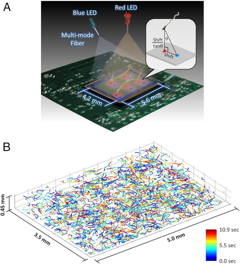
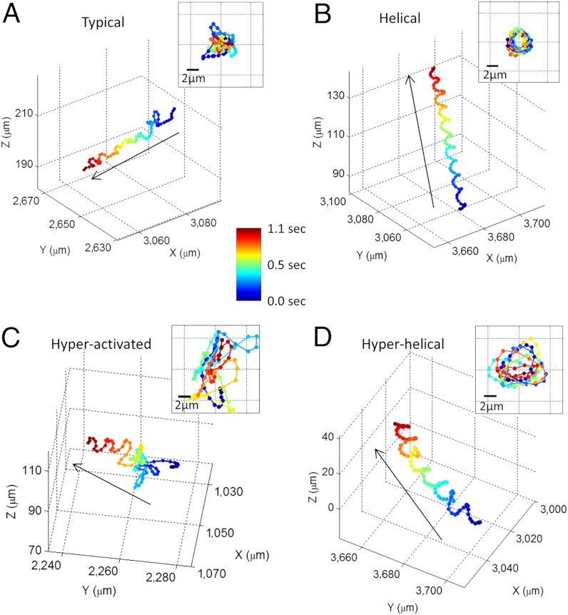
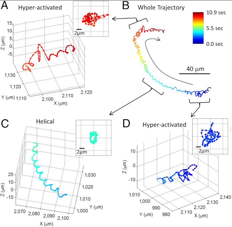
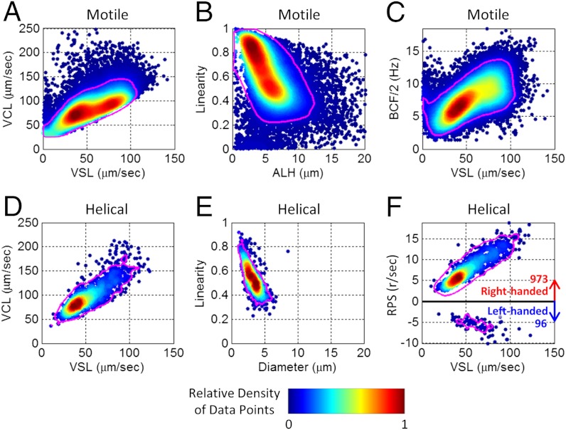
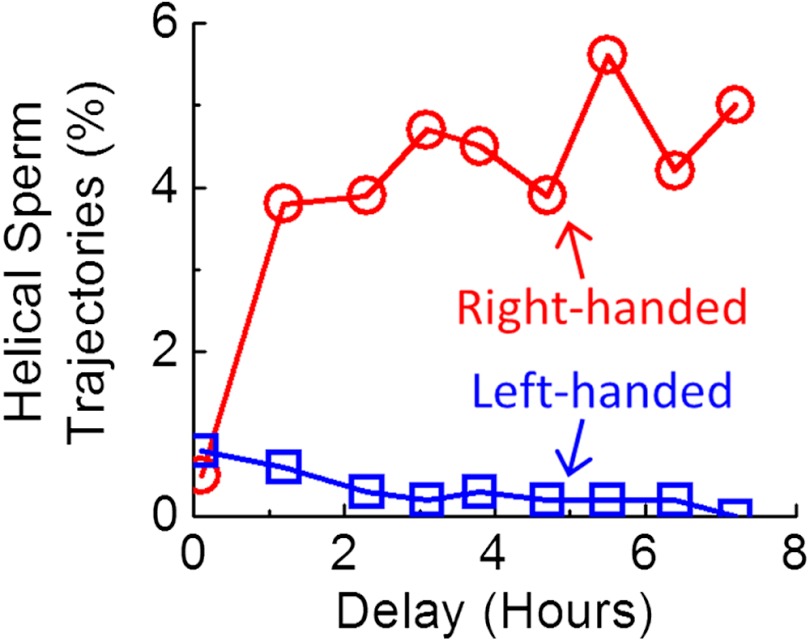
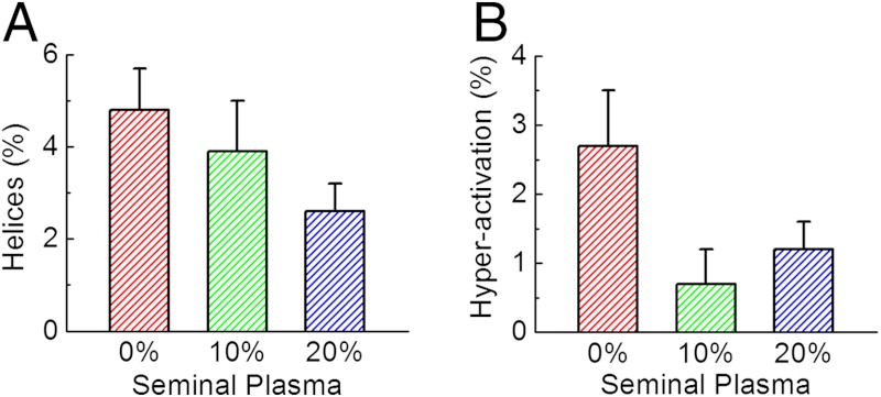
Dynamic tracking of human sperms across a large volume is a challenging task. To provide a high-throughput solution to this important need, here we describe a lensfree on-chip imaging technique that can track the three-dimensional (3D) trajectories of > 1,500 individual human sperms within an observation volume of approximately 8-17 mm(3). This computational imaging platform relies on holographic lensfree shadows of sperms that are simultaneously acquired at two different wavelengths, emanating from two partially-coherent sources that are placed at 45° with respect to each other. This multiangle and multicolor illumination scheme permits us to dynamically track the 3D motion of human sperms across a field-of-view of > 17 mm(2) and depth-of-field of approximately 0.5-1 mm with submicron positioning accuracy. The large statistics provided by this lensfree imaging platform revealed that only approximately 4-5% of the motile human sperms swim along well-defined helices and that this percentage can be significantly suppressed under seminal plasma. Furthermore, among these observed helical human sperms, a significant majority (approximately 90%) preferred right-handed helices over left-handed ones, with a helix radius of approximately 0.5-3 μm, a helical rotation speed of approximately 3-20 rotations/s and a linear speed of approximately 20-100 μm/s. This high-throughput 3D imaging platform could in general be quite valuable for observing the statistical swimming patterns of various other microorganisms, leading to new insights in their 3D motion and the underlying biophysics.


Conflict of interest statement

The authors declare no conflict of interest.

Figures

Fig. 1.
Fig. 1.
Dual-view lensfree 3D tracking of human sperms. (A) The schematic diagram of the imaging system. Two partially-coherent light sources (red and blue LEDs at 625 nm and 470 nm, respectively) are butt-coupled to multimode fibers (0.4 mm core diameter each) to simultaneously illuminate the sperms at two different angles (red at 0° and blue at 45°). A CMOS sensor chip records the dual-view lensfree holograms that encode the position information of each sperm. The 3D location of each sperm is determined by the centroids of its head images reconstructed in the vertical (red) and oblique (blue) channels. This schematic diagram is not drawn to scale. (B) The reconstructed 3D sperm trajectories. 1,575 human sperms inside a volume of 7.9 μL were tracked at a frame rate of 92 FPS. The time position of each track point is encoded by its color (see the color bar).
Fig. 2.
Fig. 2.
Four major categories of human sperm swimming patterns. (A) The typical pattern. (B) The helical pattern. (C) The hyperactivated pattern. (D) The hyperhelical pattern. The inset in each panel represents the front view of the straightened trajectory of the sperm (Methods). The arrows indicate the directions of the sperms’ forward movement. The time position of each track point is encoded by its color (see the color bar). The helices shown in (B) and (D) are both right-handed. See Movies S1S4 for the time evolution of the sperm trajectories shown in (AD), respectively. Some other examples of human sperm trajectories are also provided in Figs. S1S4.
Fig. 3.
Fig. 3.
A 10.9-s long trajectory showing the transitions between different swimming patterns of a human sperm. (A), (C), and (D) illustrate digitally extracted segments (approximately 1 s long each) of the whole sperm trajectory shown in (B). See Movie S5 for the time evolution of this trajectory. More sample trajectories with different pattern transitions are also provided in Fig. S4. The inset in each panel is the front view of the straightened trajectory of the sperm. The time position of each track point is encoded by its color (see the color bar).
Fig. 4.
Fig. 4.
Dynamic swimming parameters of 24,090 motile human sperms and 1,069 helical trajectories. Color bar represents the relative density of data points in each graph. Magenta lines enclose 90% of the motile/helical tracks presented in each panel. A helix with RPS > 0 (RPS < 0) is defined as right-handed (left-handed). VSL: straight-line velocity. VCL: curvilinear velocity. ALH: amplitude of lateral head displacement. BCF: beat-cross frequency. RPS: rotation speed. The unit r/s: revolutions per second. These measurements were made in baseline medium (artificial HTF) after > 2 h of incubation as described in SI Text.
Fig. 5.
Fig. 5.
Time evolution of helical sperm trajectories after resuspension in artificial human tubal fluid (HTF). After approximately 2–3 h of incubation in HTF, the percentage of right-handed helical trajectories significantly increased to approximately 4–5% of motile human sperms, while the percentage of left-handed ones did not show a major change, remaining < 0.5% of motile sperms.
Fig. 6.
Fig. 6.
Quenching of human sperms’ helical (A) and hyperactivated (B) trajectories as a function of increased seminal plasma concentration in culture media. Each of the mean ± SD bars in (A) and (B) was based on 14 measurements of two specimens (seven with each) obtained from different anonymous donors.

Comment in

References

    1. Phillips DM. Comparative analysis of mammalian sperm motility. J Cell Biol. 1972;53:561–573. - PMC - PubMed
    1. Rikmenspoel R. Movement of Sea Urchin Sperm Flagella. J Cell Biol. 1978;76:310–322. - PMC - PubMed
    1. Serres C, Feneux D, Jouannet P, David G. Influence of the flagellar wave development and propagation on the human sperm movement in seminal plasma. Gamete Res. 1984;9:183–195.
    1. Ishijima S, Oshio S, Mohri H. Flagellar movement of human spermatozoa. Gamete Res. 1986;13:185–197.
    1. Mortimer ST, Swan MA. Variable kinematics of capacitating human spermatozoa. Hum Reprod. 1995;10:3178–3182. - PubMed

Publication types

LinkOut - more resources