. 2012 Oct 2;109(40):16101-6.
doi: 10.1073/pnas.1211023109. Epub 2012 Sep 17.

Synthesis of site-specific antibody-drug conjugates using unnatural amino acids

Affiliations

Synthesis of site-specific antibody-drug conjugates using unnatural amino acids

Jun Y Axup et al. Proc Natl Acad Sci U S A. .

Abstract

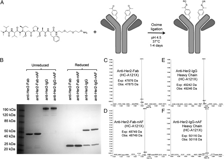
Antibody-drug conjugates (ADCs) allow selective targeting of cytotoxic drugs to cancer cells presenting tumor-associated surface markers, thereby minimizing systemic toxicity. Traditionally, the drug is conjugated nonselectively to cysteine or lysine residues in the antibody. However, these strategies often lead to heterogeneous products, which make optimization of the biological, physical, and pharmacological properties of an ADC challenging. Here we demonstrate the use of genetically encoded unnatural amino acids with orthogonal chemical reactivity to synthesize homogeneous ADCs with precise control of conjugation site and stoichiometry. p-Acetylphenylalanine was site-specifically incorporated into an anti-Her2 antibody Fab fragment and full-length IgG in Escherichia coli and mammalian cells, respectively. The mutant protein was selectively and efficiently conjugated to an auristatin derivative through a stable oxime linkage. The resulting conjugates demonstrated excellent pharmacokinetics, potent in vitro cytotoxic activity against Her2(+) cancer cells, and complete tumor regression in rodent xenograft treatment models. The synthesis and characterization of homogeneous ADCs with medicinal chemistry-like control over macromolecular structure should facilitate the optimization of ADCs for a host of therapeutic uses.


Conflict of interest statement

The authors declare no conflict of interest.

Figures

Fig. 1.
Fig. 1.
Site-specific conjugation of alkoxy-amine–derivatized auristatin to anti-Her2 Fab and IgG with pAcPhe. (A) The noncleavable auristatin derivatized with a terminal alkoxy-amine is coupled by oxime ligation to antibodies through pAcPhe residues. (B) SDS/PAGE gel of anti–Her2-Fab (HC-A121X) and anti–Her2-IgG (HC-A121X) before and after coupling to auristatin. The unreduced Fab migrates at ∼48 kDa and the reduced (50 mM DTT) Fab migrates as a single band at ∼24 kDa. The IgG includes natural heterogeneous N-linked glycosylation; the reduced light chain is ∼24 kDa and heavy chain is ∼55 kDa (includes glycan). The 4–12% Tris-Glycine gel (Invitrogen) has Benchmark prestained protein ladder in the first lane and was stained with Coomassie. (C–F) ESI-MS of Fab (HC-A121X) and IgG (HC-A121X) before and after conjugation to auristatin. Fab spectra (C and D) correspond to the fully intact fragment, but IgG spectra (E and F) show only the heavy chain after deglycosylation with PNGase and reduction with 10 mM DTT. All masses are as expected with a ∼874-Da difference between the conjugated and unconjugated heavy chains, corresponding to conjugation of one noncleavable auristatin per Fab or full-length heavy chain. No unreacted antibody was observed by SDS/PAGE or ESI-MS, suggesting >95% coupling efficiency.
Fig. 2.
Fig. 2.
In vitro cytotoxicity assays. (A) Anti–Her2-IgG(HC-A121X)-nAF (EC50 0.37 ± 0.38 nM) is more cytotoxic than the unconjugated auristatin-linker (EC50 1.5 ± 1.7 nM) and almost 100-times more cytotoxic than Fab(LC-K169X)-nAF (EC50 21.3 ± 13.7 nM) on MDA-MB-435/Her2+ cells. (B) IgG(HC-A121X)-nAF has little effect on MDA-MB-435/Her2 cells but the Fab-nAF is cytotoxic at >40 nM. The EC50 of the auristatin-linker alone on MDA-MB-435/Her cells is 0.92 ± 2.4 nM. (C) The cytotoxicity of nAF conjugated to sites LC-S202X (EC50 2.1 ± 2.8 nM) and HC-A121X (EC50 1.8 ± 0.33 nM) on the Fab were slightly greater than the cytotoxicity of Fab(LC-K169X)-nAF (EC50 8.3 ± 3.4 nM) on SK-BR-3 cells. Percent viability is normalized between no compound and 5 μM taxol controls. Error bars represent SD of three replicates.
Fig. 3.
Fig. 3.
In vivo efficacy and pharmacokinetics. (A) In vivo efficacy studies were performed with two 5 mg/kg i.v. doses of anti–Her2-IgG(HC-A121X)-nAF, anti–Her2-IgG alone, or DPBS against MDA-MB-435/Her2+ tumors in C.B-1.7/SCID mice. Anti–Her2-IgG(HC-A121X)-nAF (■) shows reduction of MDA-MB-435/Her2+ F-luc tumors 14 d after treatment (n = 8 mice/group; significant, P < 0.01). Tumors were implanted in the fourth mammary fat pad and sizes were monitored by longitudinal noninvasive bioluminescence imaging (IVIS 200; Caliper Life Science). (B) Dose effects were observed with a single injection of 5 mg/kg, 1 mg/kg, and 0.2 mg/kg (n = 8 mice/group). The 5-mg/kg group (■) had undetectable tumor after 14 d (significant, P < 0.01), the 1-mg/kg group (□) decreased the tumor, and 0.2 mg/kg (○) showed no difference from DPBS control. (C) MDA-MB-435/Her2 cells were treated in a similar manner with a single dose of 5 mg/kg anti–Her2-IgG-nAF. No regression of tumor was observed, and the growth curve was similar to that of DPBS control. (D) Pharmacokinetics study of serum concentration versus time of anti–Her2-IgG-nAF and anti–Her2-IgG in male Sprague–Dawley rats (n = 5 rats/group). Compound was injected at 1 mg/kg intravenously at time 0 and blood was collected at regular intervals for 14 d. Serum concentration was determined by capturing antibody with ErbB2 receptor protein and detecting with biotinylated anti–κ-antibody using 96-well ElectroChemiLuminescent technology (Meso Scale Discovery). The IgG-nAF (■) was not different from unconjugated mutant IgG alone (○). Datapoints represent mean and error bars represent SEM.

Similar articles

Cited by

References

    1. Wu AM, Senter PD. Arming antibodies: Prospects and challenges for immunoconjugates. Nat Biotechnol. 2005;23:1137–1146. - PubMed
    1. Polakis P. Arming antibodies for cancer therapy. Curr Opin Pharmacol. 2005;5:382–387. - PubMed
    1. Lambert JM. Drug-conjugated monoclonal antibodies for the treatment of cancer. Curr Opin Pharmacol. 2005;5:543–549. - PubMed
    1. Vater CA, Goldmacher VS. Antibody-cytotoxic compound conjugates for oncology. Macromolecular Anticancer Therapeutics. 2010 Part 4:331–369.
    1. Kovtun YV, Goldmacher VS. Cell killing by antibody-drug conjugates. Cancer Lett. 2007;255:232–240. - PubMed

Publication types

MeSH terms