DEC-205 is a cell surface receptor for CpG oligonucleotides
- PMID: 22988114
- PMCID: PMC3479608
- DOI: 10.1073/pnas.1208796109
DEC-205 is a cell surface receptor for CpG oligonucleotides
Abstract
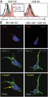
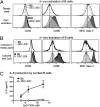
Synthetic CpG oligonucleotides (ODN) have potent immunostimulatory properties exploited in clinical vaccine trials. How CpG ODN are captured and delivered to the intracellular receptor TLR9, however, has been elusive. Here we show that DEC-205, a multilectin receptor expressed by a variety of cells, is a receptor for CpG ODN. When CpG ODN are used as an adjuvant, mice deficient in DEC-205 have impaired dendritic cell (DC) and B-cell maturation, are unable to make some cytokines such as IL-12, and display suboptimal cytotoxic T-cell responses. We reveal that DEC-205 directly binds class B CpG ODN and enhances their uptake. The CpG-ODN binding function of DEC-205 is conserved between mouse and man, although human DEC-205 preferentially binds a specific class B CpG ODN that has been selected for human clinical trials. Our findings identify an important receptor for class B CpG ODN and reveal a unique function for DEC-205.
Conflict of interest statement
The authors declare no conflict of interest.
Figures


Similar articles
-
Lymphoid follicle destruction and immunosuppression after repeated CpG oligodeoxynucleotide administration.Nat Med. 2004 Feb;10(2):187-92. doi: 10.1038/nm987. Epub 2004 Jan 25. Nat Med. 2004. PMID: 14745443
-
CD205-TLR9-IL-12 axis contributes to CpG-induced oversensitive liver injury in HBsAg transgenic mice by promoting the interaction of NKT cells with Kupffer cells.Cell Mol Immunol. 2017 Aug;14(8):675-684. doi: 10.1038/cmi.2015.111. Epub 2016 Apr 4. Cell Mol Immunol. 2017. PMID: 27041637 Free PMC article.
-
Robust T-cell stimulation by Epstein-Barr virus-transformed B cells after antigen targeting to DEC-205.Blood. 2013 Feb 28;121(9):1584-94. doi: 10.1182/blood-2012-08-450775. Epub 2013 Jan 7. Blood. 2013. PMID: 23297134 Free PMC article.
-
CD93 is a cell surface lectin receptor involved in the control of the inflammatory response stimulated by exogenous DNA.Immunology. 2019 Oct;158(2):85-93. doi: 10.1111/imm.13100. Epub 2019 Jul 23. Immunology. 2019. PMID: 31335975 Free PMC article.
-
Bacterial DNA and immunostimulatory CpG oligonucleotides trigger maturation and activation of murine dendritic cells.Eur J Immunol. 1998 Jun;28(6):2045-54. doi: 10.1002/(SICI)1521-4141(199806)28:06<2045::AID-IMMU2045>3.0.CO;2-8. Eur J Immunol. 1998. PMID: 9645386
Cited by
-
Bacteria in cancer therapy: A new generation of weapons.Cancer Med. 2022 Dec;11(23):4457-4468. doi: 10.1002/cam4.4799. Epub 2022 May 6. Cancer Med. 2022. PMID: 35522104 Free PMC article. Review.
-
The Emerging Role of STING in Insect Innate Immune Responses and Pathogen Evasion Strategies.Front Immunol. 2022 May 10;13:874605. doi: 10.3389/fimmu.2022.874605. eCollection 2022. Front Immunol. 2022. PMID: 35619707 Free PMC article. Review.
-
Use of Dendritic Cell Receptors as Targets for Enhancing Anti-Cancer Immune Responses.Cancers (Basel). 2019 Mar 24;11(3):418. doi: 10.3390/cancers11030418. Cancers (Basel). 2019. PMID: 30909630 Free PMC article. Review.
-
Stories From the Dendritic Cell Guardhouse.Front Immunol. 2019 Dec 11;10:2880. doi: 10.3389/fimmu.2019.02880. eCollection 2019. Front Immunol. 2019. PMID: 31921144 Free PMC article. Review.
-
Towards Targeted Delivery Systems: Ligand Conjugation Strategies for mRNA Nanoparticle Tumor Vaccines.J Immunol Res. 2015;2015:680620. doi: 10.1155/2015/680620. Epub 2015 Dec 24. J Immunol Res. 2015. PMID: 26819957 Free PMC article. Review.
References
-
- Carbone FR, Belz GT, Heath WR. Transfer of antigen between migrating and lymph node-resident DCs in peripheral T-cell tolerance and immunity. Trends Immunol. 2004;25:655–658. - PubMed
-
- Joffre O, Nolte MA, Spörri R, Reis e Sousa C. Inflammatory signals in dendritic cell activation and the induction of adaptive immunity. Immunol Rev. 2009;227:234–247. - PubMed
-
- Kumar H, Kawai T, Akira S. Pathogen recognition by the innate immune system. Int Rev Immunol. 2011;30:16–34. - PubMed
-
- Wagner H. The sweetness of the DNA backbone drives Toll-like receptor 9. Curr Opin Immunol. 2008;20:396–400. - PubMed
Publication types
MeSH terms
Substances
LinkOut - more resources
Full Text Sources
Other Literature Sources
Molecular Biology Databases
Research Materials

