. 2012 Dec 1;590(23):6141-55.
doi: 10.1113/jphysiol.2012.240838. Epub 2012 Sep 17.

Association of TMEM16A chloride channel overexpression with airway goblet cell metaplasia

Affiliations

Association of TMEM16A chloride channel overexpression with airway goblet cell metaplasia

Paolo Scudieri et al. J Physiol. .

Abstract

The TMEM16A protein has a potential role as a Ca(2+)-activated Cl(-) channel (CaCC) in airway epithelia where it may be important in the homeostasis of the airway surface fluid. We investigated the function and expression of TMEM16A in primary human bronchial epithelial cells and in a bronchial cell line (CFBE41o-). Under resting conditions, TMEM16A protein expression was relatively low. However, TMEM16A silencing with short-interfering RNAs caused a marked inhibition of CaCC activity, thus demonstrating that a low TMEM16A expression is sufficient to support Ca(2+)-dependent Cl(-) transport. Following treatment for 24-72 h with interleukin-4 (IL-4), a cytokine that induces mucous cell metaplasia, TMEM16A protein expression was strongly increased in approximately 50% of primary bronchial epithelial cells, with a specific localization in the apical membrane. IL-4 treatment also increased the percentage of cells expressing MUC5AC, a marker of goblet cells. Interestingly, MUC5AC was detected specifically in cells expressing TMEM16A. In particular, MUC5AC was found in 15 and 60% of TMEM16A-positive cells when epithelia were treated with IL-4 for 24 or 72 h, respectively. In contrast, ciliated cells showed expression of the cystic fibrosis transmembrane conductance regulator Cl(-) channel but not of TMEM16A. Our results indicate that TMEM16A protein is responsible for CaCC activity in airway epithelial cells, particularly in cells treated with IL-4, and that TMEM16A upregulation by IL-4 appears as an early event of goblet cell differentiation. These findings suggest that TMEM16A expression is particularly required under conditions of mucus hypersecretion to ensure adequate secretion of electrolytes and water.


Figures
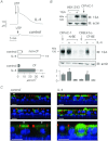
Figure 1
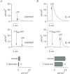
Figure 1. TMEM16A protein expression in cultured human bronchial epithelial cells
A, representative short-circuit current recordings (top) and summary of data (bottom) showing upregulation of Ca2+-dependent Cl secretion by IL-4 (10 ng ml−1 for 24 h). Before stimulation with apical UTP (100 μm), cells were previously exposed to amiloride and CFTRinh-172 (10 μm in the apical solution for both compounds) to inhibit the epithelial sodium channel (ENaC) and CFTR activity, respectively. Bars report the amplitude of the current induced by UTP (n= 6–10, **P < 0.01). B, detection of TMEM16A protein by Western blot in lysates from HEK-293 (with and without TMEM16A transfection), CFPAC-1, CFBE41o– and primary cultured bronchial epithelial cells from non-CF and CF individuals (N-BE and CF-BE, with and without IL-4 treatment). The bar graph shows a summary of the densitometric analysis performed for experiments as above. Each bar reports the relative density of TMEM16A band normalized to actin band for each sample (n= 5, *P < 0.05). C, representative xz images taken with a confocal microscope to show immunofluorescence for TMEM1A (green) and ZO-1 (red) in bronchial epithelial cells with and without IL-4 treatment. Nuclei are also stained with DAPI (blue). The blue signal below the cells arises from the porous membrane autofluorescence. The images were taken with a 40× or a 63× (bottom two images) objective. Arrows show untreated cells with apical TMEM16A localization. Scale bars: 10 μm. Data are representative of four preparations with non-CF and three preparations from CF cells.
Figure 2
Figure 2. Inhibition of CaCC activity by anti-TMEM16A siRNA and channel inhibitors
A–C, CaCC activity measured as normalized quenching rate (QR) of HS-YFP fluorescence in CFBE41o– cells. Cells were transfected with siRNAs against indicated targets obtained from Invitrogen (A), Riboxx (B) and Dharmacon (C). **P < 0.01. D, inhibition of CaCC activity (QR) in CFBE41o– cells by various concentrations of T16inh-A01 and CaCCinh-A01. Graphs show data from four separate experiments.
Figure 3
Figure 3. Inhibition of CaCC currents by anti-TMEM16A siRNA
Whole-cell membrane currents recorded with the patch-clamp technique in CFBE41o– cells transfected with control and anti-TMEM16A siRNAs. Experiments were performed with a free Ca2+ concentration of 305 nm (A) and 8 nm (B) in the pipette (intracellular) solution. Each panel shows representative currents recorded from a cell transfected with control (non-targeting, NT) siRNA and from a cell transfected with anti-TMEM16A siRNA. Current–voltage relationships summarizing the results obtained from similar experiments (mean ± SEM, n= 11–17) are depicted below corresponding traces. TMEM16A silencing caused a significant decrease of currents at all positive membrane potentials (P < 0.01) in experiments performed with high Ca2+ in the pipette solution (A). With low intracellular Ca2+, the effect of TMEM16A knockdown was not detectable (B). C, whole-cell membrane currents (left) and current–voltage relationship (right) from a representative experiment (out of three total experiments) performed on a cell transfected with a non-targeting siRNA. Currents were recorded with high and low Cl concentration in the extracellular solution. D, as C but from a cell transfected with anti-TMEM16A siRNA (n= 3).
Figure 4
Figure 4. Inhibition of Ca2+-dependent Cl secretion by TMEM16A inhibitor
Short-circuit currents recordings from human bronchial epithelial cells under control conditions (A) or after treatment with IL-4 (B). Cells were stimulated with UTP (100 μm) with and without T16inh-A01 (10 μm). The figure shows representative traces and the summary of the peak of UTP-dependent current. Experiments were carried out in the presence of amiloride and CFTRinh-172 (10 μm each). In the presence of T16inh-A01, the peak of UTP current was significantly decreased (n= 5 per condition, *P < 0.05).
Figure 5
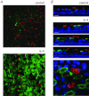
Figure 5. Expression of TMEM16A in non-ciliated cells
A, confocal microscope xy images (375 × 375 μm) of human bronchial epithelial cells with and without IL-4 treatment (10 ng ml−1, 24 h) showing immunoreactivity for TMEM16A (green) and tubulin (red). B, xz and xy images of untreated and treated cells at higher magnification. Nuclei are also stained with DAPI. Scale bars: 10 μm. Size of xy image: 65 × 65 μm.
Figure 6
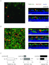
Figure 6. Expression profile of TMEM16A and MUC5AC
A and B, confocal microscope xy and xz images of human bronchial epithelial cells with and without IL-4 treatment (10 ng ml−1, 24 h) showing immunoreactivity for TMEM16A (green) and MUC5AC (red). Scale bars: 10 μm. Size of xy images: 375 × 375 μm. Nuclei are also stained with DAPI. The arrow indicates cells where MUC5AC appears to be localized in the apical membrane. C, percentage of cells expressing TMEM16A, MUC5AC and cilia with and without IL-4 for 24 h (n= 6–12, **P < 0.01, *P < 0.05).
Figure 7
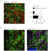
Figure 7. Differential expression of TMEM16A and CFTR in goblet and ciliated cells
A, representative image (left) showing a high level of TMEM16A and MUC5AC co-expression in primary bronchial epithelial cells treated with IL-4 for 72 h (compare with 24 h effect in Fig. 6B). Panel A also shows a bar graph (right) demonstrating the increase in cells co-expressing MUC5AC and TMEM16A at 72 h vs. 24 h (n= 6, **P < 0.01). B, representative images indicating differential expression of TMEM16A (green) and CFTR (red). Cells were treated with IL-4 for 72 h. The image on the left shows that the two proteins are expressed in different cells with little overlap. The image on the right, taken from the same field, shows also the staining for cilia (acetylated tubulin, magenta). Comparison of the two images shows perfect overlap between CFTR and cilia staining. This result is also evident from the xz image shown in the inset. Size of xy images: 375 × 375 μm.

Comment in

  • Time for TMEM?
    Fuller CM. Fuller CM. J Physiol. 2012 Dec 1;590(23):5931-2. doi: 10.1113/jphysiol.2012.245563. J Physiol. 2012. PMID: 23204100 Free PMC article. No abstract available.

References

    1. Billig GM, Pál B, Fidzinski P, Jentsch TJ. Ca2+-activated Cl− currents are dispensable for olfaction. Nat Neurosci. 2011;14:763–769. - PubMed
    1. Boucher RC. New concepts of the pathogenesis of cystic fibrosis lung disease. Eur Respir J. 2004;23:146–158. - PubMed
    1. Caputo A, Caci E, Ferrera L, Pedemonte N, Barsanti C, Sondo E, Pfeffer U, Ravazzolo R, Zegarra-Moran O, Galietta LJ. TMEM16A, a membrane protein associated with calcium-dependent chloride channel activity. Science. 2008;322:590–594. - PubMed
    1. Cui L, Aleksandrov L, Chang XB, Hou YX, He L, Hegedus T, Gentzsch M, Aleksandrov A, Balch WE, Riordan JR. Domain interdependence in the biosynthetic assembly of CFTR. J Mol Biol. 2007;365:981–994. - PubMed
    1. Curran DR, Cohn L. Advances in mucous cell metaplasia: a plug for mucus as a therapeutic focus in chronic airway disease. Am J Respir Cell Mol Biol. 2010;42:268–275. - PMC - PubMed

Publication types

LinkOut - more resources