. 2012 Nov 2;287(45):38006-19.
doi: 10.1074/jbc.M112.400580. Epub 2012 Sep 17.

Combined effects of agitation, macromolecular crowding, and interfaces on amyloidogenesis

Affiliations

Combined effects of agitation, macromolecular crowding, and interfaces on amyloidogenesis

Chiu Fan Lee et al. J Biol Chem. .

Abstract

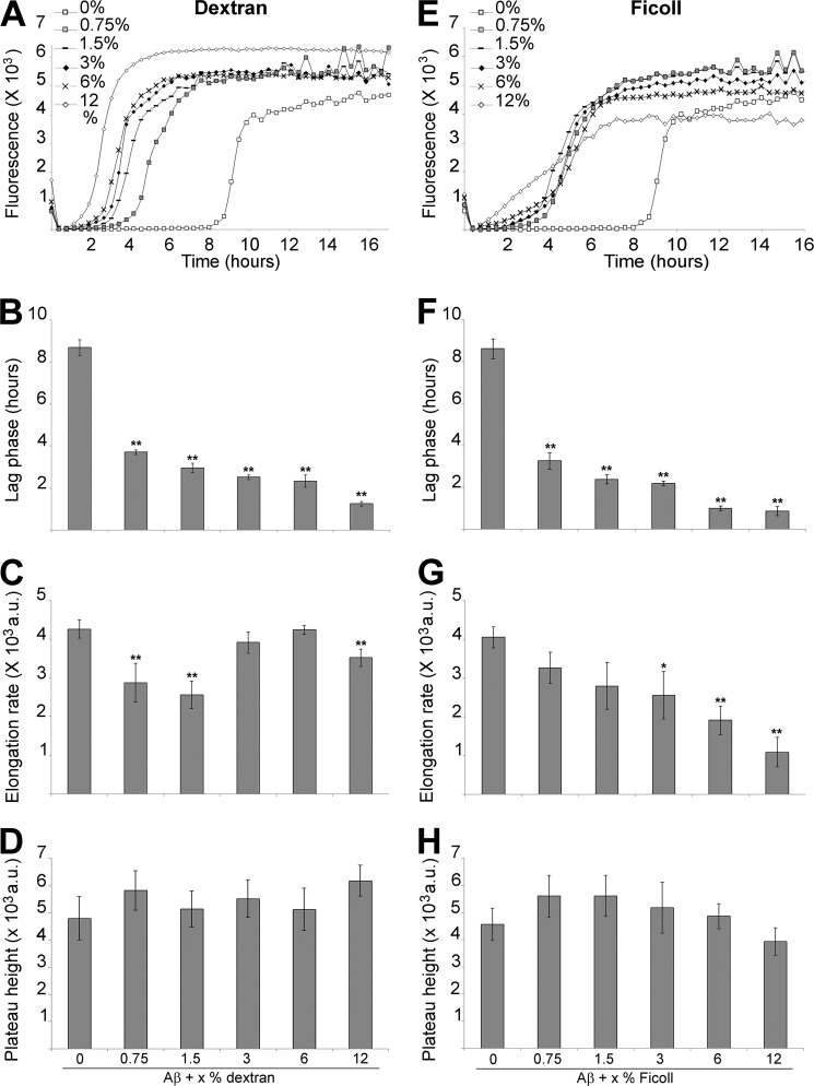
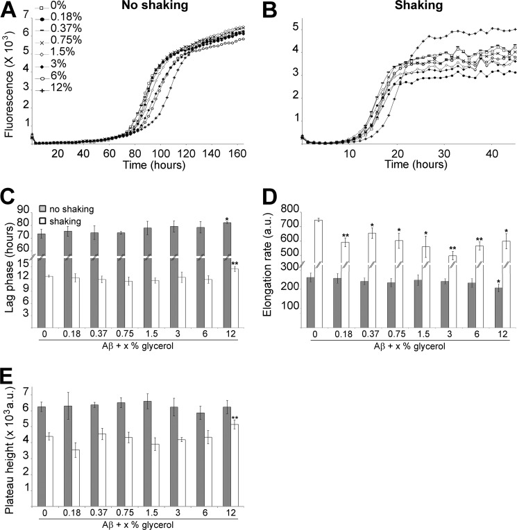
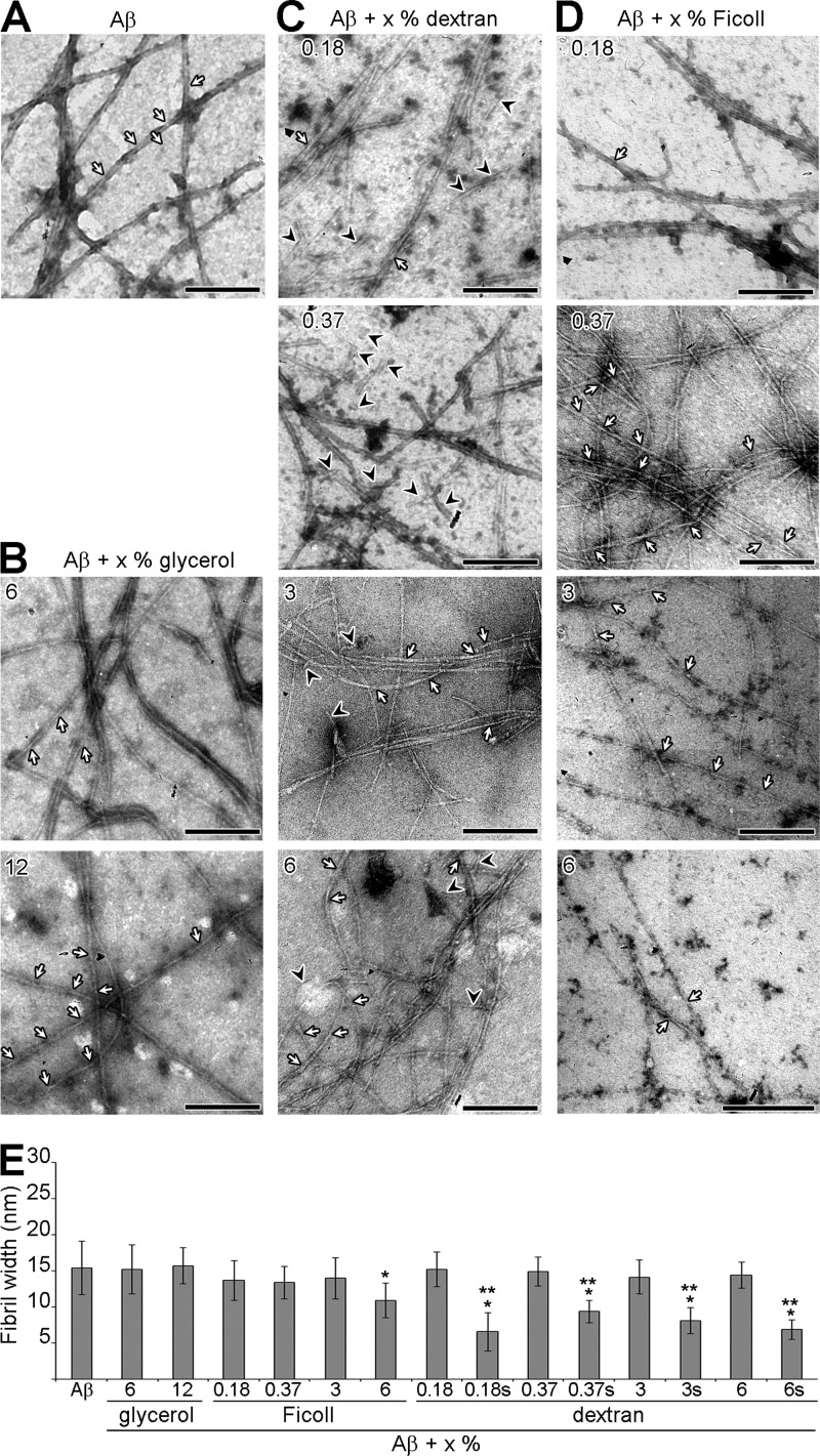
Amyloid formation and accumulation is a hallmark of protein misfolding diseases and is associated with diverse pathologies including type II diabetes and Alzheimer's disease (AD). In vitro, amyloidogenesis is widely studied in conditions that do not simulate the crowded and viscous in vivo environment. A high volume fraction of most biological fluids is occupied by various macromolecules, a phenomenon known as macromolecular crowding. For some amyloid systems (e.g. α-synuclein) and under shaking condition, the excluded volume effect of macromolecular crowding favors aggregation, whereas increased viscosity reduces the kinetics of these reactions. Amyloidogenesis can also be catalyzed by hydrophobic-hydrophilic interfaces, represented by the air-water interface in vitro and diverse heterogeneous interfaces in vivo (e.g. membranes). In this study, we investigated the effects of two different crowding polymers (dextran and Ficoll) and two different experimental conditions (with and without shaking) on the fibrilization of amyloid-β peptide, a major player in AD pathogenesis. Specifically, we demonstrate that, during macromolecular crowding, viscosity dominates over the excluded volume effect only when the system is spatially non homogeneous (i.e. an air-water interface is present). We also show that the surfactant activity of the crowding agents can critically influence the outcome of macromolecular crowding and that the structure of the amyloid species formed may depend on the polymer used. This suggests that, in vivo, the outcome of amyloidogenesis may be affected by both macromolecular crowding and spatial heterogeneity (e.g. membrane turn-over). More generally, our work suggests that any factors causing changes in crowding may be susceptibility factors in AD.


Figures

FIGURE 1.
FIGURE 1.
The effect of high concentrations of crowding agents on Aβ fibrilization under non-agitating conditions. 30 μm Aβ, in 165 μm ThT and PBS, was incubated under non-agitating conditions in absence or presence of 0.75 to 12% dextran (A–D) or Ficoll (E–H). Changes in ThT fluorescence were monitored (A and E) with the lag phase of fibrilization (B and F), the elongation rate (C and G) and plateau height (D and H) depicted. **, p < 0.03 and *, p < 0.05 when compared with Aβ without crowding agents. a.u.: arbitrary units. The mean of at least three independent assays is shown. Error bars represent ± S.E.
FIGURE 2.
FIGURE 2.
The effect of low concentrations of crowding agents on Aβ fibrilization under non-agitating conditions. 30 μm Aβ, in 165 μm ThT and PBS, was incubated under non-agitating conditions in absence or presence of 0.18 and 0.37% dextran or Ficoll. Changes in ThT fluorescence were monitored (A) with the lag phase of fibrilization (B), the elongation rate (C) and plateau height (D) depicted. **, p < 0.03 and *, p < 0.05 when compared with Aβ without crowding agents. a.u.: arbitrary units. The mean of at least three independent assays is shown. Error bars represent ± S.E.
FIGURE 3.
FIGURE 3.
The effect of crowding agents on Aβ fibrilization under agitating conditions. 30 μm Aβ, in 165 μm ThT and PBS, was incubated under agitating conditions in absence or presence of 0.75 to 12% dextran (A–D) or Ficoll (E–H). Changes in ThT fluorescence were monitored (A and E) with the lag phase of fibrilization (B and F), the elongation rate (C and G) and plateau height (D and H) depicted. ** p < 0.03 and * p < 0.05 when compared with Aβ without crowding agents. a.u.: arbitrary units. The mean of at least three independent assays is shown. Error bars represent ± S.E.
FIGURE 4.
FIGURE 4.
The effect of glycerol on Aβ fibrilization. 30 μm Aβ, in 165 μm ThT and PBS, was incubated in absence or presence of 0.18 to 12% glycerol under non-agitating (A) or agitating conditions (B). Changes in ThT fluorescence were monitored (A and B) with the lag phase of fibrilization (C), the elongation rate (D) and plateau height (E) depicted. **, p < 0.03 and *, p < 0.05 when compared with Aβ without crowding agents. a.u.: arbitrary units. The mean of at least three independent assays is shown. Error bars represent ± S.E.
FIGURE 5.
FIGURE 5.
Glycerol, dextran, and Ficoll have different surface activities and affect differently the recruitment of Aβ to the AWI. Dynamic measurement of the surface tension for solutions of 12% glycerol, dextran, or Ficoll (A), with the surface tension at 350 s depicted (B). The surface tension for a solution of 30 μm Aβ, in absence or presence of 12% glycerol or crowding agents was also monitored over time (C), with the surface tension at the start of the reaction and at 350 s (D), the rate of decrease in surface tension (E) and the time required for this decrease (F, delay) depicted. The inset in E shows, highlighted by a gray box, the area on the curves used to calculate the rate of decrease. The graphs in A and C show 4 point moving averages of the raw data. a.u.: arbitrary units. The mean of at least three independent assays is shown. Error bars represent ± S.E. ** indicates p < 0.006 when compared with Ficoll in B and when compared with Aβ alone in D to F.
FIGURE 6.
FIGURE 6.
Theoretical model linking macromolecular crowding and kinetics of amyloidogenesis. A, schematic showing the adsorbed layer at the AWI, where all the amyloid species (from monomers to fibrils) are located. The monomers in the bulk have to diffuse to the adsorbed layer in order to fibrilize. B, schematic showing the adsorbed layer of amyloid species (from monomers to fibrils) at the AWI and dextran in the bulk solution (left panel). Since dextran does not interact with the adsorbed layer, the crowding agent only affects the viscosity of the solution and as a result, the dynamics of fibrilization is slowed down (right panel). C, schematic showing the adsorbed layer of amyloid species (from monomers to fibrils) at the AWI and Ficoll at the AWI and in the bulk solution (left panel). Since Ficoll is surface active, it interacts with the adsorbed layer and as a result, decreases the kinetics of fibrilization and the amount of fibrils at the AWI (right panel). See “Experimental Procedures” on how the curves were generated.
FIGURE 7.
FIGURE 7.
The effect of crowding agents on the morphology of Aβ fibrils under non-agitating conditions. A–D, electron micrographs of negatively stained Aβ fibrilization reactions. Aβ fibrilization was performed under non-agitating conditions in absence (A) or presence of different percentages of glycerol (B), dextran (C), or Ficoll (D) until the reaction reached plateau. The white arrows indicate helical twists in the fibrils, and the black arrowheads indicate short fibrilar species. The scale bar represents 200 nm. E, the width of Aβ fibrils (n = 12), in absence or presence of glycerol, dextran, or Ficoll, was measured from electron micrographs of negatively stained reactions. s indicates short fibrilar species (as defined in the text). * indicates p < 0.004 when compared with Aβ, ** indicates p < 6 × 10⁻⁷ when compared with Aβ + x% dextran.
FIGURE 8.
FIGURE 8.
The effect of crowding agents on the morphology of Aβ fibrils under agitating conditions. Electron micrographs of negatively stained Aβ fibrilization reactions. Aβ fibrilization was performed under agitating conditions in absence or presence of 12% glycerol, dextran, or Ficoll until the reaction reached plateau. The white arrows indicate helical twists in the fibrils, the black arrowheads indicate short fibrilar species, and the white arrowheads indicate short fibrils. The scale bar represents 200 nm. The width of Aβ fibrils (n = 12), in absence or presence of glycerol, dextran, or Ficoll, was measured from electron micrographs of negatively stained reactions (right panel). short indicates short fibrilar species (as defined in the text). ** indicates p < 0.02.

References

    1. Stefani M., Dobson C. M. (2003) Protein aggregation and aggregate toxicity: new insights into protein folding, misfolding diseases and biological evolution. J. Mol. Med. 81, 678–699 - PubMed
    1. Haass C., Selkoe D. J. (2007) Soluble protein oligomers in neurodegeneration: lessons from the Alzheimer's amyloid beta-peptide. Nat. Rev. Mol. Cell Biol. 8, 101–112 - PubMed
    1. Selkoe D. J. (2004) Cell biology of protein misfolding: the examples of Alzheimer's and Parkinson's diseases. Nat. Cell Biol. 6, 1054–1061 - PubMed
    1. Harper J. D., Lansbury P. T., Jr. (1997) Models of amyloid seeding in Alzheimer's disease and scrapie: mechanistic truths and physiological consequences of the time-dependent solubility of amyloid proteins. Annu. Rev. Biochem. 66, 385–407 - PubMed
    1. Lee C. F. (2009) Self-assembly of protein amyloids: A competition between amorphous and ordered aggregation Phys. Rev. E. 80, 031922 - PubMed

Publication types

LinkOut - more resources