

Mechanisms for kinase-mediated dimerization of the epidermal growth factor receptor
- PMID: 22988250
- PMCID: PMC3488093
- DOI: 10.1074/jbc.M112.414391
Mechanisms for kinase-mediated dimerization of the epidermal growth factor receptor
Abstract
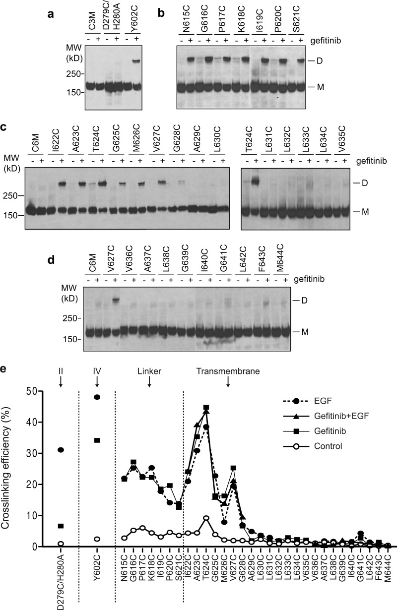
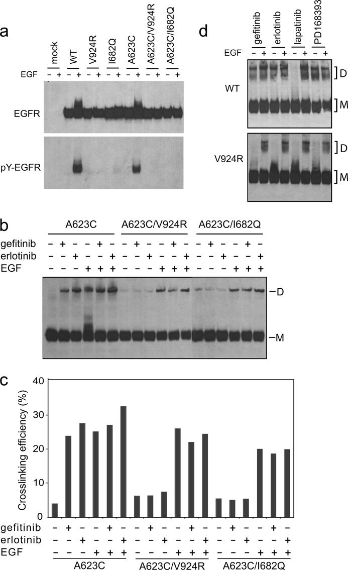
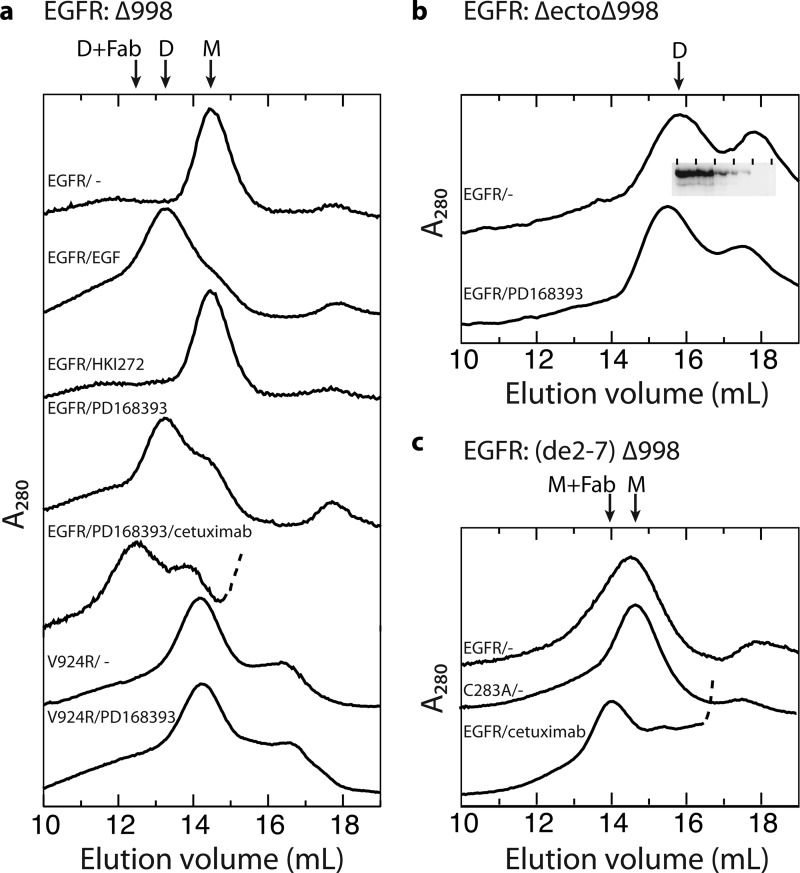
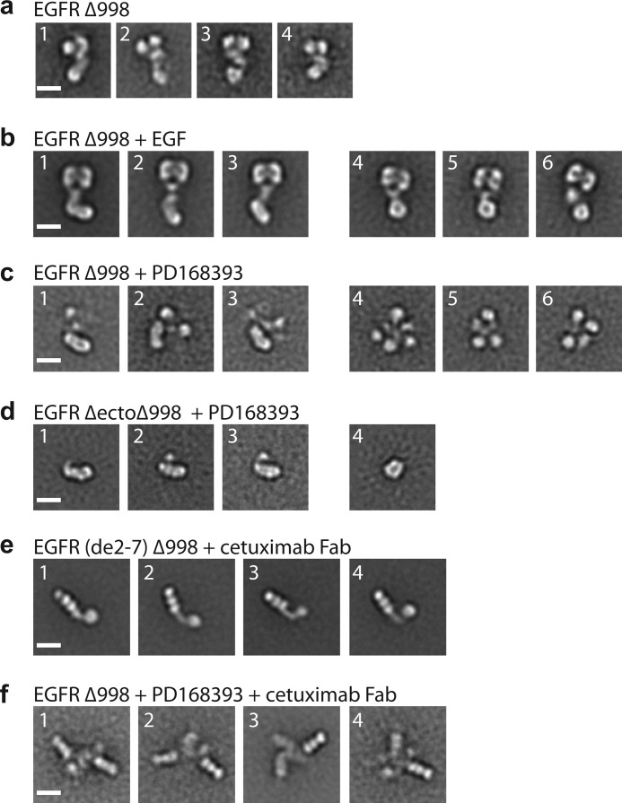
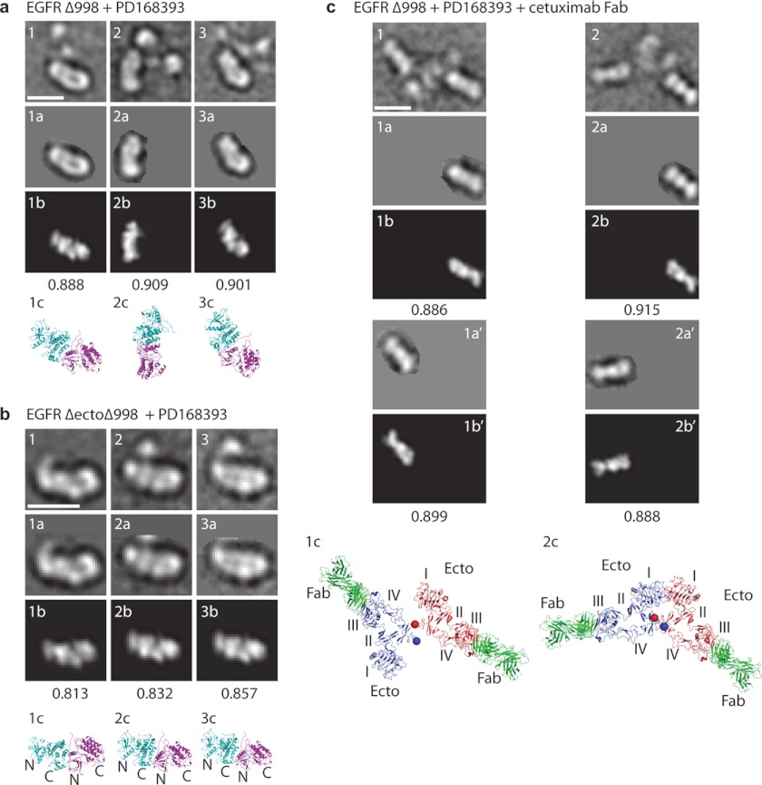
We study a mechanism by which dimerization of the EGF receptor (EGFR) cytoplasmic domain is transmitted to the ectodomain. Therapeutic and other small molecule antagonists to the kinase domain that stabilize its active conformation, but not those that stabilize an inactive conformation, stabilize ectodomain dimerization. Inhibitor-induced dimerization requires an asymmetric kinase domain interface associated with activation. EGF and kinase inhibitors stimulate formation of identical dimer interfaces in the EGFR transmembrane domain, as shown by disulfide cross-linking. Disulfide cross-linking at an interface in domain IV in the ectodomain was also stimulated similarly; however, EGF but not inhibitors stimulated cross-linking in domain II. Inhibitors similarly induced noncovalent dimerization in nearly full-length, detergent-solubilized EGFR as shown by gel filtration. EGFR ectodomain deletion resulted in spontaneous dimerization, whereas deletion of exons 2-7, in which extracellular domains III and IV are retained, did not. In EM, kinase inhibitor-induced dimers lacked any well defined orientation between the ectodomain monomers. Fab of the therapeutic antibody cetuximab to domain III confirmed a variable position and orientation of this domain in inhibitor-induced dimers but suggested that the C termini of domain IV of the two monomers were in close proximity, consistent with dimerization in the transmembrane domains. The results provide insights into the relative energetics of intracellular and extracellular dimerization in EGFR and have significance for physiologic dimerization through the asymmetric kinase interface, bidirectional signal transmission in EGFR, and mechanism of action of therapeutics.
Figures

Similar articles
-
Simultaneous visualization of the extracellular and cytoplasmic domains of the epidermal growth factor receptor.Nat Struct Mol Biol. 2011 Aug 7;18(9):984-9. doi: 10.1038/nsmb.2092. Nat Struct Mol Biol. 2011. PMID: 21822280 Free PMC article.
-
Structural evidence for loose linkage between ligand binding and kinase activation in the epidermal growth factor receptor.Mol Cell Biol. 2010 Nov;30(22):5432-43. doi: 10.1128/MCB.00742-10. Epub 2010 Sep 13. Mol Cell Biol. 2010. PMID: 20837704 Free PMC article.
-
Kinase-mediated quasi-dimers of EGFR.FASEB J. 2010 Dec;24(12):4744-55. doi: 10.1096/fj.10-166199. Epub 2010 Aug 3. FASEB J. 2010. PMID: 20682838 Free PMC article.
-
Small molecule inhibitors targeting the EGFR/ErbB family of protein-tyrosine kinases in human cancers.Pharmacol Res. 2019 Jan;139:395-411. doi: 10.1016/j.phrs.2018.11.014. Epub 2018 Nov 27. Pharmacol Res. 2019. PMID: 30500458 Review.
-
The ErbB/HER family of protein-tyrosine kinases and cancer.Pharmacol Res. 2014 Jan;79:34-74. doi: 10.1016/j.phrs.2013.11.002. Epub 2013 Nov 20. Pharmacol Res. 2014. PMID: 24269963 Review.
Cited by
-
A highly efficient peptide substrate for EGFR activates the kinase by inducing aggregation.Biochem J. 2013 Aug 1;453(3):337-44. doi: 10.1042/BJ20130537. Biochem J. 2013. PMID: 23734957 Free PMC article.
-
Fluorescence Imaging of Epidermal Growth Factor Receptor Tyrosine Kinase Inhibitor Resistance in Non-Small Cell Lung Cancer.Cancers (Basel). 2022 Jan 28;14(3):686. doi: 10.3390/cancers14030686. Cancers (Basel). 2022. PMID: 35158954 Free PMC article. Review.
-
Magnetic nanoparticles as mediators of ligand-free activation of EGFR signaling.PLoS One. 2013 Jul 23;8(7):e68879. doi: 10.1371/journal.pone.0068879. Print 2013. PLoS One. 2013. PMID: 23894364 Free PMC article.
-
Dimerization of the Trk receptors in the plasma membrane: effects of their cognate ligands.Biochem J. 2018 Nov 30;475(22):3669-3685. doi: 10.1042/BCJ20180637. Biochem J. 2018. PMID: 30366959 Free PMC article.
-
The RTK Interactome: Overview and Perspective on RTK Heterointeractions.Chem Rev. 2019 May 8;119(9):5881-5921. doi: 10.1021/acs.chemrev.8b00467. Epub 2018 Dec 27. Chem Rev. 2019. PMID: 30589534 Free PMC article. Review.
References
-
- Hynes N. E., Lane H. A. (2005) ERBB receptors and cancer. The complexity of targeted inhibitors. Nat. Rev. Cancer 5, 341–354 - PubMed
-
- Arteaga C. L. (2003) ErbB-targeted therapeutic approaches in human cancer. Exp. Cell Res. 284, 122–130 - PubMed
-
- Ferguson K. M., Berger M. B., Mendrola J. M., Cho H. S., Leahy D. J., Lemmon M. A. (2003) EGF activates its receptor by removing interactions that autoinhibit ectodomain dimerization. Mol. Cell 11, 507–517 - PubMed
-
- Ogiso H., Ishitani R., Nureki O., Fukai S., Yamanaka M., Kim J. H., Saito K., Sakamoto A., Inoue M., Shirouzu M., Yokoyama S. (2002) Crystal structure of the complex of human epidermal growth factor and receptor extracellular domains. Cell 110, 775–787 - PubMed
-
- Garrett T. P., McKern N. M., Lou M., Elleman T. C., Adams T. E., Lovrecz G. O., Zhu H. J., Walker F., Frenkel M. J., Hoyne P. A., Jorissen R. N., Nice E. C., Burgess A. W., Ward C. W. (2002) Crystal structure of a truncated epidermal growth factor receptor extracellular domain bound to transforming growth factor α. Cell 110, 763–773 - PubMed
Publication types
MeSH terms
Substances
Grants and funding
LinkOut - more resources
Full Text Sources
Other Literature Sources
Research Materials
Miscellaneous
