. 2012 Nov;32(22):4674-83.
doi: 10.1128/MCB.01067-12. Epub 2012 Sep 17.

A phosphatidylinositol 3-kinase-Pax3 axis regulates Brn-2 expression in melanoma

Affiliations

A phosphatidylinositol 3-kinase-Pax3 axis regulates Brn-2 expression in melanoma

Elise Bonvin et al. Mol Cell Biol. 2012 Nov.

Abstract

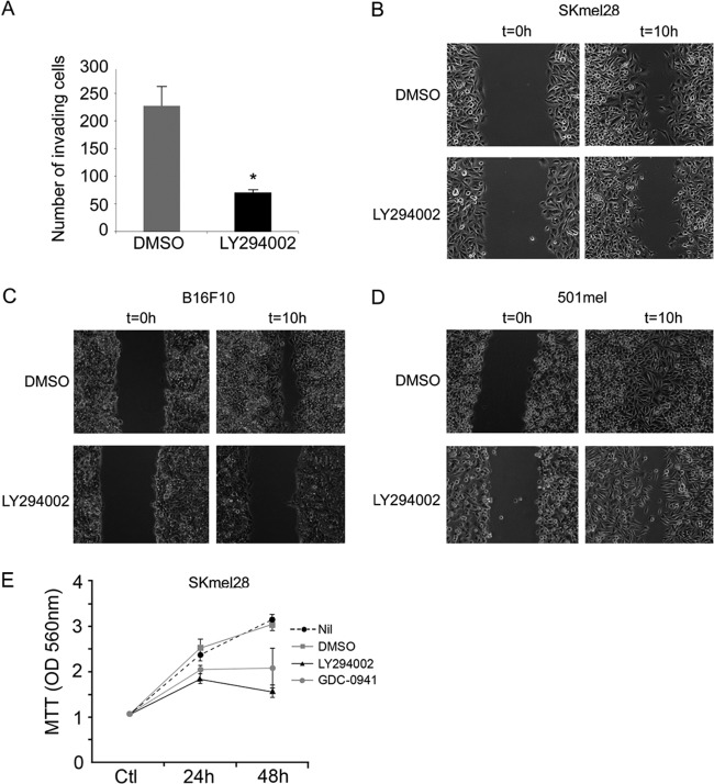
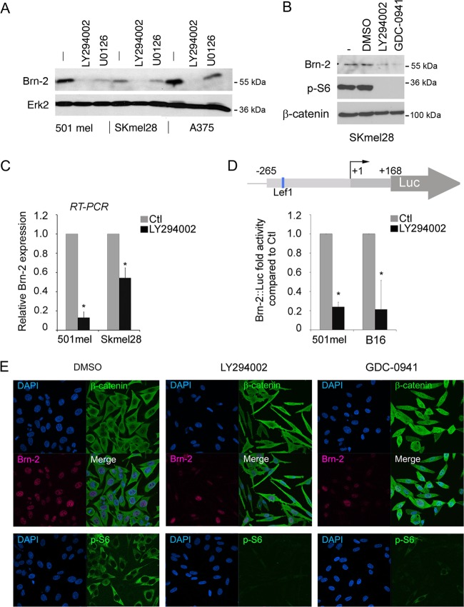
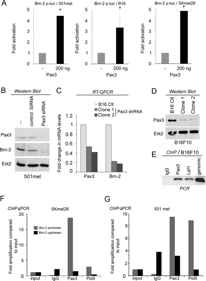
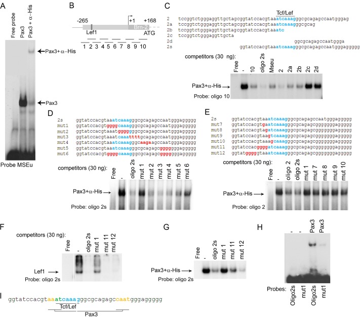
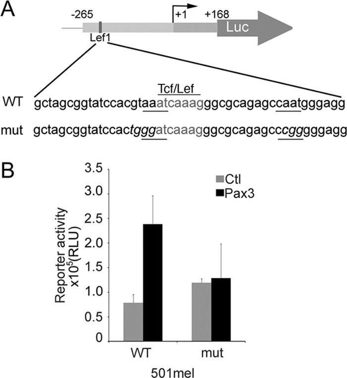
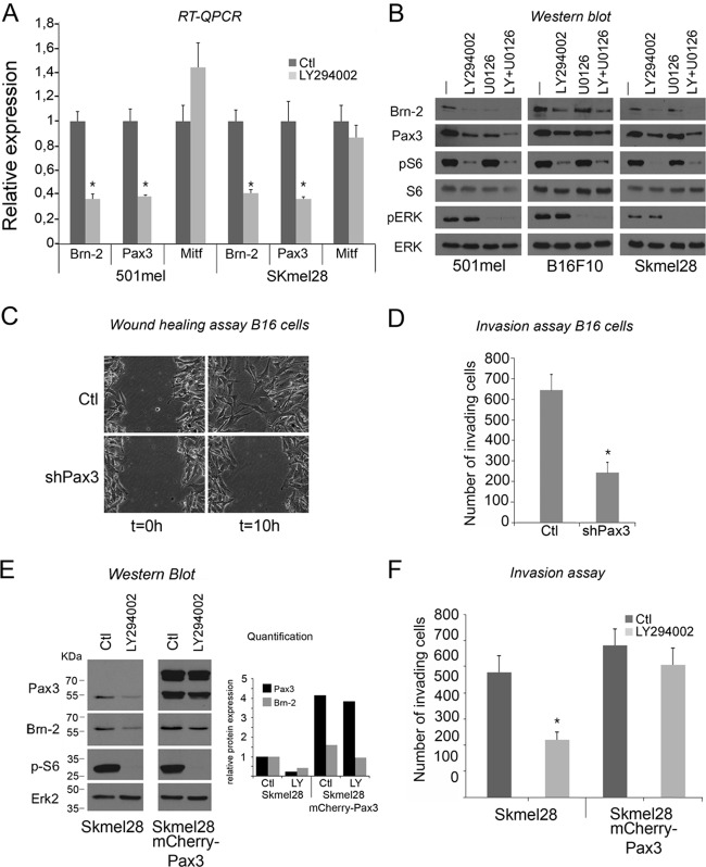
Deregulation of transcription arising from mutations in key signaling pathways is a hallmark of cancer. In melanoma, the most aggressive and lethal form of skin cancer, the Brn-2 transcription factor (POU3F2) regulates proliferation and invasiveness and lies downstream from mitogen-activated protein kinase (MAPK) and Wnt/β-catenin, two melanoma-associated signaling pathways. In vivo Brn-2 represses expression of the microphthalmia-associated transcription factor, MITF, to drive cells to a more stem cell-like and invasive phenotype. Given the key role of Brn-2 in regulating melanoma biology, understanding the signaling pathways that drive Brn-2 expression is an important issue. Here, we show that inhibition of phosphatidylinositol 3-kinase (PI3K) signaling reduces invasiveness of melanoma cells in culture and strongly inhibits Brn-2 expression. Pax3, a transcription factor regulating melanocyte lineage-specific genes, directly binds and regulates the Brn-2 promoter, and Pax3 expression is also decreased upon PI3K inhibition. Collectively, our results highlight a crucial role for PI3K in regulating Brn-2 and Pax3 expression, reveal a mechanism by which PI3K can regulate invasiveness, and imply that PI3K signaling is a key determinant of melanoma subpopulation diversity. Together with our previous work, the results presented here now place Brn-2 downstream of three melanoma-associated signaling pathways.


Figures

Fig 1
Fig 1
LY294002 treatment decreases invasion and migration of melanoma cell lines. (A) Skmel28 invasive melanoma cells were treated with 20 μM LY294002. After 24 h, the number of LY294002-treated invading cells was counted and compared to the control (DMSO). Columns represent means from three experiments; bars indicate SD; *, P < 0.05. (B to D) LY294002 treatment decreases the wound-healing ability of the SKmel28 (B), B16 (C), and 501mel (D) melanoma cell lines in a scratch/wound assay. (E) Proliferation MTT assay on Skmel28 cells over 48 h in the presence or absence of PI3K inhibitors. Values represent means of optical density values measured at 560 nm ± standard deviations (SD).
Fig 2
Fig 2
PI3K inhibition decreases Brn-2 protein expression. (A) Western blot of 501mel, SKmel28, and A375 melanoma cells treated with LY294002 (25 μM) for 24 h. MEK inhibition (U0126; 10 μM) was used as a control, and Erk2 was used to ensure equal loading. (B) Western blot performed on SKmel28 cells treated with two different PI3K inhibitors (LY294002 at 25 μM or GDC-0941 at 10 μM) for 24 h using anti-Brn-2 and anti-p-S6 antibodies. β-Catenin was used as a loading control. (C) Relative expression of Brn-2 mRNA as determined using quantitative real-time RT-PCR from the indicated melanoma cell lines. Values were normalized to those for GAPDH and ratios were established compared to DMSO treatment. (D) Schematic of the Brn-2 promoter showing the binding site for Lef1 (top). Shown is a luciferase assay of 501mel and SKmel28 cells transfected with a Brn-2-luciferase reporter and treated with LY294002 or DMSO. Columns represent means from three experiments; bars indicate SD; *, P < 0.05. (E) PI3K inhibition does not affect β-catenin cellular localization. Immunofluorescence performed on SKmel28 treated with LY294002, GDC-0941, or DMSO (Ctl) using a monoclonal mouse anti-β-catenin (green), a polyclonal goat anti-Brn2, and a polyclonal rabbit anti-p-S6 antibody. Nuclei were counterstained using DAPI.
Fig 3
Fig 3
Pax3 regulates Brn-2 transcription. (A) Luciferase assay of B16, SKmel28, and 501mel cells transfected with a Brn-2-luciferase reporter together with a Pax3 expression vector or control vector as indicated. Data from a representative experiment are presented as fold changes compared to the control (set to 1). Columns represent means from four experiments; bars indicate SD; *, P < 0.05. (B) Western blot using anti-Brn-2 or anti-Pax3 antibodies of 501mel transfected with an siRNA of Pax3 or scrambled (control) as indicated. Erk2 was used as a loading control. (C) Relative expression of Pax3 and Brn-2 mRNA as determined using RT-qPCR from two independent clones of B16F10 depleted for Pax3 using shRNA compared to the WT B16F10. Values were normalized to GAPDH and are shown as fold changes. (D) Western blot of Pax3 protein in parental and shPax3 B16-expressing clones 1 and 2. ERK was used as a loading control. (E) Chromatin immunoprecipitation (ChIP) of B16 cells using an anti-Pax3 or anti-Lef1 antibody as a control. Amplification of the Brn-2 promoter was detected by semiquantitative PCR using primers specific for the Brn-2 promoter. (F and G) ChIP of Pax3 and RNA PolII followed by qPCR at the Brn-2 promoter or an unrelated region 1.8 kb upstream using IgG as a control in the indicated cell lines.
Fig 4
Fig 4
Pax3 directly binds the Brn-2 promoter. (A) Band shift using the MSEu probe from the Tyrp1 promoter together with bacterially expressed and purified Pax3-PDHD-His tagged with or without α-His antibody. (B) Schematic of the Brn-2 promoter showing the Tcf/Lef1 binding site. The lines under the Brn-2 promoter represent the locations of 10 oligonucleotides used as probes or competitors in our band shift assays. (C) Band shift assay using an excess of probe (oligonucleotide 10) together with bacterially expressed and purified Pax3-PDHD-His. Thirty nanograms of the indicated competitors corresponding to a region of the proximal promoter of Brn-2 was used. The described Tcf/Lef1 binding site is highlighted in blue. (D) Binding of Pax3 to the probe oligonucleotide 2s was challenged using 30 ng of the indicated competitors. Blue, Lef1 binding site; red, mutated bases of the potential Pax3 binding site. (E) Binding of Pax3 to the probe oligonucleotide 2 was challenged using 30 ng of the indicated competitors. (F) Band shift using bacterially purified GST-Lef1 together with oligonucleotide 2s and 30 ng of unlabeled oligonucleotide 2s corresponding to the probe and mutant 1, 11, and 12 as competitors. (G) Binding of Pax3 to the probe oligonucleotide 2 was challenged using 30 ng of the indicated competitors. (H) Binding of Pax3 to WT oligonucleotide 2s or Mut1 probes as indicated. (I) Scheme representing the Pax3 binding sites (yellow) adjacent to Lef1 (blue) on the Brn-2 promoter. The bases shared by both Pax3 and Lef1 are represented in green.
Fig 5
Fig 5
(A) Scheme representing the WT Brn-2 promoter reporter construct with the Lef/Tcf site in gray, the two putative Pax3 binding element underlined, and the mutated bases represented in italics. (B) Luciferase assay was performed on 501mel cells to confirm the loss of responsiveness of the Brn-2 promoter mutated for the Pax3 binding site after transfection of Pax3 or an empty vector (Ctl). Values reported are means from 2 independent experiments performed in triplicate; error bars represent SD.
Fig 6
Fig 6
Pax3 is implicated in melanoma cell migration and invasiveness. (A) Relative Brn-2, Pax3, and Mitf mRNA levels of 501mel and SKmel28 cells were determined by RT-qPCR. Results represent the means from three independent experiments with error bars indicating standard deviations. (B) Western blot of 501mel, B16, and Skeml28 cells after 24 h of treatment with LY294002 using anti-Brn-2 and anti-Pax3 antibodies. Anti-p-S6 was used as a control of LY294002 efficiency, and total S6 (tS6) and Erk2 were used for the loading control. p-Erk was used for PI3K inhibition specificity. (C) Pax3 knockdown decreases the wound-healing ability of the B16 cell line in a scratch/wound assay. (D) Pax3 knockdown decreases invasiveness potential of B16 cells. After 24 h, the number B16ShPax3 clone 1 invading cells was counted and compared to the control (parental cell line). (E) Brn-2 expression is sustained upon LY294002 exposure when Pax3 is overexpressed. Brn-2 and Pax-3 expression analysis was assessed by Western blotting of Skmel28 parental or Skmel28 cells expressing mCherry-Pax3 protein. Anti-p-S6 was used as a control of LY294002 efficiency, and total S6 (tS6) and Erk2 were used for the loading control. Quantification was achieved using ImageJ software. (F) LY294002 does not decrease invasion of Skmel28 melanoma cells when Pax3 is overexpressed. After 24 h, the number of Skmel28 parental cells or those expressing mCherry-Pax3 treated with LY294002 was counted and compared to the control (DMSO). Columns represent means from three experiments; bars indicate SD; *, P < 0.05.

References

    1. Arnheiter H. 2010. The discovery of the microphthalmia locus and its gene, Mitf. Pigment Cell Melanoma Res. 23:729–735 - PMC - PubMed
    1. Arozarena I, et al. 2011. Oncogenic BRAF induces melanoma cell invasion by downregulating the cGMP-specific phosphodiesterase PDE5A. Cancer Cell 19:45–57 - PubMed
    1. Aziz SA, et al. 2009. Phosphatidylinositol-3-kinase as a therapeutic target in melanoma. Clin. Cancer Res. 15:3029–3036 - PMC - PubMed
    1. Boyle GM, et al. 2011. Melanoma cell invasiveness is regulated by miR-211 suppression of the BRN2 transcription factor. Pigment Cell Melanoma Res. 24:525–537 - PubMed
    1. Carreira S, et al. 2005. Mitf cooperates with Rb1 and activates p21Cip1 expression to regulate cell cycle progression. Nature 433:764–769 - PubMed

Publication types

MeSH terms

Substances