. 2012 Nov;32(22):4691-704.
doi: 10.1128/MCB.06267-11. Epub 2012 Sep 17.

Interaction of cyclin-dependent kinase 12/CrkRS with cyclin K1 is required for the phosphorylation of the C-terminal domain of RNA polymerase II

Affiliations

Interaction of cyclin-dependent kinase 12/CrkRS with cyclin K1 is required for the phosphorylation of the C-terminal domain of RNA polymerase II

S-W Grace Cheng et al. Mol Cell Biol. 2012 Nov.

Abstract

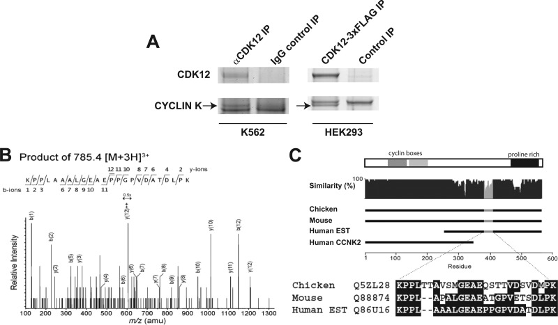
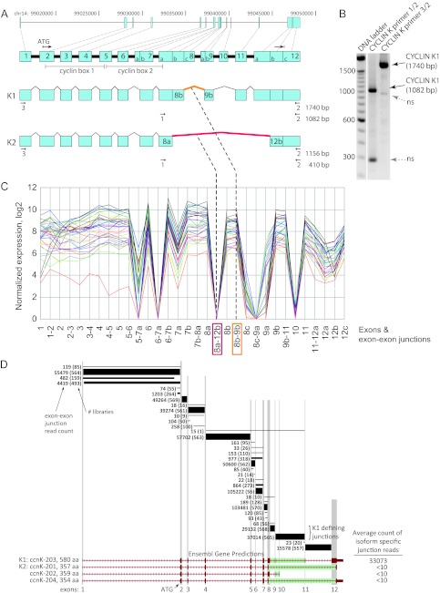
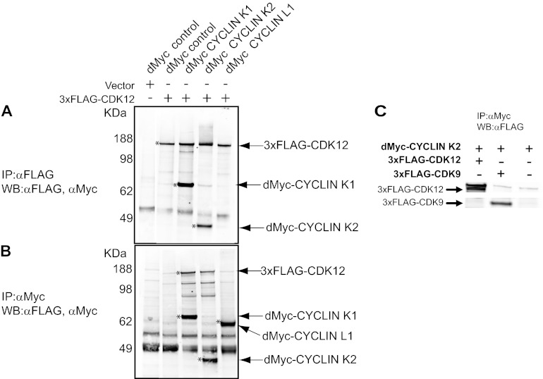
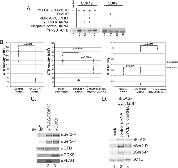
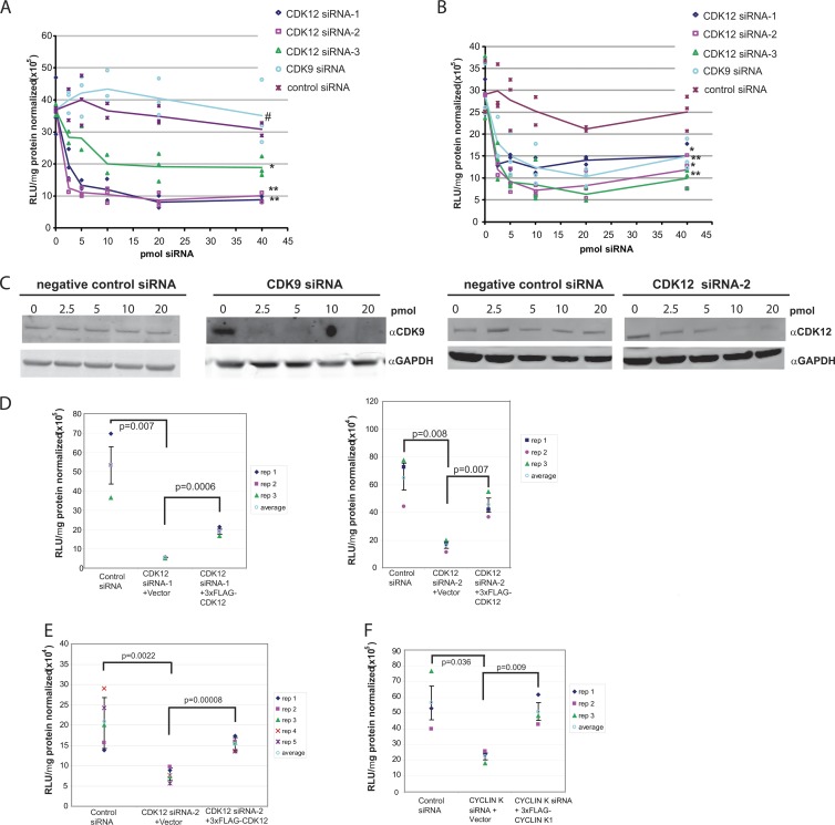
CrkRS (Cdc2-related kinase, Arg/Ser), or cyclin-dependent kinase 12 (CKD12), is a serine/threonine kinase believed to coordinate transcription and RNA splicing. While CDK12/CrkRS complexes were known to phosphorylate the C-terminal domain (CTD) of RNA polymerase II (RNA Pol II), the cyclin regulating this activity was not known. Using immunoprecipitation and mass spectrometry, we identified a 65-kDa isoform of cyclin K (cyclin K1) in endogenous CDK12/CrkRS protein complexes. We show that cyclin K1 complexes isolated from mammalian cells contain CDK12/CrkRS but do not contain CDK9, a presumed partner of cyclin K. Analysis of extensive RNA-Seq data shows that the 65-kDa cyclin K1 isoform is the predominantly expressed form across numerous tissue types. We also demonstrate that CDK12/CrkRS is dependent on cyclin K1 for its kinase activity and that small interfering RNA (siRNA) knockdown of CDK12/CrkRS or cyclin K1 has similar effects on the expression of a luciferase reporter gene. Our data suggest that cyclin K1 is the primary cyclin partner for CDK12/CrkRS and that cyclin K1 is required to activate CDK12/CrkRS to phosphorylate the CTD of RNA Pol II. These properties are consistent with a role of CDK12/CrkRS in regulating gene expression through phosphorylation of RNA Pol II.


Figures

Fig 1
Fig 1
Identification of a novel isoform of cyclin K in CDK12/CrkRS protein complexes. (A) Colloidal Coomassie stain of immunoprecipitated endogenous CDK12/CrkRS complexes from K562 cells and 3×FLAG-CDK12/CrkRS complexes from HEK293A cells. Cyclin K peptides were found in a 65- to 80-kDa gel slice. (B) MS-MS spectra of a cyclin K1 peptide. (C) Sequence of a human C-terminal cyclin K1 peptide found by MS-MS aligned to the corresponding chicken and mouse cyclin K peptides. The predicted cyclin boxes and a proline-rich domain of the cyclin K1 isoform are shown in the bar above the alignment.
Fig 2
Fig 2
Cyclin K1 is the predominant cyclin K mRNA isoform. (A) Cyclin K gene exon arrangement and splicing. The exon arrangement of the CCNK locus on chromosome 14 is depicted. Splicing pathways that produce a 43-kDa isoform (cyclin K2) and a 64-kDa isoform (cyclin K1) are shown; subdivisions in exons 7, 8, 9, and 12 represent different known splice acceptors and donors observed in the mRNA and EST data. The numbered arrows depict primers and RT-PCR product sizes that were used to discriminate between the cyclin K1 and K2 isoforms. The 8b-9b (orange) and 8a-12b (red) exon-exon junctions differentiate the expression of the cyclin K1 and K2 isoforms, respectively. (B) Cyclin K1 mRNA is present in HeLa cells. RT-PCR of HeLa poly(A)-enriched RNA was performed with primer pairs 1/2 and 3/2 that were designed to differentiate between cDNAs for cyclin K1 and K2 (see panel A). RT-PCR products corresponding to the predicted size for cyclin K1 are indicated. Sequencing of the ∼300-bp and ∼950-bp bands showed that they are not cyclin K related, and they are denoted as nonspecific (ns). (C) RNA-Seq analysis of known cyclin K exons and exon junctions. The normalized expression of observed combinations of known splice donors and acceptor sites (Fig. 2A) for the CCNK locus for 38 human cancer and cell line mRNA libraries is shown. (D) Detection of cyclin K exon-exon junctions by HMMSplicer analysis of RNA-Seq data. Exon-exon junction RNA-Seq read counts (552,300 total) from 570 human cancer and cell mRNA libraries that mapped to the CCNK locus using HMMSplicer are shown. The junctions putatively connect the exons (vertical gray bars) for the Ensembl-predicted gene transcripts (red, bottom). Total read count among the 570 libraries for each junction read is shown; in parentheses is the number of libraries where the junction read was observed. The thickness of each line approximates the relative abundance of the junctions. In the exon 8 region, three different splice donor sites (Fig. 2A) are used between four Ensembl transcripts. The green bars represent the junctions or junction combinations that would uniquely define each of the Ensembl-predicted isoforms. The number of counts observed for each of the isoform-defining junctions is shown in the right-hand column. For the K1 isoform (CCNK-203), the isoform-specific read count, 33,073, is the mean of the counts of the two K1-defining junctions (29,132 and 37,014).
Fig 3
Fig 3
Immunoprecipitation-Western blotting validation of CDK12-cyclin K interaction. (A and B) Western blot analyses of anti-FLAG-immunoprecipitated complexes probed with anti-FLAG and anti-Myc antibodies (A) and of anti-Myc-immunoprecipitated complexes probed with anti-FLAG and anti-Myc antibodies (B). An asterisk denotes the protein indicated by the arrow on the right. dMyc, 2×Myc. (C) dMyc-cyclin K2 was immunoprecipitated with anti-Myc, and the blots probed with anti-FLAG antibody. Both 3×FLAG-CDK12 and 3×FLAG-CDK9 interact with dMyc-cyclin K2.
Fig 4
Fig 4
CDK12 is dependent on cyclin K to phosphorylate the C-terminal domain of RNA Pol II. (A) Cyclin K expression modulates CTD phosphorylation by CDK12. 3×FLAG-CDK12 complexes isolated from cells stably expressing 3×FLAG-CDK12 that were modulated with cyclin K siRNA designed against the 3′ UTR of the CCNK gene showed a significant decrease in RNA Pol II CTD phosphorylation compared to the results for the negative control (lane 3 versus lane 2). CTD phosphorylation was restored with the expression of dMyc-cyclin K from an exogenous plasmid (lane 4). Lane 1, a negative-control anti-FLAG IP from a control cell line not expressing 3×FLAG-CDK12; lanes 2 to 9, anti-FLAG-CDK12 or endogenous CDK9 complexes isolated from a cell line stably expressing 3×FLAG-CDK12; lane 5, cells were mock transfected with an empty dMyc vector and without siRNA. (B) Graphical representation of the results of 3 replicate experiments showing CDK12 dependence on cyclin K to phosphorylate RNA Pol II CTD. The vertical range and horizontal mark are the standard deviation and the mean, respectively. (C) Immunoprecipitated CDK12 and CDK9 phosphorylate Ser2 and Ser5 of the CTD heptad in vitro. “IP” denotes the Western analysis of the immunoprecipitated complexes used in the CTD assays. (D) siRNA knockdown of cyclin K in CD K12 immunoprecipitates showed a decrease in Ser2 and Ser5 phosphorylation relative to the results of a control siRNA knockdown.
Fig 5
Fig 5
CDK12 modulates CMV and SV40 promoter-driven transcription. (A and B) SiRNA dose-dependent knockdown by 3 different CDK12 siRNAs and a CDK9 siRNA of transcription driven by CMV (A) or SV40 (B). Transcription is measured by Rluc (A) or Flu (B) activity. Lines represent the averages of 3 replicates. Significance was calculated for the siRNA treatments versus the negative control using the 5 to 40 pmol data with the Mann-Whitney U test [two-sided; P(2)], as follows: in panel A, * indicates P(2) < 0.0002 (CDK12 siRNA-3), ** indicates P(2) < 0.0001 (CDK12 siRNA-1 and siRNA-2), and # indicates P(2) = 0.1135; in panel B, * indicates P(2) < 0.00011 (CDK12 siRNA-1 and siRNA-3 and ** indicates P(2) <0.0001 (CDK12 siRNA-2 and CDK9 siRNA). (C) Western blots showing examples of CDK9 siRNA and CDK12 siRNA-2 knockdown of CDK9 and CDK12 protein expression, respectively. (D) CDK12 siRNAs reduce SV40-mediated transcription. CDK12 siRNA knockdown of SV40-driven transcriptional activities was rescued by ectopic expression of CDK12. (E) CDK12 siRNA-2 reduces CMV-mediated transcription. CDK12 siRNA knockdown of CMV transcriptional activities was also rescued by ectopic expression of CDK12. P values indicated are significant (P < 0.05) and reproducible across different CDK12 siRNA molecules. (F) Transcription from the CMV promoter was sensitive to cyclin K expression, demonstrating that CDK12 was dependent on cyclin K for transcriptional activity (P < 0.004). (D to F) The vertical range is the standard deviation.

References

    1. Ahn SH, Kim M, Buratowski S. 2004. Phosphorylation of serine 2 within the RNA polymerase II C-terminal domain couples transcription and 3′ end processing. Mol. Cell 13:67–76 - PubMed
    1. Baek K, Brown RS, Birrane G, Ladias JA. 2007. Crystal structure of human cyclin K, a positive regulator of cyclin-dependent kinase 9. J. Mol. Biol. 366:563–573 - PMC - PubMed
    1. Balk SP, Knudsen KE. 2008. AR, the cell cycle, and prostate cancer. Nucl. Recept. Signal. 6:e001 doi:10.1621/nrs.06001 - DOI - PMC - PubMed
    1. Bartkowiak B, et al. 2010. CDK12 is a transcription elongation-associated CTD kinase, the metazoan ortholog of yeast Ctk1. Genes Dev. 24:2303–2316 - PMC - PubMed
    1. Baumli S, et al. 2008. The structure of P-TEFb (CDK9/cyclin T1), its complex with flavopiridol and regulation by phosphorylation. EMBO J. 27:1907–1918 - PMC - PubMed

Publication types

MeSH terms