. 2012 Sep 18:6:306.
doi: 10.1186/1752-1947-6-306.

Neuroimaging characteristics and growth pattern on magnetic resonance imaging in a 52-year-old man presenting with pituicytoma: a case report

Affiliations

Neuroimaging characteristics and growth pattern on magnetic resonance imaging in a 52-year-old man presenting with pituicytoma: a case report

Yasushi Kosuge et al. J Med Case Rep. .

Abstract

Introduction: Pituicytoma is a rare neoplasm of the neurohypophysis. To the best of our knowledge there have been no reports of pituicytoma in which long-term magnetic resonance imaging observation was performed. We calculated the doubling time of the tumor volume and described the growth pattern of a pituicytoma.

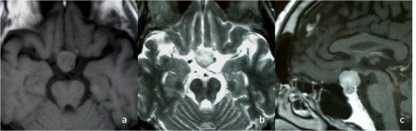
Case presentation: A 52-year-old Japanese man with a history of decreased libido was found to have a sellar and suprasellar mass. He underwent transsphenoidal surgery, but only a small specimen was obtained because of intraoperative bleeding. The tentative histological diagnosis was schwannoma. He noticed bitemporal hemianopsia 7 years later. A follow-up magnetic resonance imaging disclosed a tumor volume doubling time of 3830 days. Transcranial gross-total tumor resection was performed. The lesion consisted of elongated and plump tumor cells that were arranged in a fascicular or storiform pattern and were positive for S-100 protein and focally positive for glial fibrillary acidic protein. The final histological diagnosis was pituicytoma.

Conclusion: Pituicytoma is a slow-growing tumor, but the growth rate may change during follow-up.


Figures

Figure 1
Figure 1
Initial magnetic resonance imaging. A) T1-weighted magnetic resonance image showing an isointense pituitary mass. B) T2-weighted magnetic resonance image showing a hyperintense mass.
Figure 2
Figure 2
Magnetic resonance imaging at 7 years after initial surgery. AC) Axial, coronal and sagittal contrast-enhanced magnetic resonance image showing enlargement of the tumor.
Figure 3
Figure 3
Microscopic findings. A) Microphotograph of an hematoxylin and eosin-stained section showing bipolar spindle cells arranged in a storiform pattern. B) S-100 immunohistochemical stain. C,D) Immunohistochemical stains were focally positive for glial fibrillary acidic protein and positive for vimentin.
Figure 4
Figure 4
A semi-logarithmic graph showing serial changes of the tumor volume. The tumor volume doubling time (TVDT) was 3830 days during the follow-up period of 7 years, with TVDTs of 20267 days over the first 5 years and 1369 days over the last 2 years.

References

    1. Ulm AJ, Yachnis AT, Brat DJ, Rhoton AL Jr. Pituicytoma: report of two cases and clues regarding histogenesis. Neurosurgery. 2004;54:753–758. doi: 10.1227/01.NEU.0000108983.50966.B7. - DOI - PubMed
    1. Wolfe SQ, Bruce J, Morcos JJ. Pituicytoma: case report. Neurosurgery. 2008;63:E173–E174. doi: 10.1227/01.NEU.0000335084.93093.C8. - DOI - PubMed
    1. Brat DJ, Scheithauer BW, Fuller GN, Tihan T. Newly codified glial neoplasms of the 2007 WHO classification of tumors of the central nervous system: angiocentric glioma, pilomyxoid astrocytoma and pituicytoma. Brain Pathol. 2007;17:319–324. doi: 10.1111/j.1750-3639.2007.00082.x. - DOI - PMC - PubMed
    1. Secci F, Merciadri P, Rossi DC, D'Andrea A, Zona G. Pituicytomas: radiological findings, clinical behavior and surgical management. Acta Neurochir (Wien) 2012;154:649–657. doi: 10.1007/s00701-011-1235-7. - DOI - PubMed
    1. Nakasu Y, Nakasu S, Saito A, Horiguchi S, Kameya T. Pituicytoma. Two case reports. Neurol Med Chir (Tokyo) 2006;46:152–156. doi: 10.2176/nmc.46.152. - DOI - PubMed

LinkOut - more resources