. 2012 Nov;40(21):10904-15.
doi: 10.1093/nar/gks843. Epub 2012 Sep 18.

Binding of two DNA molecules by type II topoisomerases for decatenation

Affiliations

Binding of two DNA molecules by type II topoisomerases for decatenation

Rupesh Kumar et al. Nucleic Acids Res. 2012 Nov.

Abstract

Topoisomerases (topos) maintain DNA topology and influence DNA transaction processes by catalysing relaxation, supercoiling and decatenation reactions. In the cellular milieu, division of labour between different topos ensures topological homeostasis and control of central processes. In Escherichia coli, DNA gyrase is the principal enzyme that carries out negative supercoiling, while topo IV catalyses decatenation, relaxation and unknotting. DNA gyrase apparently has the daunting task of undertaking both the enzyme functions in mycobacteria, where topo IV is absent. We have shown previously that mycobacterial DNA gyrase is an efficient decatenase. Here, we demonstrate that the strong decatenation property of the enzyme is due to its ability to capture two DNA segments in trans. Topo IV, a strong dedicated decatenase of E. coli, also captures two distinct DNA molecules in a similar manner. In contrast, E. coli DNA gyrase, which is a poor decatenase, does not appear to be able to hold two different DNA molecules in a stable complex. The binding of a second DNA molecule to GyrB/ParE is inhibited by ATP and the non-hydrolysable analogue, AMPPNP, and by the substitution of a prominent positively charged residue in the GyrB N-terminal cavity, suggesting that this binding represents a potential T-segment positioned in the cavity. Thus, after the GyrA/ParC mediated initial DNA capture, GyrB/ParE would bind efficiently to a second DNA in trans to form a T-segment prior to nucleotide binding and closure of the gate during decatenation.


Figures
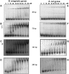
Figure 1.
Figure 1.
DNA-binding properties of M. smegmatis and E. coli DNA gyrase. EMSA carried out with increasing concentrations of MsGyr (M. smegmatis) or EcGyr (E. coli) gyrase with radioactively labelled DNA molecules of different lengths. DNA binding with MsGyr (A–D) and EcGyr (E–H). The reactions were carried out at 25°C and then resolved on 5% native polyacrylamide gels using 0.5× TBE buffer and visualized using a phosphorimager. The arrows indicate two DNA-bound complexes in (B) and (C). Details of the reactions are given in ‘Materials and Methods’ section. For sequences of the DNA molecules refer to Supplementary Table S1.
Figure 2.
Figure 2.
EMSA of M. smegmatis and E. coli gyrase with 72- and 240-bp DNA. DNA gyrase (50 nM) was incubated with DNA and 2 µg/ml MFX at 37°C for 10 min and then shifted back to ice before adding the second DNA. The 72- and 240-bp DNA used were the same as in Figure 1. Lane 1—MsGyr + 240 bp, Lane 2—MsGyr + 240 bp→72 bp (where the arrow-head indicates order of addition), Lane 3—MsGyr + 72 bp+ MFX→240 bp, Lane 4—MsGyr + 240 bp+ MFX→72 bp, Lane 5—Free 72- and 240-bp DNA, Lane 6-EcGyr + 72 bp, Lane 7-EcGyr + 240 bp, Lane 8—EcGyr + 72 bp+ MFX→240 bp. The complexes were resolved on a 5% native polyacrylamide gel and visualized using a phosphor imager. The pictograms indicate the likely mode of binding of the DNAs to the gyrase holoenzymes.
Figure 3.
Figure 3.
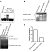
DNA binding by GyrB. (A) Crosslinking of DNA gyrase holoenzymes and individual subunits from M. smegmatis and E. coli with 72-bp DNA. An amount of 100 nM of GyrA subunit and 1 µM of each of the GyrB subunits were used for individual subunit crosslinking. The holoenzymes were reconstituted by mixing the 100 nM of GyrA subunit with 200 nM of the respective GyrB subunits. DNA crosslinking with gyrase and its individual subunits was carried out as described in ‘Materials and Methods’ section and the products were resolved on an 8% SDS–PAGE. Lane 1—DNA only, Lane 2—EcGyr, Lane 3—MsGyr, Lane 4—EcGyrA, Lane 5—MsGyrA, Lane 6—EcGyrB and Lane 7—MsGyrB; (B) EMSA with 72-bp DNA using MsGyrB and EcGyrB (0.1 and 0.2 µM) subunits. The DNA-bound complexes were resolved on a 5% native polyacrylamide gel; (C) Filter-binding assay with 20 nM each of MsGyrB, EcGyrB and E. coli ParE (EcParE) using 72-bp DNA. Enzyme was pre-incubated with 2 mM AMPPNP before addition of DNA (where indicated); (D) DNA (72 bp)-mediated stimulation of ATPase activity. ATPase assays were carried out with MsGyr (100 nM) or its GyrB subunit (100 nM) in the absence and presence of 72-bp DNA (400 nM). Lane 1—MsGyrB alone, Lane 2—MsGyrB + DNA, Lane 3—MsGyr alone, Lane 4—MsGyr + DNA and Lane 5—MsGyr + Novobiocin (10 µg/ml). The error bars represent standard deviation across three measurements.
Figure 4.
Figure 4.
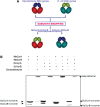
Swapping of subunits between MsGyr and EcGyr forms active heterotetramers. (A) Schematic representation of the experiment. MsGyrB—blue; MsGyrA—red; EcGyrB—green; EcGyrA—turquoise; (B) Glutaraldehyde crosslinking to show heterotetramer formation by EcGyrA + EcGyrB, MsGyrA + MsGyrB and MsGyrA + EcGyrB and EcGyrA + MsGyrB. The subunits were mixed and incubated on ice for 30 min. Crosslinking reaction with glutaraldehyde (final concentration—0.01%) was carried out at 37°C for 30 min. Native and crosslinked proteins were separated on 8% SDS–PAGE.
Figure 5.
Figure 5.
Topoisomerase activity of interspecies heterotetramers. (A) Supercoiling assay with the interspecies heterotetramers and the parent holoenzymes (25 nM). An amount of 300 ng relaxed pUC18 plasmid was used as substrate for the supercoiling assay. The topoisomers were resolved on a 1.2% agarose gel; (B) Decatenation activity of the parent and interspecies heterotetramers (25 nM) using 300 ng of kinetoplast DNA as substrate. The details of the reaction are given in ‘Materials and Methods’ section; (C) EMSA carried out with increasing concentrations (nM) of reconstituted hybrid heterotetramers (EAMB (EcGyrA + MsGyrB), MAEB (MsGyrA + EcGyrB) and MsGyr. 72-bp DNA was used as substrate. The DNA-bound complexes were resolved on a 5% native polyacrylamide gel. Arrows indicate the two complexes formed.
Figure 6.
Figure 6.
DNA binding and ATPase activity of E. coli topoIV and its ParE subunit (A) EMSA with E. coli topo IV holoenzyme (5–40 nM) using 72-bp DNA. The arrows indicate two DNA-bound complexes; (B) EMSA with increasing concentrations of ParE (5–100 nM) subunit using 72-bp DNA. The reaction conditions were similar to those used for Figure 1; (C) ATP hydrolysis by the topo IV holoenzyme and ParE (100 nM of each) subunit in the absence and presence of 72-bp (100 nM) DNA; (D) ATPase activity of wild-type and R284Q mutant ParE43 fragment (2 μM dimer), in the presence and absence of linear pBR322 at a ratio of 25-bp DNA per protein dimer. The activity is expressed as the apparent kcat of the protein dimer in the presence of 2 mM ATP. The error bars represent the standard deviation of three measurements.
Figure 7.
Figure 7.
DNA-binding activity of the wild-type (wt) and R321A GyrB subunit. (A) EMSA with 200 and 400 nM each of wt MsGyrB and its R321A mutant. The reaction conditions were similar to those used for Figure 2B. The 72-bp DNA was used as binding substrate; (B) DNA supercoiling of the mycobacterial gyrase reconstituted by mixing the wt or the R321A GyrB with GyrA subunit. An amount of 25 nM of M. smegmatis gyrase was reconstituted by mixing 25 nM of GyrA with 50 nM of either wt or R321A GyrB. The details of the reaction are given in ‘Materials and Methods’ section; (C) Decatenation assay with the holoenzyme reconstituted by mixing 25 nM of GyrA with 50 nM of either wt or R321A GyrB. An amount of 300 ng of kDNA was used as substrate. The decatenated minicircles were resolved on a 1.2% agarose gel; (D) Quantitation of the decatenation (released minicircles) by the wt and R321A GyrB containing holoenzyme from M. smegmatis (in Figure 7C) using Fujifilm multigauge V2.3.

Similar articles

Cited by

References

    1. Akrigg A, Cook PR. DNA gyrase stimulates transcription. Nucleic Acids Res. 1980;8:845–854. - PMC - PubMed
    1. De Wyngaert M, Hinkle DC. Involvement of DNA gyrase in replication and transcription of bacteriophage T7 DNA. J. Virol. 1979;29:529–535. - PMC - PubMed
    1. Filutowicz M. Requirement of DNA gyrase for the initiation of chromosome replication in Escherichia coli K-12. Mol. Gen. Genet. 1980;177:301–309. - PubMed
    1. Masse E, Drolet M. Relaxation of transcription-induced negative supercoiling is an essential function of Escherichia coli DNA topoisomerase I. J. Biol. Chem. 1999;274:16654–16658. - PubMed
    1. Zechiedrich EL, Cozzarelli NR. Roles of topoisomerase IV and DNA gyrase in DNA unlinking during replication in Escherichia coli. Genes Dev. 1995;9:2859–2869. - PubMed

Publication types

MeSH terms