. 2012 Nov 2;287(45):38231-43.
doi: 10.1074/jbc.M112.402289. Epub 2012 Sep 18.

Solution NMR structure of the Ca2+-bound N-terminal domain of CaBP7: a regulator of golgi trafficking

Affiliations

Solution NMR structure of the Ca2+-bound N-terminal domain of CaBP7: a regulator of golgi trafficking

Hannah V McCue et al. J Biol Chem. .

Abstract

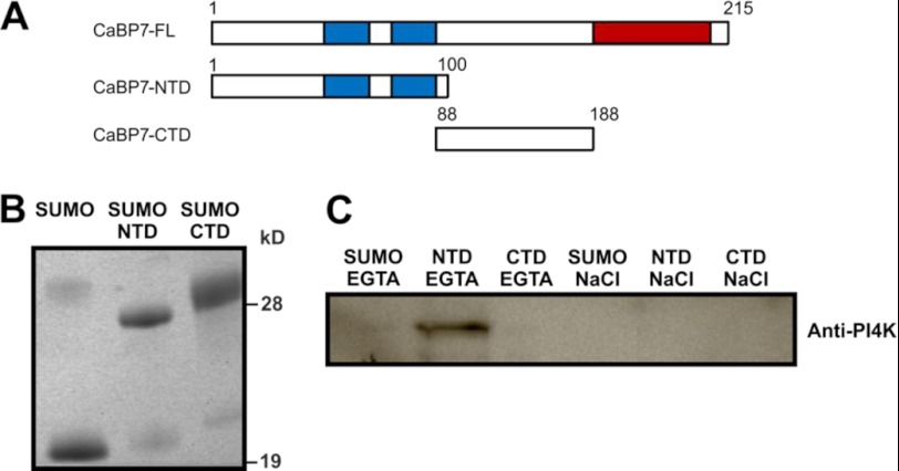
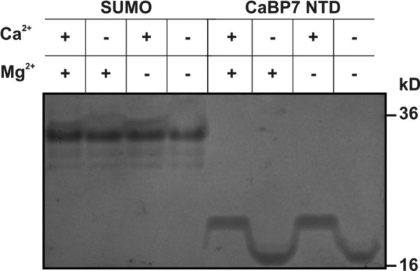
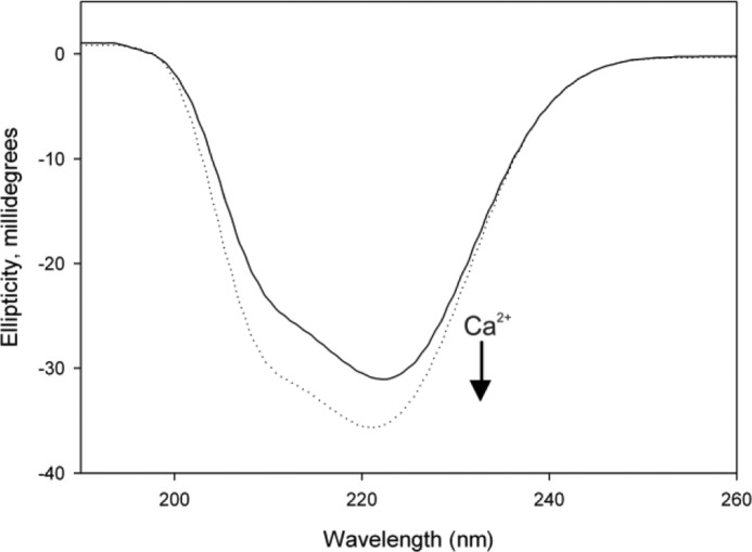
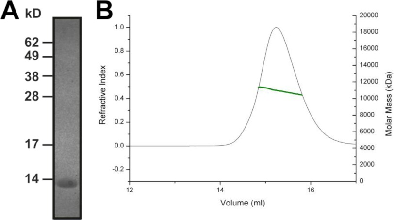
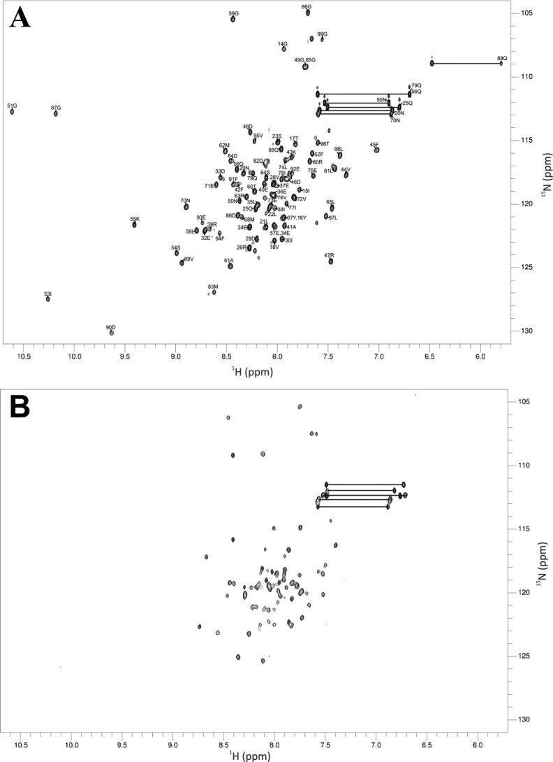
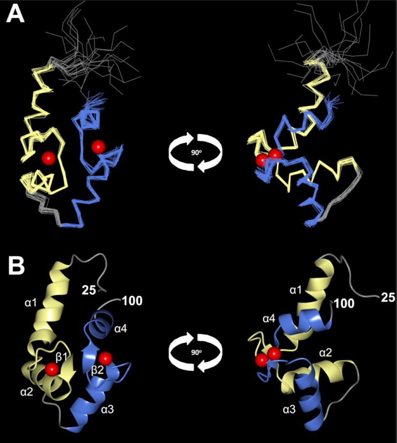
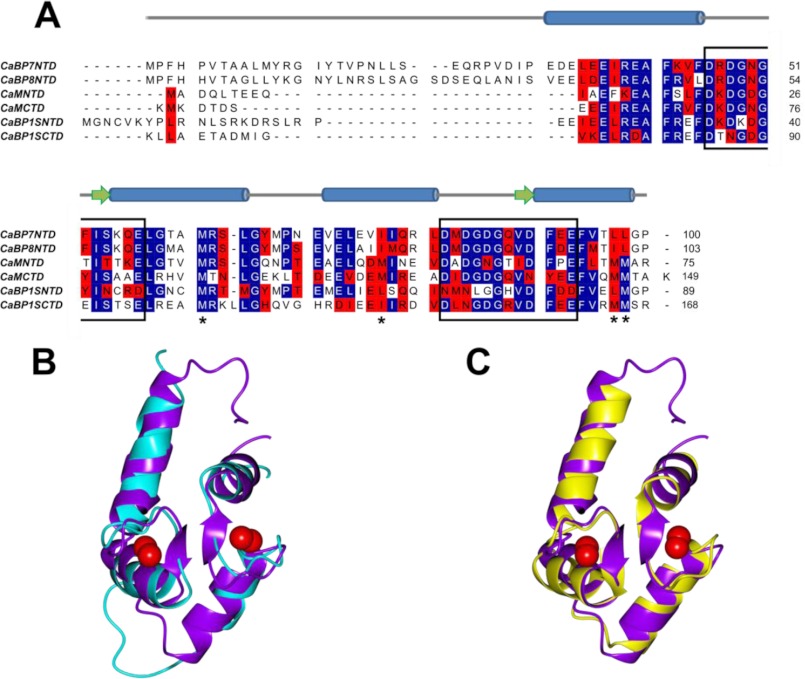
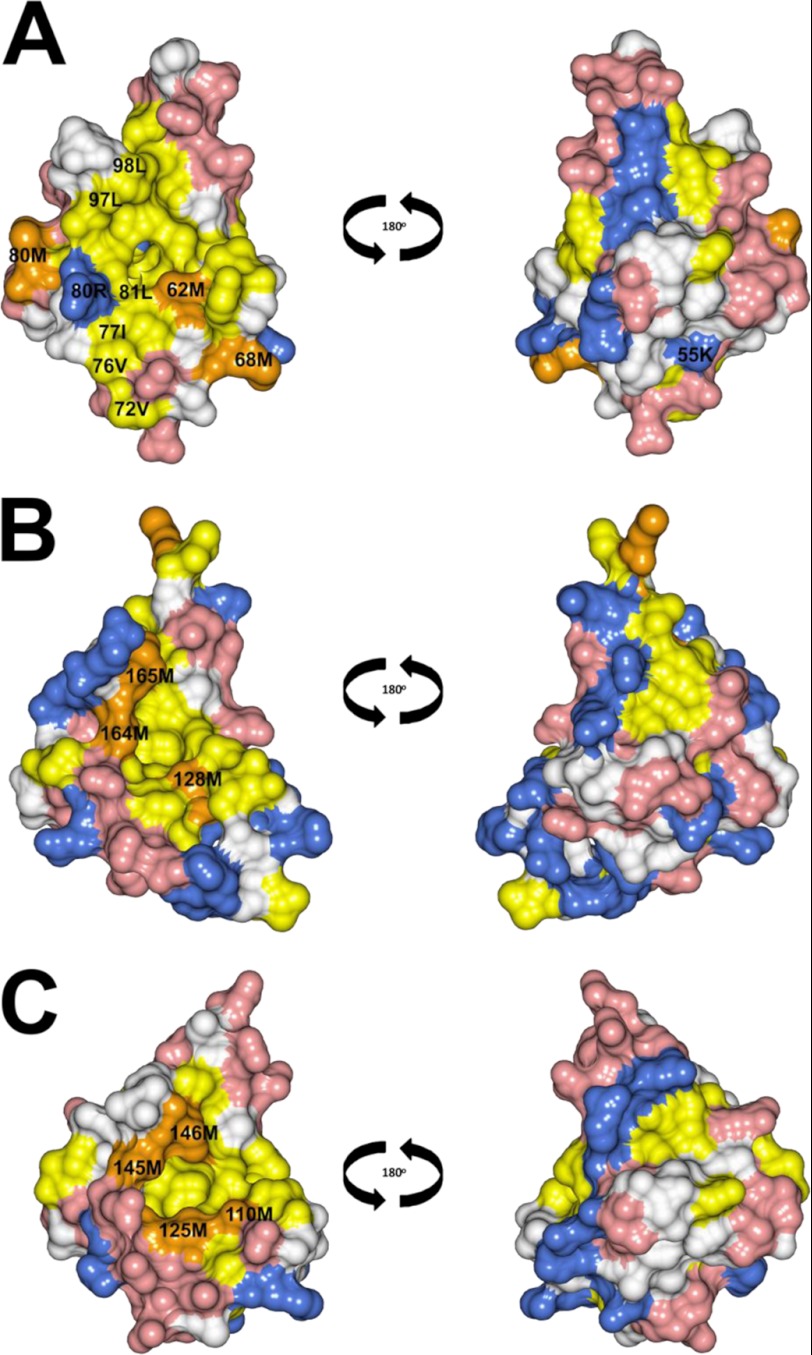
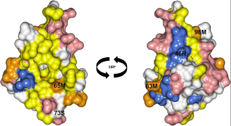
Calcium-binding protein 7 (CaBP7) is a member of the calmodulin (CaM) superfamily that harbors two high affinity EF-hand motifs and a C-terminal transmembrane domain. CaBP7 has been previously shown to interact with and modulate phosphatidylinositol 4-kinase III-β (PI4KIIIβ) activity in in vitro assays and affects vesicle transport in neurons when overexpressed. Here we show that the N-terminal domain (NTD) of CaBP7 is sufficient to mediate the interaction of CaBP7 with PI4KIIIβ. CaBP7 NTD encompasses the two high affinity Ca(2+) binding sites, and structural characterization through multiangle light scattering, circular dichroism, and NMR reveals unique properties for this domain. CaBP7 NTD binds specifically to Ca(2+) but not Mg(2+) and undergoes significant conformational changes in both secondary and tertiary structure upon Ca(2+) binding. The Ca(2+)-bound form of CaBP7 NTD is monomeric and exhibits an open conformation similar to that of CaM. Ca(2+)-bound CaBP7 NTD has a solvent-exposed hydrophobic surface that is more expansive than observed in CaM or CaBP1. Within this hydrophobic pocket, there is a significant reduction in the number of methionine residues that are conserved in CaM and CaBP1 and shown to be important for target recognition. In CaBP7 NTD, these residues are replaced with isoleucine and leucine residues with branched side chains that are intrinsically more rigid than the flexible methionine side chain. We propose that these differences in surface hydrophobicity, charge, and methionine content may be important in determining highly specific interactions of CaBP7 with target proteins, such as PI4KIIIβ.


Figures

FIGURE 1.
FIGURE 1.
Affinity pull-down assay of PI4KIIIβ from bovine brain cytosol fraction using either the NTD or CTD of CaBP7 immobilized through a His6 tag on cobalt-charged metal affinity resin. A, schematic diagram of CaBP7. Blue boxes indicate active EF-hand domains. Red boxes indicate the C-terminal TMD. B, Coomassie-stained SDS-polyacrylamide gel showing equivalent expression levels of SUMO, SUMO-CaBP7 NTD, and SUMO-CaBP7 CTD used in pull-down assays. C, Western blot showing an immunoreactive band for PI4KIIIβ, which is only extracted by SUMO-CaBP7 NTD and is eluted specifically by an EGTA wash. Bound proteins were eluted from resin using a 5 mm EGTA wash (first three lanes) followed by a 1 m NaCl wash (last three lanes).
FIGURE 2.
FIGURE 2.
Coomassie-stained native polyacrylamide gel showing the electrophoretic gel mobility of SUMO and CaBP7 NTD in the presence or absence of an excess of Ca2+ or Mg2+. Samples were resolved on an 18% acrylamide gel in loading buffer containing 2 mm EGTA and 2 mm EDTA with either 10 mm CaCl2 or 10 mm MgCl2. The presence or absence of Ca2+ or Mg2+ is indicated by a plus or minus sign above each lane of the gel image.
FIGURE 3.
FIGURE 3.
The effect of Ca2+ on far-UV CD of apo- and Ca2+-bound CaBP7 NTD. Changes to CaBP7 NTD secondary structure were monitored by far-UV CD spectroscopy. The solid line represents the trace for apo-CaBP7 NTD, and the dashed line represents the trace for Ca2+-bound CaBP7 NTD. To prepare apo-CaBP7 NTD, 5 mm EGTA and 5 mm EDTA were added to the sample and then removed using a desalting column. Final samples were analyzed in 20 mm HEPES, pH 6.5, 150 mm NaCl, 30 mm n-Octyl-β-d-glucopyranoside at 25 °C. Each spectrum is representative of 10 averaged scans and is normalized to the spectrum of buffer alone.
FIGURE 4.
FIGURE 4.
CaBP7 NTD is monomeric. A, Coomassie-stained SDS-polyacrylamide gel of CaBP7 NTD after purification and cleavage of a His-SUMO tag. The protein migrates at a rate consistent with the theoretical molecular mass of 11.4 kDa. B, SECMALLS characterization of Ca2+-bound CaBP7 NTD. The refractive index of the sample is plotted against elution volume. The molar mass distribution is indicated by the green line. The data illustrate that CaBP7 NTD forms a stable monomer in 20 mm HEPES, pH 6.5, 150 mm NaCl at 20 °C. There is a single peak with an average molar mass of 11.1 ± 0.11 kDa.
FIGURE 5.
FIGURE 5.
1H-15N HSQC spectra of CaBP7 NTD. A, assigned 1H-15N HSQC spectrum of Ca2+-bound CaBP7 NTD; B, 1H-15N HSQC spectrum of apo-CaBP7 NTD. Sample heterogeneity of apo-CaBP7 NTD is seen from the unequal distribution of peak intensities. Asn and Gln side chain NH2 groups are indicated by peaks joined with a solid black line. Ca2+-bound CaBP7 NTD was prepared in 20 mm HEPES, pH 6.5, 150 mm NaCl, 30 mm n-octyl-β-d-glucopyranoside. Apo CaBP7 NTD was prepared by the addition of 5 mm EGTA and 5 mm EDTA to the sample followed by buffer exchange into 20 mm HEPES, pH 6.5, 150 mm NaCl, 30 mm n-octyl-β-d-glucopyranoside. Spectra were acquired at 303 K.
FIGURE 6.
FIGURE 6.
Main chain structures of Ca2+-bound CaBP7 NTD determined by solution NMR (PDB code 2LV7). Shown are superposition of the 20 lowest energy structures (A) and a ribbon representation of the lowest energy structure (B) seen from the front (left) and side (right). N-terminal residues (residues 1–25) are unstructured and are not shown. EF-hand 1 is shown in yellow, and EF-hand 2 is shown in blue. Red spheres represent bound Ca2+.
FIGURE 7.
FIGURE 7.
Comparison of Ca2+-bound CaBP7 NTD, CaM CTD, and CaBP1 CTD sequence and main chain structure. A, sequence alignment of CaBP7 NTD with CaBP8 NTD and the N- and C-terminal domains of CaM and CaBP1. Identical amino acids are highlighted in blue, and similar amino acids are highlighted in red. Black boxes indicate the 12-amino acid Ca2+-binding loop of each EF-hand. Asterisks below the sequences indicate methionine residues that are conserved in CaM NTD and CTD. These are Met-36, -51, -71, and -72 in the N-terminal domain and Met-110, -125, -145, and -146 in the C-terminal domain. The NMR-derived secondary structure elements of CaBP7 NTD are indicated above the alignment. Cylinders, α-helices; arrows, β-sheets. B and C, ribbon representation of Ca2+-bound CaBP7 NTD (PDB code 2LV7, residues 30–100) lowest energy conformer (purple) superposed with Ca2+-bound CaBP1 CTD (cyan, PDB code 2LAP) (B) and Ca2+-bound CaM CTD (yellow, PDB code 1CLL, residues 80–147) (C). Red spheres represent bound Ca2+.
FIGURE 8.
FIGURE 8.
Space-filling representations of Ca2+-bound CaBP7 NTD (PDB code 2LV7, residues 30–100) (A), Ca2+-bound CaBP1 (PDB code 2LAP) (B), and Ca2+-bound CaM (PDB code 1CLL, residues 80–147) (C). The front face is shown on the left, and the back face is shown on the right. Acidic residues (Asp and Glu) and basic residues (Arg, His, and Lys) are shown in red and blue, respectively. Hydrophobic residues (Ile, Leu, Phe, Trp, Val, and Tyr) are shown in yellow with the exception of Met residues, which are highlighted in orange.
FIGURE 9.
FIGURE 9.
Space-filling representation of CaBP8 homology model. The front face is shown on the left, and the back face is shown on the right. Acidic residues (Asp and Glu) and basic residues (Arg, His, and Lys) are shown in red and blue, respectively. Hydrophobic residues (Ile, Leu, Phe, Trp, Val, and Tyr) are shown in yellow with the exception of Met residues, which are highlighted in orange. Solvent-exposed amino acids that are significantly different from the corresponding residues in CaBP7 are labeled. The predicted surface of CaBP8 is very similar to CaBP7 (Fig. 8A). The homology model was predicted using the Modeler software package (74) and residues 30–100 of the calculated Ca2+-bound CaBP7 NTD structure as a template.

Similar articles

Cited by

References

    1. McCue H. V., Haynes L. P., Burgoyne R. D. (2010) Bioinformatic analysis of CaBP/calneuron proteins reveals a family of highly conserved vertebrate Ca2+-binding proteins. BMC Res. Notes 3, 118. - PMC - PubMed
    1. Haeseleer F., Sokal I., Verlinde C. L., Erdjument-Bromage H., Tempst P., Pronin A. N., Benovic J. L., Fariss R. N., Palczewski K. (2000) Five members of a novel Ca2+-binding protein (CABP) subfamily with similarity to calmodulin. J. Biol. Chem. 275, 1247–1260 - PMC - PubMed
    1. Seidenbecher C. I., Langnaese K., Sanmartí-Vila L., Boeckers T. M., Smalla K. H., Sabel B. A., Garner C. C., Gundelfinger E. D., Kreutz M. R. (1998) Caldendrin, a novel neuronal calcium-binding protein confined to the somato-dendritic compartment. J. Biol. Chem. 273, 21324–21331 - PubMed
    1. Wu Y. Q., Lin X., Liu C. M., Jamrich M., Shaffer L. G. (2001) Identification of a human brain-specific gene, calneuron 1, a new member of the calmodulin superfamily. Mol. Genet. Metab. 72, 343–350 - PubMed
    1. Mikhaylova M., Sharma Y., Reissner C., Nagel F., Aravind P., Rajini B., Smalla K. H., Gundelfinger E. D., Kreutz M. R. (2006) Neuronal Ca2+ signaling via caldendrin and calneurons. Biochim. Biophys. Acta 1763, 1229–1237 - PubMed

Publication types

MeSH terms

Substances

LinkOut - more resources