. 2012 Oct 26;287(44):36830-6.
doi: 10.1074/jbc.M112.409276. Epub 2012 Sep 18.

Crystal and solution structures of Plasmodium falciparum erythrocyte-binding antigen 140 reveal determinants of receptor specificity during erythrocyte invasion

Affiliations

Crystal and solution structures of Plasmodium falciparum erythrocyte-binding antigen 140 reveal determinants of receptor specificity during erythrocyte invasion

Daniel H Lin et al. J Biol Chem. .

Abstract

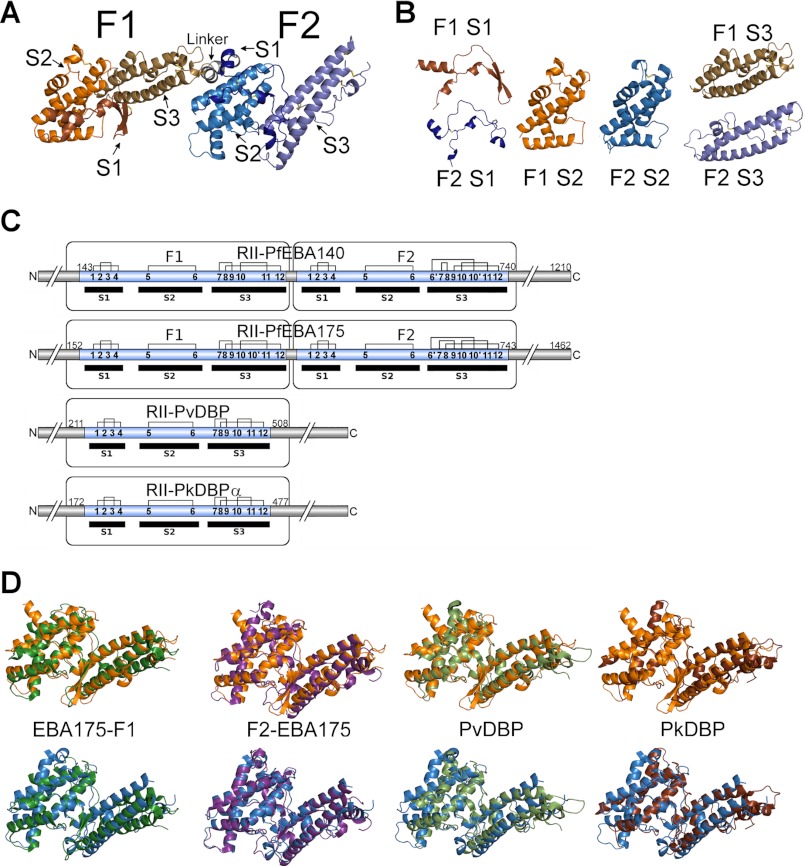
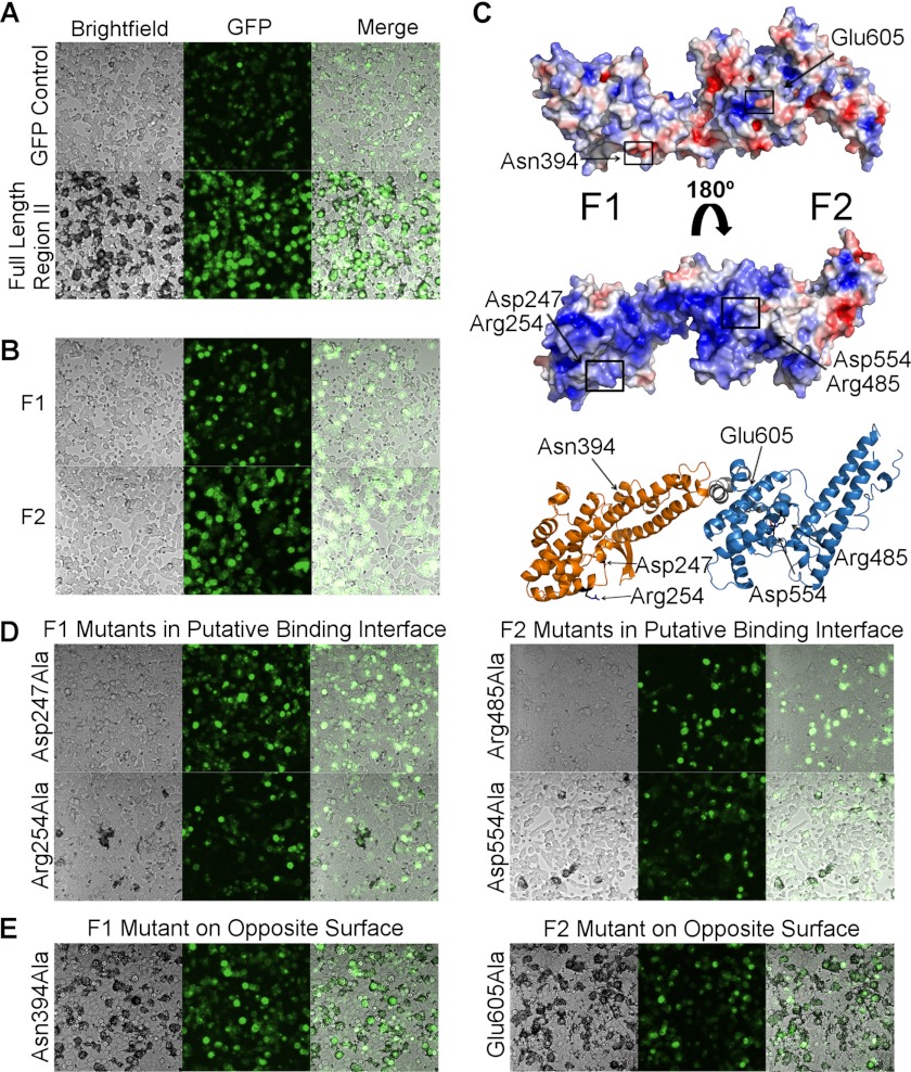
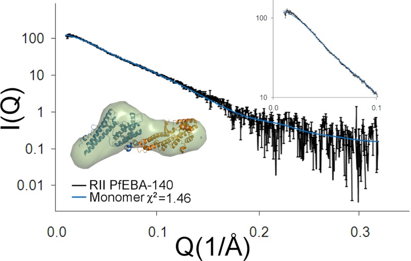
Erythrocyte-binding antigen 140 (PfEBA-140) is a critical Plasmodium falciparum erythrocyte invasion ligand that engages glycophorin C on host erythrocytes during malaria infection. The minimal receptor-binding region of PfEBA-140 contains two conserved Duffy binding-like (DBL) domains, a fold unique to Plasmodium species. Here, we present the crystal structure of the receptor-binding region of PfEBA-140 at 2.4 Å resolution. The two-domain binding region is present as a monomer in the asymmetric unit, and the structure reveals novel features in PfEBA-140 that are likely determinants of receptor specificity. Analysis by small-angle x-ray scattering demonstrated that the minimal binding region is monomeric in solution, consistent with the crystal structure. Erythrocyte binding assays showed that the full-length binding region containing the tandem DBL domains is required for erythrocyte engagement, suggesting that both domains contain critical receptor contact sites. The electrostatic surface of PfEBA-140 elucidates a basic patch that constitutes a putative high-affinity binding interface spanning both DBL domains. Mutation of residues within this interface results in severely diminished erythrocyte binding. This study provides insight into the structural basis and mechanism of PfEBA-140 receptor engagement and forms a basis for future studies of this critical interaction. In addition, the solution and crystal structures allow the first identification of likely determinants of erythrocyte receptor specificity for P. falciparum invasion ligands. A complete understanding of the PfEBA-140 erythrocyte invasion pathway will aid in the design of invasion inhibitory therapeutics and vaccines.


Figures

FIGURE 1.
FIGURE 1.
Structure of RII PfEBA-140. A, overall structure of RII PfEBA-140 as well as the location of individual subdomains within each DBL domain. F1 subdomain 1 is shown in bronze, subdomain 2 in orange, and subdomain 3 in dark orange. F2 subdomain 1 is shown in dark blue, subdomain 2 in blue, and subdomain 3 in light blue. The short helical linker between the two domains is shown in gray. B, structures of the individual subdomains in RII PfEBA-140. Coloring is the same as described for A. C, representation of the disulfide bridge pattern of characterized EBL ligands displaying the two altered disulfides in RII PfEBA-140. D, overlay of Plasmodium invasion ligand DBL domains with each DBL domain of RII PfEBA-140. F1 PfEBA-140 in shown in orange (upper), and F2 PfEBA-140 is shown in blue (lower). F1 PfEBA-175 is shown in dark green, F2 PfEBA-175 in purple, PvDBP in light green, and PkDBP in brown.
FIGURE 2.
FIGURE 2.
PfEBA-140 erythrocyte binding requires both DBL domains. A, RII PfEBA-140 was expressed on the surface of HEK-293 mammalian cells and tested for erythrocyte binding. GFP was utilized to assess proper surface expression. A construct expressing only GFP did not bind erythrocytes (upper panel). The full-length RII construct, identical to the construct used for crystallization, was capable of extensively engaging erythrocytes (lower panel). Bound erythrocytes appear black around the mammalian cells. B, the individual DBL domains of PfEBA-140 were also tested for erythrocyte binding. The F1 (upper panel) and F2 (lower panel) DBL domains could not independently engage erythrocytes. C, the electrostatic surface of PfEBA-140 RII elucidates a putative high-affinity binding interface. The surface of the protein shown in the upper panel has no clear region of concentrated charged residues (blue, positive charge; red, negative charge). However, when rotated 180°, a region of positive charge that forms an arch between the two DBL domains is illuminated (middle panel). Surface charge potential is colored from +3.5 to −3.5 eV. The lower panel displays the face of the protein-containing the basic patch identified as a putative binding interface. Residues mutated to alanine and tested for erythrocyte binding are identified with arrows and shown in black. D, alanine mutations within the basic patch disrupted erythrocyte binding. E, in contrast to residues within the basic patch, alanine mutations on the opposite face of RII PfEBA-140 did not disrupt erythrocyte binding.
FIGURE 3.
FIGURE 3.
Structural determinants of receptor specificity within the EBL family. A, overlay of PfEBA-140 (with the F1 domain in orange and the F2 domain in blue) with PfEBA-175 (with the F1 domain in green and the F2 domain in purple). The conserved F1 β-sheet motifs are boxed. The inset displays two monomers of PfEBA-140 overlaid onto the PfEBA-175 dimer observed in the crystal structure (17). B, overlay of the F1 and F2 PfEBA-140 DBL domains (left panel). The altered helical element in F2 is boxed in black. A close-up view of this F2 helical element overlaid onto the F1 β-sheet is shown (right panel). C, view of the altered helical orientation of F1 RII PfEBA-140 subdomain 3 (orange). This orientation occurs due to the absence of a glycine, which is conserved in other EBL members, at residue 325 in PfEBA-140. The kink observed in F1 PfEBA-175 (green) is shown for comparison.
FIGURE 4.
FIGURE 4.
Oligomeric state of RII PfEBA-140. RII PfEBA-140 is monomeric in solution in the absence of receptor. Shown here are the experimental and theoretical SAXS plots of scattering intensity versus scattering momentum. The inset displays an ab initio construct of PfEBA-140 (light blue) overlaid onto the crystal structure (with the F1 domain in orange and the F2 domain in blue).

Similar articles

Cited by

References

    1. Cowman A. F., Crabb B. S. (2006) Invasion of red blood cells by malaria parasites. Cell 124, 755–766 - PubMed
    1. Mayer D. C., Cofie J., Jiang L., Hartl D. L., Tracy E., Kabat J., Mendoza L. H., Miller L. H. (2009) Glycophorin B is the erythrocyte receptor of Plasmodium falciparum erythrocyte-binding ligand EBL-1. Proc. Natl. Acad. Sci. U.S.A. 106, 5348–5352 - PMC - PubMed
    1. Orlandi P. A., Klotz F. W., Haynes J. D. (1992) A malaria invasion receptor, the 175-kilodalton erythrocyte-binding antigen of Plasmodium falciparum, recognizes the terminal Neu5Ac(α2–3)Gal sequences of glycophorin A. J. Cell Biol. 116, 901–909 - PMC - PubMed
    1. Lobo C. A., Rodriguez M., Reid M., Lustigman S. (2003) Glycophorin C is the receptor for the Plasmodium falciparum erythrocyte-binding ligand PfEBP-2 (baebl). Blood 101, 4628–4631 - PubMed
    1. Gilberger T. W., Thompson J. K., Triglia T., Good R. T., Duraisingh M. T., Cowman A. F. (2003) A novel erythrocyte-binding antigen 175 paralogue from Plasmodium falciparum defines a new trypsin-resistant receptor on human erythrocytes. J. Biol. Chem. 278, 14480–14486 - PubMed

Publication types

Substances

Associated data

LinkOut - more resources