. 2012 Nov 1;120(18):3829-36.
doi: 10.1182/blood-2012-07-440743. Epub 2012 Sep 18.

Minihepcidins prevent iron overload in a hepcidin-deficient mouse model of severe hemochromatosis

Affiliations

Minihepcidins prevent iron overload in a hepcidin-deficient mouse model of severe hemochromatosis

Emilio Ramos et al. Blood. .

Abstract

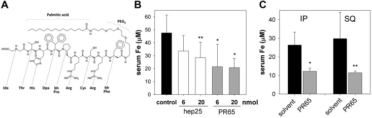
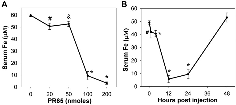
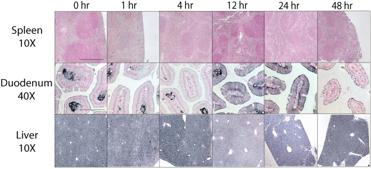
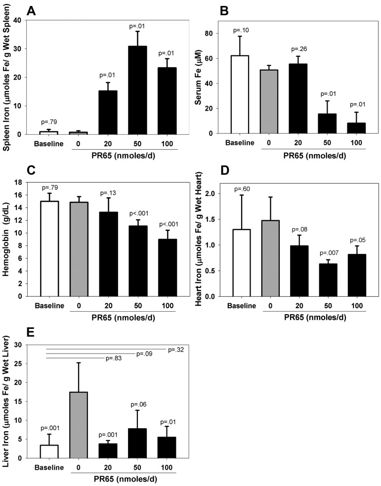
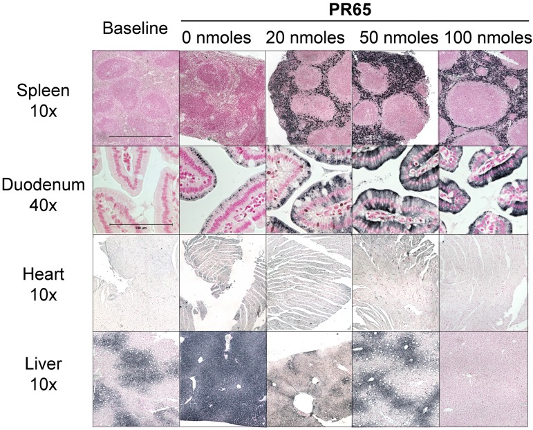
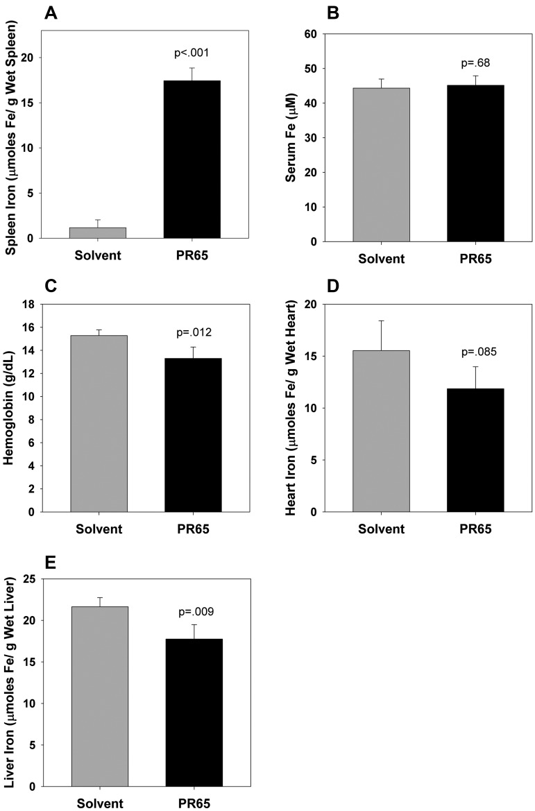
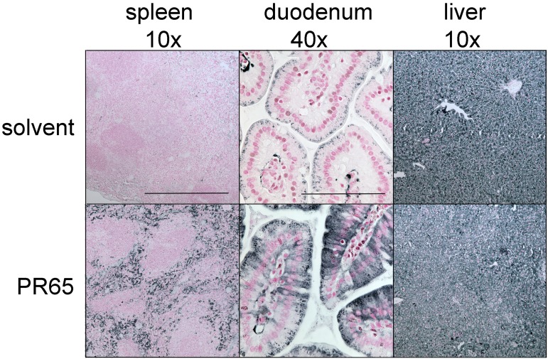
The deficiency of hepcidin, the hormone that controls iron absorption and its tissue distribution, is the cause of iron overload in nearly all forms of hereditary hemochromatosis and in untransfused iron-loading anemias. In a recent study, we reported the development of minihepcidins, small drug-like hepcidin agonists. Here we explore the feasibility of using minihepcidins for the prevention and treatment of iron overload in hepcidin-deficient mice. An optimized minihepcidin (PR65) was developed that had superior potency and duration of action compared with natural hepcidin or other minihepcidins, and favorable cost of synthesis. PR65 was administered by subcutaneous injection daily for 2 weeks to iron-depleted or iron-loaded hepcidin knockout mice. PR65 administration to iron-depleted mice prevented liver iron loading, decreased heart iron levels, and caused the expected iron retention in the spleen and duodenum. At high doses, PR65 treatment also caused anemia because of profound iron restriction. PR65 administration to hepcidin knockout mice with pre-existing iron overload had a more moderate effect and caused partial redistribution of iron from the liver to the spleen. Our study demonstrates that minihepcidins could be beneficial in iron overload disorders either used alone for prevention or possibly as adjunctive therapy with phlebotomy or chelation.


Figures

Figure 1
Figure 1
Minihepcidin PR65 and its activity in wild-type mice. (A) Structural formula of PR65. Ida indicates iminodiacetic acid; Dpa, diphenylalanine; bhPro, β-homo proline; and bhPhe, β-homo phenylalanine. (B) Serum iron in wild-type C57BL/6 mice 4 hours after intraperitoneal injection of solvent, native hepcidin or PR65 (n = 4-8 in each group; **P = .01, *P = .005). (C) Serum iron in wild-type C57BL/6 mice 4 hours after intraperitoneal or subcutaneous injection of 20 nmol of PR65 (n = 4 in each group; *P = .007, **P = .04). In panels B and C, the bars represent mean values and error bars standard deviations.
Figure 2
Figure 2
The hypoferremic effect of PR65 in iron-loaded hepcidin knockout mice. (A) Twenty-four hours after a subcutaneous injection, PR65 induced a dose-dependent decrease in serum iron. Mean values and standard deviations are shown, n = 3-5 mice per point (#P = .005 and P = .004, *P < .001). (B) The time course of hypoferremia induced by a subcutaneous injection of 100 nmol of PR65. Mean and standard deviations are shown, n = 4-6 mice per point (#P = .008, *P < .001).
Figure 3
Figure 3
Changes in iron distribution in PR65-treated hepcidin knockout mice. Tissue iron was visualized by enhanced Perls stain at 0 to 48 hours after subcutaneous injection of PR65 (100 nmol). Representative images are shown. Horizontal bars indicate 400 μm (10×) and 100 μm (40×). Top row: Spleen iron was scant and its distribution did not change appreciably during the 48 hours. Middle row: Iron in the villus stroma was evident in solvent-treated and 1 to 4 hours PR65–treated mice, indicating active ferroportin-mediated efflux of iron from basolateral membranes of enterocytes. From 12 to 24 hours, iron was retained in enterocytes consistent with (mini)hepcidin-induced ferroportin degradation. Forty-eight hours after injection iron was no longer retained by enterocytes. Bottom row: As expected, the livers were iron-loaded at baseline and no changes in the pattern of iron staining were seen within 48 hours of PR65 treatment.
Figure 4
Figure 4
PR65 prevented iron loading in iron-depleted hepcidin knockout mice. All mice were placed on an iron-deficient diet (4 ppm iron) from ages 5 to 6 weeks until 12 weeks. The “baseline” group (n = 7) was examined at 12 weeks of age (white bars). The rest of the mice were fed an iron-loading diet (300 ppm) for 2 more weeks while receiving daily subcutaneous injections of solvent (gray bars, n = 6) or PR65 at 20, 50, or 100 nmol per day (black bars, n = 4 per dose). The mice were analyzed 24 hours after the last injection. Compared with solvent, PR65 injections resulted in: (A) iron retention in the spleen, (B) a dose-dependent decrease in serum iron, (C) a corresponding dose-dependent decrease in Hb levels, (D) a decrease in heart iron at higher doses, and (E) decreased liver iron. Liver iron content in PR65-injected mice did not significantly differ from that in the baseline group of mice, indicating that little to no new iron was absorbed or deposited in the liver during the 2-week treatment. Graphs show means and standard deviations. Student t test was used to compare the mean of each condition to that of solvent treatment (P value over bars). In panel E, mean of each condition was also compared with the baseline (P values at lines over bars).
Figure 5
Figure 5
Cellular distribution of iron after 2 weeks of PR65 injections for the prevention of iron overload. Representative images are shown. Horizontal bars indicate 400 μm (10 ×) and 100 μm (40 ×). Iron accumulation was seen in the splenic red pulp of PR65–treated mice but not solvent-treated mice. Similarly, iron accumulation in duodenal enterocytes was seen only in PR65-treated mice. Compared with heart iron staining of solvent-injected mice, there was less iron accumulation in the heart of animals injected with 50 and 100 nmol of PR65, consistent with the quantitative method in Figure 4. Liver iron loading in mice treated with 20 and 50 nmol of PR65 was similar to that of the baseline group and much less than the iron loading in the solvent-treated group. At the highest PR65 dose, liver iron was lower than at baseline indicating that mice were able to mobilize liver iron despite high minihepcidin activity.
Figure 6
Figure 6
Two-week PR65 treatment of iron-loaded hepcidin knockout mice caused modest redistribution of iron. Hepcidin knockout mice were kept on a 300 ppm iron diet for their entire lifespan. Starting at 12 weeks of age, one group of mice was injected subcutaneously with solvent (n = 4) and the other with 50 nmol of PR65 (n = 4) daily for 2 weeks. Iron and hematologic parameters were measured 24 hours after the last injection. In PR65-treated mice compared with solvent-treated mice: (A) spleen iron increased more than 15-fold confirming PR65 activity; (B) serum iron concentrations were similar 24 hours after the last injection; (C) hemoglobin decreased by 2 g/dL indicating iron restriction to erythropoiesis; (D) heart iron tended to decrease, though the difference was not statistically significant at the number of mice tested; and (E) liver iron decreased by approximately 20%.
Figure 7
Figure 7
Cellular distribution of iron after 2 weeks of PR65 injections for the treatment of established iron overload. Tissue sections correspond to the animals analyzed in Figure 6, with representative images shown. Horizontal bars indicate 400 μm (10 ×) and 100 μm (40 ×). Enhanced Perls stain confirmed that splenic macrophages and duodenal enterocytes retained iron in PR65-treated but not in solvent-treated mice. Compared with solvent-treated controls, less intense iron staining was observed in the liver of mice treated with PR65. No consistent differences between solvent and PR65-treated mice were seen in sections of the heart (not shown).

References

    1. Ganz T, Nemeth E. Hepcidin and iron homeostasis. Biochim Biophys Acta. 2012;1823(9):1434–1443. - PMC - PubMed
    1. Nemeth E, Tuttle MS, Powelson J, et al. Hepcidin regulates cellular iron efflux by binding to ferroportin and inducing its internalization. Science. 2004;306(5704):2090–2093. - PubMed
    1. Brissot P, Bardou-Jacquet E, Jouanolle AM, Loreal O. Iron disorders of genetic origin: a changing world. Trends Mol Med. 2011;17(12):707–713. - PubMed
    1. Musallam KM, Cappellini MD, Wood JC, Taher AT. Iron overload in non-transfusion-dependent thalassemia: a clinical perspective. Blood Rev. 2012;26(Suppl 1):S16–S19. - PubMed
    1. Viatte L, Nicolas G, Lou DQ, et al. Chronic hepcidin induction causes hyposideremia and alters the pattern of cellular iron accumulation in hemochromatotic mice. Blood. 2006;107(7):2952–2958. - PubMed

Publication types

MeSH terms