Review
. 2013 Feb;25(1):85-91.
doi: 10.1016/j.ceb.2012.08.013. Epub 2012 Sep 16.

Cell polarisation and the immunological synapse

Affiliations
Review

Cell polarisation and the immunological synapse

Karen L Angus et al. Curr Opin Cell Biol. 2013 Feb.

Abstract

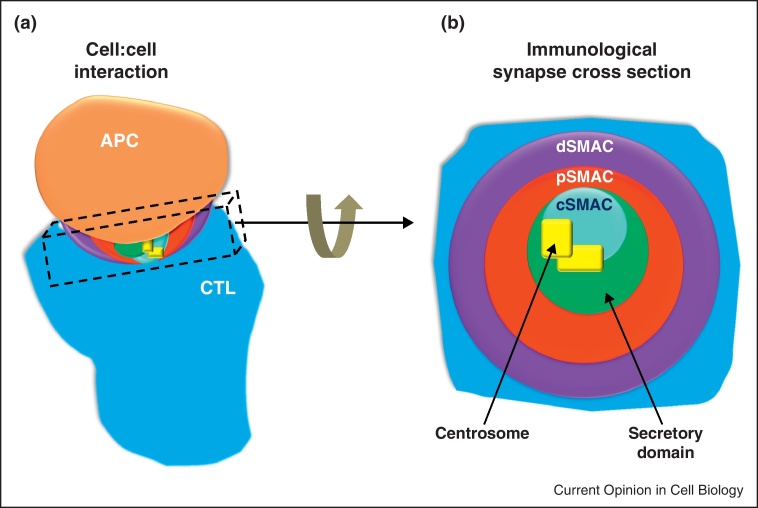
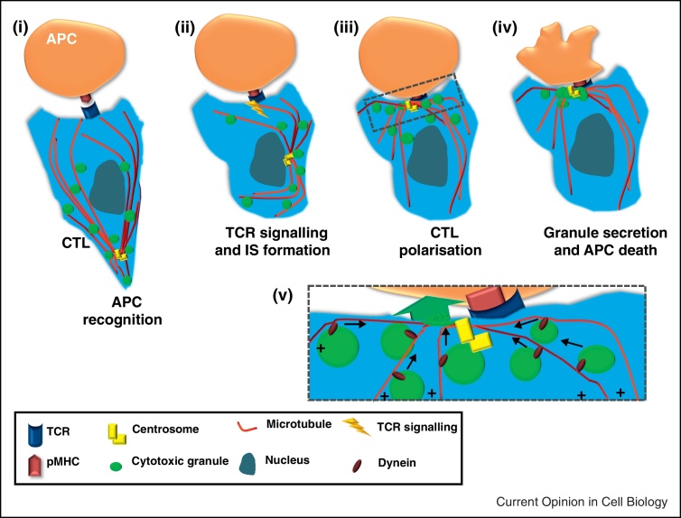
Directed secretion by immune cells requires formation of the immunological synapse at the site of cell-cell contact, concomitant with a dramatic induction of cell polarity. Recent findings provide us with insights into the various steps that are required for these processes: for example, the first identification of a protein at the centrosome that regulates its relocation to the plasma membrane; the use of super-resolution imaging techniques to reveal a residual actin network at the immunological synapse that may permit secretory granule exocytosis; and the drawing of parallels between primary cilia and IS architecture. Here we discuss these and other novel findings that have advanced our understanding of the complex process of immunological synapse formation and subsequent induced cell polarity in immune cells.


Figures

Figure 1
Figure 1
The immunological synapse (IS) in cytotoxic T cells (CTL). The IS forms at the site of cell contact between CTL and APC (a), with a series of supramolecular activation clusters (SMAC) forming as receptors segregate into a characteristic bullseye pattern when viewed en face (b) the central SMAC (cSMAC) with clustered T cell receptors (TCRs) involved in target recognition; the peripheral SMAC (pSMAC) with integrins involved in adhesion and the distal SMAC (dSMAC) with excluded phosphatases (CD45) and actin. Polarised secretion from CTL is directed by the centrosome (see Fig. 2) [13], which contacts the cSMAC forming the secretory domain around this point [49].
Figure 2
Figure 2
Cell polarity in CTL upon IS formation. (i) Migrating CTL recognise potential target APCs by engagement of peptide-major histocompatibility complex (pMHC) on the APC with cognate T cell receptor (TCR) on the CTL. (ii) Once TCR has engaged with pMHC, signalling within the CTL begins to occur, inducing the formation of the IS and polarisation of the CTL. (iii) The centrosome (which is the microtubule organising centre in CTL) moves from the uropod of the cell where it is found in migrating CTL and repositions itself at the point of TCR signalling. Cytotoxic granules move along microtubules in a minus-end direction towards the polarised centrosome. (iv) Secretion of cytotoxic granules at the secretory domain of the IS induces cell death in the recognised target APC. (v) Zoomed in from box in (iii) showing dynein moving granules in a minus-end direction along microtubules towards the centrosome where they are secreted towards the target cell.
Figure 3
Figure 3
Fluorescence microscopy for study of the IS. (a) An immune cell conjugate, shown here as a CTL and an APC, is trapped using optical tweezers and reorientated. The IS now lies in the focal plane of the confocal microscope allowing high-resolution imaging. The optical tweezers are shown as yellow light surrounding the conjugate with the orange lines representing force generating units required for reorientation. (b) As demonstrated by Biggs [5], a grid can be fabricated with pits into which APCs can be seeded. Upon addition of immune cells to the plate, for example CTL, the size constraints of the pit cause the IS to form in the microscope focal plane, increasing the resolution obtainable. This method allows many conjugates to be prepared in the correct orientation compared to the optical tweezer method due to the number of pits on the array. (c) Immune cells will conjugate to a planar lipid bilayer containing specific activating proteins, as if recognising an APC. In this schematic, a CTL has interacted with a planar lipid bilayer, consequently forming an IS and high-resolution imaging is obtained by total internal reflection microscopy. An evanescent wave, generated upon reflection of light at the coverslip, ascends only 100 nm into the CTL meaning background fluorescence is much reduced and high-resolution of IS components is obtained as only fluorophores in this small illuminated region are activated. (d) A comparison of a widefield deconvoluted confocal image the IS of a T cell activated with poly-l-lysine and anti-CD3 to that obtained using super-resolution structured illumination microscopy (SIM). Images are of the same cell, showing actin in white and the centrosome in blue, achieved by use of Phalloidin-Alexa Fluor 488 (Sigma) and anti-pericentrin (abcam ab4448). SIM reveals more detail at the IS centre as well as at the dSMAC. Scale bars are 2 μm. Imaging was performed using DeltaVision OMX 3D-SIM System V3 (Applied Precision) at the Gurdon Institute, Cambridge, UK. All data capture used an Olympus 100 × 1.4NA oil objective, 405 and 488 nm laser illumination and standard excitation and emission filter sets. 3D-SIM images were sectioned using 125 nm Z-step size. Raw 3-phase images were rendered and reconstructed in 3D by softWoRx 5.0.0 (Applied Precision) software. Images obtained and provided by Nele Dieckmann and Nicola Lawrence, Cambridge, UK.

References

    1. Grakoui A., Bromley S.K., Sumen C., Davis M.M., Shaw A.S., Allen P.M., Dustin M.L. The immunological synapse: a molecular machine controlling T cell activation. Science. 1999;285:221–227. - PubMed
    1. Monks C., Freiberg B., Kupfer H., Sciaky N., Kupfer A. Three-dimensional segregation of supramolecular activation clusters in T cells. Nature. 1998;395:82–86. - PubMed
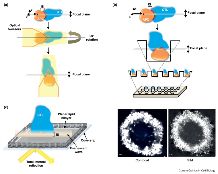
    1. Purbhoo M.A., Liu H., Oddos S., Owen D.M., Neil M.A., Pageon S.V., French P.M., Rudd C.E., Davis D.M. Dynamics of subsynaptic vesicles and surface microclusters at the immunological synapse. Sci Signal. 2010;3:ra36. - PubMed
    2. This paper elegantly employs optical tweezers to observe the immunological synapse in high resolution and demonstrates the delivery of signalling components from intracellular vesicles to the immunological synapse.

    1. Billadeau D.D. T cell activation at the immunological synapse: vesicles emerge for LATer signaling. Sci Signal. 2010;3:pe16. - PubMed
    1. Biggs M.J., Milone M.C., Santos L.C., Gondarenko A., Wind S.J. High-resolution imaging of the immunological synapse and T-cell receptor microclustering through microfabricated substrates. J R Soc Interface. 2011;8:1462–1471. - PMC - PubMed

Publication types

MeSH terms

LinkOut - more resources