

The effect of maternal vitamin D concentration on fetal bone
- PMID: 22990090
- PMCID: PMC3485609
- DOI: 10.1210/jc.2012-2538
The effect of maternal vitamin D concentration on fetal bone
Abstract
Context: Vitamin D deficiency during pregnancy may be associated with suboptimal fetal growth, but direct evidence is lacking.
Objectives: The aim of the study was to validate a method for fetal femur volume (FV) measurement using three-dimensional ultrasound and to detect correlations between FV and maternal vitamin D concentration.
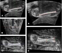
Design, setting, and participants: A novel method for assessing FV consists of three ultrasound measurements-femur length, proximal metaphyseal diameter (PMD), and midshaft diameter-and a volume equation; this was validated by comparing ultrasound to computed tomography measurements in six pregnancies after mid-trimester termination. This method was then applied in a cohort of healthy pregnant women participating in the Southampton Women Survey. Fetal three-dimensional ultrasound and maternal 25-hydroxyvitamin D [25(OH)D] levels were performed at 34 wk; dual-energy x-ray absorptiometry of the newborn was performed shortly after birth. Univariate and multiple linear regression analyses were performed between maternal characteristics and fetal outcomes.
Main outcome measures: We performed ultrasound measurements of the fetal femur.
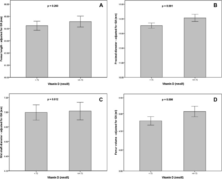
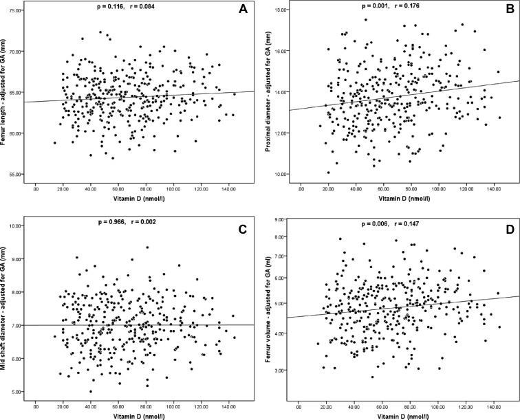
Results: In 357 pregnant participants, serum 25(OH)D correlated significantly with FV (P = 0.006; r = 0.147) and PMD (P = 0.001; r = 0.176); FV also demonstrated positive univariate correlations with maternal height (P < 0.001; r = 0.246), weight (P = 0.003; r = 0.160), triceps skinfold thickness (P = 0.013; r = 0.134), and a borderline negative effect from smoking (P = 0.061). On multiple regression, independent predictors of FV were the maternal height and triceps skinfold thickness; the effect of 25(OH)D on FV was attenuated, but it remained significant for PMD.
Conclusion: Using a novel method for assessing FV, independent predictors of femoral size were maternal height, adiposity, and serum vitamin D. Future trials should establish whether pregnancy supplementation with vitamin D is beneficial for the fetal skeleton, using FV and PMD as fetal outcome measures.
Figures
References
-
- Javaid MK, Cooper C. 2002. Prenatal and childhood influences on osteoporosis. Best Pract Res Clin Endocrinol Metab 16:349–367 - PubMed
-
- Godfrey K, Walker-Bone K, Robinson S, Taylor P, Shore S, Wheeler T, Cooper C. 2001. Neonatal bone mass: influence of parental birthweight, maternal smoking, body composition, and activity during pregnancy. J Bone Miner Res 16:1694–1703 - PubMed
-
- Chitty LS, Altman DG, Henderson A, Campbell S. 1994. Charts of fetal size: 4. Femur length. Br J Obstet Gynaecol 101:132–135 - PubMed
Publication types
MeSH terms
Substances
Grants and funding
LinkOut - more resources
Full Text Sources
Other Literature Sources
Medical
