. 2012 Nov 5;31(21):4204-20.
doi: 10.1038/emboj.2012.262. Epub 2012 Sep 18.

Endocytic tubules regulated by Rab GTPases 5 and 11 are used for envelopment of herpes simplex virus

Affiliations

Endocytic tubules regulated by Rab GTPases 5 and 11 are used for envelopment of herpes simplex virus

Michael Hollinshead et al. EMBO J. .

Abstract

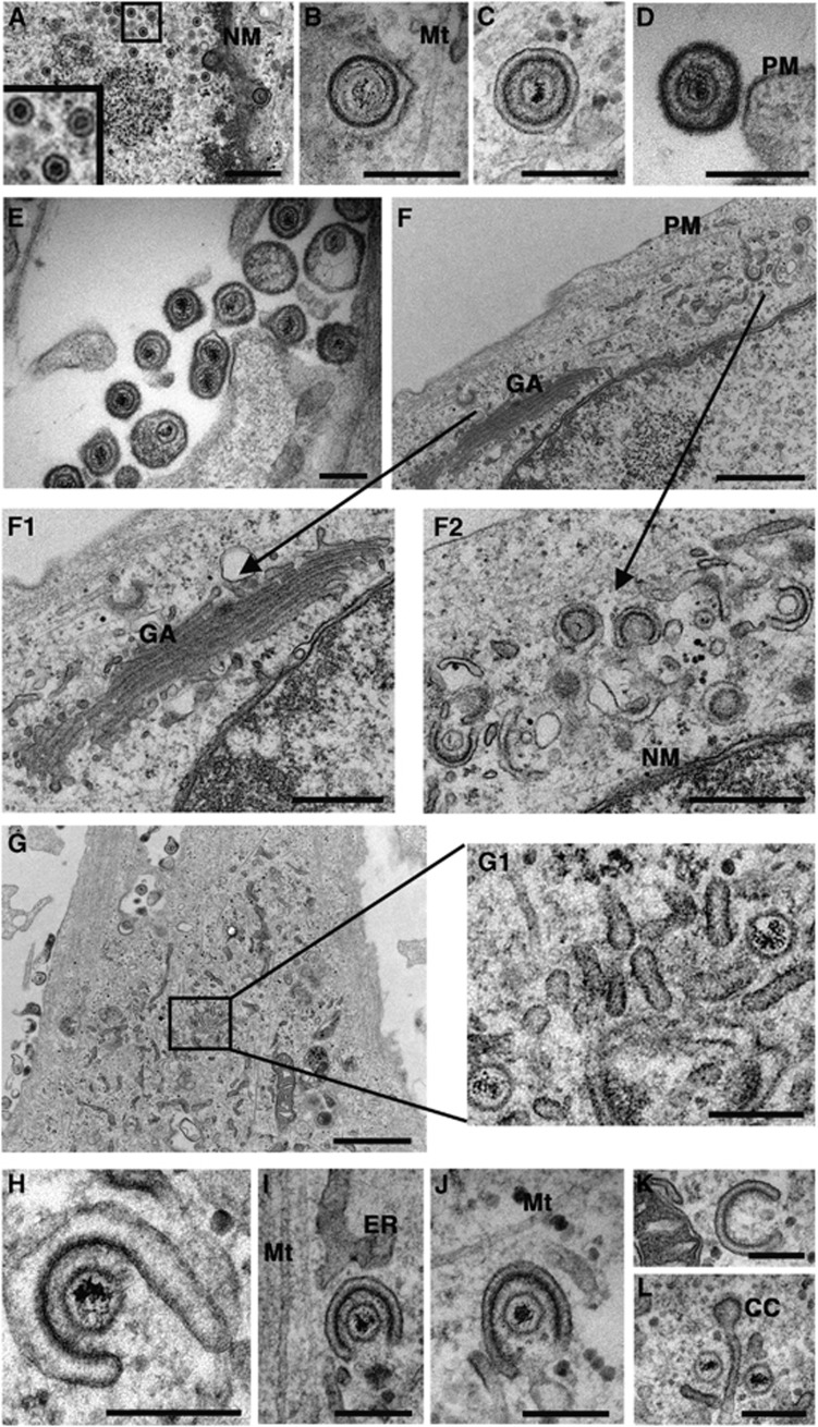
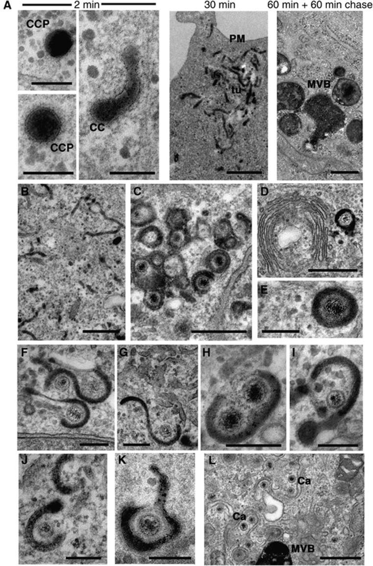
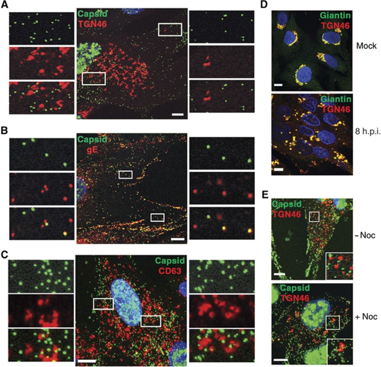
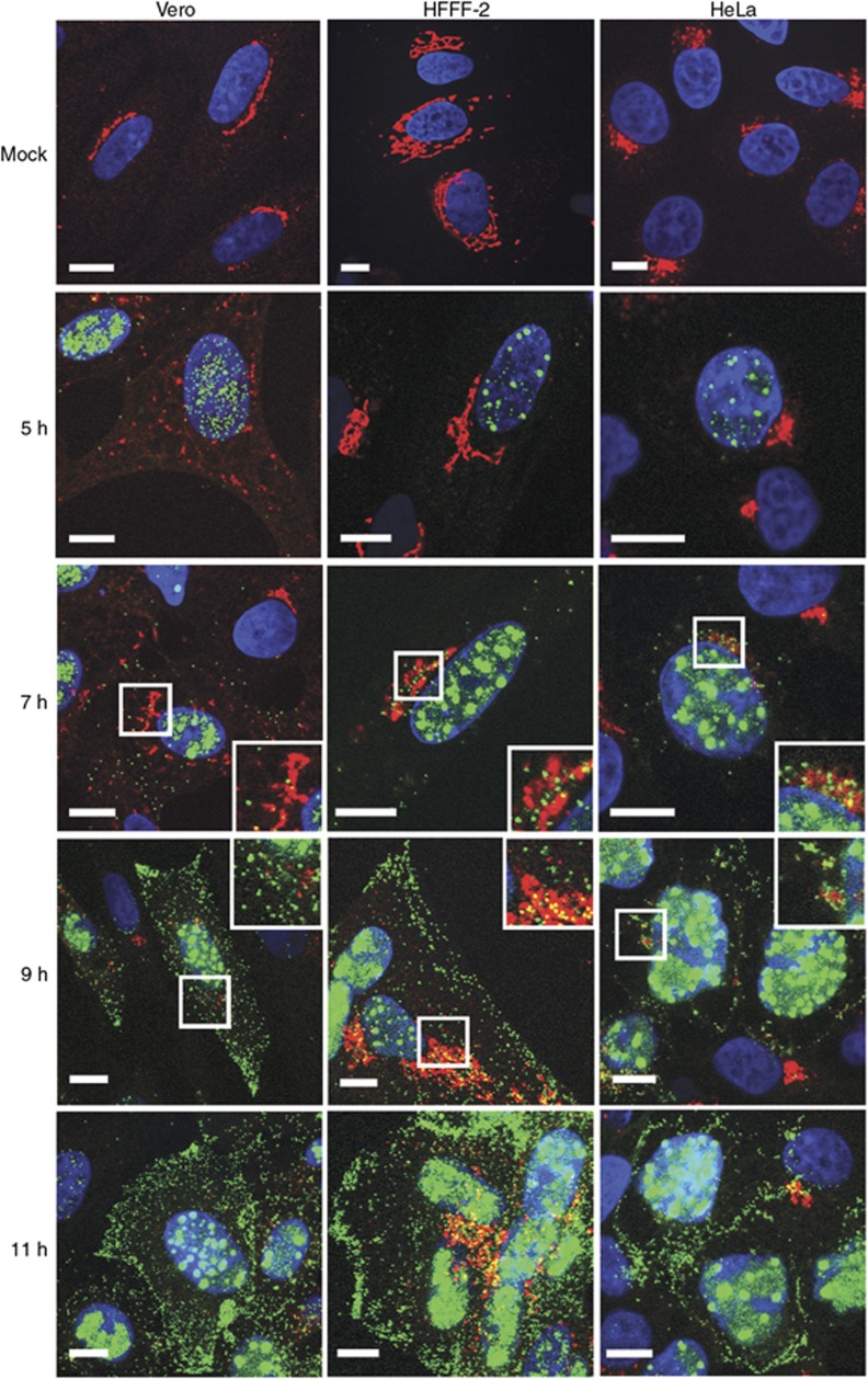
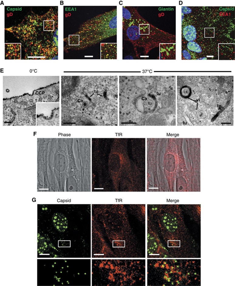
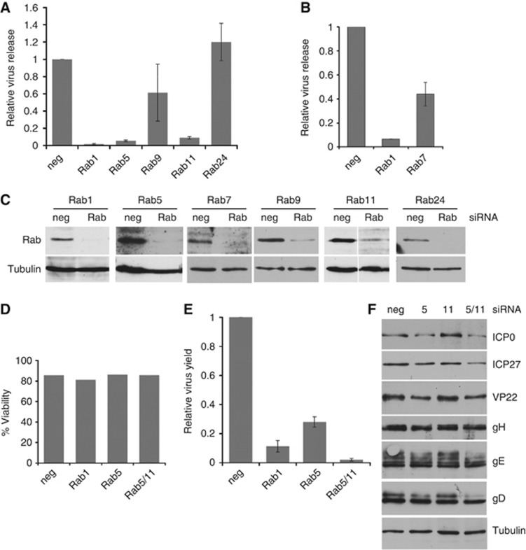
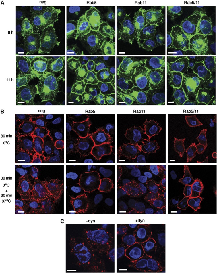
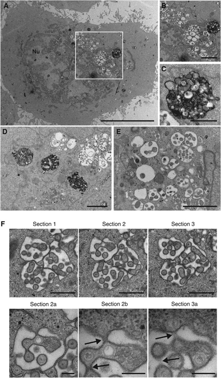
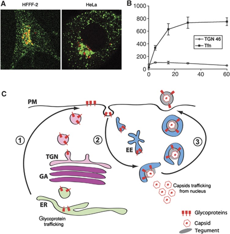
Enveloped viruses employ diverse and complex strategies for wrapping at cellular membranes, many of which are poorly understood. Here, an ultrastructural study of herpes simplex virus 1 (HSV1)-infected cells revealed envelopment in tubular membranes. These tubules were labelled by the fluid phase marker horseradish peroxidase (HRP), and were observed to wrap capsids as early as 2 min after HRP addition, indicating that the envelope had recently cycled from the cell surface. Consistent with this, capsids did not colocalise with either the trans-Golgi network marker TGN46 or late endosomal markers, but showed coincidence with the transferrin receptor. Virus glycoproteins were retrieved from the plasma membrane (PM) to label wrapping capsids, a process that was dependent on both dynamin and Rab5. Combined depletion of Rab5 and Rab11 reduced virus yield to <1%, resulting in aberrant localisation of capsids. These results suggest that endocytosis from the PM into endocytic tubules provides the main source of membrane for HSV1, and reveal a new mechanism for virus exploitation of the endocytic pathway.


Conflict of interest statement

The authors declare that they have no conflict of interest.

Figures

Figure 1
Figure 1
HSV1 capsids are wrapped in tubular membranes in HFFF-2 cells. HFFF-2 cells were infected at a multiplicity of 2 with HSV1, fixed and processed 12 h after infection, and imaged by EM. Panels F1/F2, and G2 are expanded regions of panels F and G respectively. NM, nuclear membrane; GA, Golgi apparatus; PM, plasma membrane; Mt, microtubule; Ca, capsid; ER, endoplasmic reticulum; CC, clathrin coat. In (A, F1, F2) scale bar=500 nm. In (BE, G1, HL), scale bar=200 nm. In (F, G), scale bar=1 μm.
Figure 2
Figure 2
HSV1 capsids are wrapped by tubules derived from the plasma membrane. (A) Uninfected HFFF-2 cells were labelled for 2 or 30 min with 10 mg/ml HRP, or 60 min followed by 60 min chase. After fixation, cells were stained with DAB and processed for imaging by EM. (BJ) HFFF-2 cells were infected at a multiplicity of 2 with HSV1 and labelled 11 h after infection with 10 mg/ml HRP for 60 min (BI), 10 min (J), 2 min (K) or 60 min followed by 60 min chase (L). PM, plasma membrane; ER, endoplasmic reticulum; NM, nuclear membrane; GA, Golgi apparatus; MVB, multivesicular body; Mt, microtubule; CC, clathrin coat; CCP, clathrin-coated pit; Tu, HRP-positive tubule. In (A) (2 min) and (EK), scale bar=200 nm. In (A) (30 and 60 min+60 min chase) and (BD, L), scale bar=500 nm.
Figure 3
Figure 3
HSV1 capsids colocalise with glycoprotein E but not TGN46 or CD63 in HFFF-2 cells. Cells grown on coverslips were infected with the GFP-VP26 virus (green) (AC, E) or s17 (D) at a multiplicity of 2, and fixed 8 h after infection. Cells were stained with antibody against (A, E) TGN46 (red), (B) gE (red), (C) CD63 (red) or (D) TGN46 (red) and giantin (green), and nuclei stained with DAPI (blue) before imaging with a Zeiss LSM510 Meta confocal microscope. In (E), nocodazole was added at a concentration of 2 μg/ml for 30 min before fixing to depolymerise the microtubule network (+noc). Scale bar=10 μm.
Figure 4
Figure 4
Relative localisation of virus capsids and TGN46 in HSV1-infected Vero, HeLa, and HFFF-2 cells. Cells grown on coverslips were infected with the GFP-VP26 virus (green) at a multiplicity of 2, and fixed at 5, 7, 9, or 11 h after infection. Cells were stained for TGN46 (red) and nuclei stained with DAPI (blue) before imaging with a Zeiss LSM510 Meta confocal microscope. Scale bar=10 μm.
Figure 5
Figure 5
Glycoprotein D is endocytosed from the plasma membrane to HSV1 wrapping membranes. HFFF-2 cells grown on coverslips were infected with (A) the GFP-VP26 virus or (B, C) Wt HSV1 at a multiplicity of 2. Ten hours after infection, gD antibody uptake experiments were carried out by incubating cells for 30 min on ice with anti-gD monoclonal antibody followed by 30 min at 37°C to allow uptake. Cells were fixed and permeabilised then labelled with (B) anti-EEA1 (green), or (C) anti-TGN46 (green), and counterstained with Alexa 568 anti-mouse secondary antibody to detect anti-gD antibody (red) and DAPI to detect nuclei (blue). (D) HFFF-2 cells infected with the GFP-VP26 virus were fixed at 10 h and labelled with anti-EEA1 (red) and DAPI to detect nuclei (blue). (E) HFFF-2 cells were infected with HSV1 and at 10 h gD antibody uptake assay carried out as described above with the inclusion of a 30-min incubation on ice with an HRP-tagged F(ab′)2 fragment of anti-mouse IgG prior to incubation at 37°C. Cells were fixed and stained for HRP prior to processing for EM. (F) Uninfected HFFF-2 cells were labelled with anti-transferrin receptor (red). (G) HFFF-2 cells infected with GFP-VP26 virus (green) were fixed 10 h after infection and labelled with anti-transferrin receptor antibody (red). All immunofluorescence images were acquired with a Zeiss LSM510 Meta confocal microscope. In (AD, FG), scale bar=10 μm. In (E), scale bar=200 nm.
Figure 6
Figure 6
Rab GTPases 5 and 11 are required for efficient HSV1 production. (A, B) HeLa cells transfected with siRNAs as denoted were infected 48 h later with HSV1 at a multiplicity of 5, extracellular virus harvested 16 h later and titre measured. Graphs show titres in relation to those obtained for the negative siRNA (taken as 1), and values are shown as mean±standard error of the mean from three independent experiments. (C) Total cell extracts of siRNA-transfected HeLa cells were analysed by western blotting with antibodies as labelled. (D) HeLa cells transfected with siRNAs for Rab1, Rab5, or Rab11 were assessed for viability. Graph denotes viability in relation to non-transfected cells (taken as 100%). (E) HeLa cells transfected with siRNAs as denoted were infected 48 h later with HSV1 at a multiplicity of 5, and the titre of cell-associated virus was measured 16 h later. Graph shows titres in relation to those obtained for the negative siRNA (taken as 1) and values are shown as mean±standard error of the mean from three independent experiments. (F) Total cell extracts of siRNA transfected, infected HeLa cells, harvested at 16 h, were analysed by western blotting with antibodies as labelled.
Figure 7
Figure 7
Rab5 and dynamin are required for retrieval of glycoprotein D from the plasma membrane. (A) HeLa cells on coverslips transfected with siRNAs as denoted were infected 48 h later with HSV1 expressing gD-GFP (green) at a multiplicity of 2, fixed 8 or 11 h later and stained with DAPI to detect nuclei (blue). (B) HeLa cells on coverslips transfected with siRNAs as denoted were infected 48 h later with HSV1, and gD antibody uptake experiments were carried out 8 h later. After fixation, cells were stained with anti-mouse secondary antibody to detect anti-gD antibody (red) and DAPI to detect nuclei (blue). (C) HeLa cells infected with HSV1 were incubated for 30 min with dynasore 8 h later (+dyn), or left untreated (−dyn). gD antibody uptake was carried out and cells processed as for (B). Scale bar=10 μm.
Figure 8
Figure 8
Rab5 is required for HSV1 envelopment. HeLa cells transfected with siRNAs for Rab5 were infected 48 h later with HSV1 at a multiplicity of 2, and labelled for 30 min with 10 mg/ml HRP 11 h later. Cells were fixed, stained with DAB and processed for EM. (A) An overview of a representative infected, Rab5-depleted cell. (B) Magnified image of juxtanuclear membranes. (C) Magnified image of HRP containing membrane-bound structure. (D) A second example of large HRP-positive structures next to capsids clustering at membranes. (E) Distended membranes with associated capsids. (F) Three serial sections of a cell exhibiting capsids associated with dilated membranes. Sections 2a and 2b are magnified images of section 2, showing the continuum between the individual particles and the cytoplasm (arrowed) that is not obvious in section 3 or the magnified section 3a. In (A), scale bar=10 μm, (B, D), scale bar=2 μm. In (C, E, F) (sections 1–3) scale bar=1 μm. In (F) (sections 2a, 2b & 3a), scale bar=200 nm.
Figure 9
Figure 9
Endocytic membranes as a source of HSV1 envelope. (A) HFFF-2 and HeLa cells were incubated with 200 ng/ml Cy3-transferrin (green) for 30 min prior to fixing and staining for TGN46 (red). (B) HeLa cells were treated as described in (A) for times ranging from 0 to 60 min. Using LSM510 software, TGN46 and transferrin-positive areas were measured in projected Z stacks relative to area of the cell. The results are the average of five cells for each time point. (C) Model for virus envelopment via endocytosis. (1) Virus glycoproteins are processed in the Golgi/TGN and exported to the cell surface. (2) The glycoprotein-containing plasma membrane is endocytosed and transported through the early endosome to produce wrapping tubules in a pathway dependent on dynamin, and Rabs 5 and 11. (3) Glycoprotein-containing tubules wrap cytoplasmic capsids forming virions with a double membrane. Fusion of the outer membrane at the cell surface results in release of a single-membrane virion outside the cell. EE, EEA1-positive early endosome; ER, endoplasmic reticulum; GA, Golgi apparatus; PM, plasma membrane; TGN, trans-Golgi network.

Comment in

Similar articles

Cited by

References

    1. Alconada A, Bauer U, Sodeik B, Hoflack B (1999) Intracellular traffic of herpes simplex virus glycoprotein gE: characterization of the sorting signals required for its trans-Golgi network localization. J Virol 73: 377–387 - PMC - PubMed
    1. Andres G, Garcia-Escudero R, Simon-Mateo C, Vinuela E (1998) African swine fever virus is enveloped by a two-membraned collapsed cisterna derived from the endoplasmic reticulum. J Virol 72: 8988–9001 - PMC - PubMed
    1. Antinone SE, Smith GA (2006) Two modes of herpesvirus trafficking in neurons: membrane acquisition directs motion. J Virol 80: 11235–11240 - PMC - PubMed
    1. Bard F, Malhotra V (2006) The formation of TGN-to-plasma-membrane transport carriers. Annu Rev Cell Dev Biol 22: 439–455 - PubMed
    1. Barman S, Ali A, Hui EK, Adhikary L, Nayak DP (2001) Transport of viral proteins to the apical membranes and interaction of matrix protein with glycoproteins in the assembly of influenza viruses. Virus Res 77: 61–69 - PubMed

Publication types

MeSH terms