. 2013 May;6(3):498-510.
doi: 10.1038/mi.2012.89. Epub 2012 Sep 19.

Resident and pro-inflammatory macrophages in the colon represent alternative context-dependent fates of the same Ly6Chi monocyte precursors

Affiliations
Free PMC article

Resident and pro-inflammatory macrophages in the colon represent alternative context-dependent fates of the same Ly6Chi monocyte precursors

C C Bain et al. Mucosal Immunol. 2013 May.
Free PMC article

Abstract

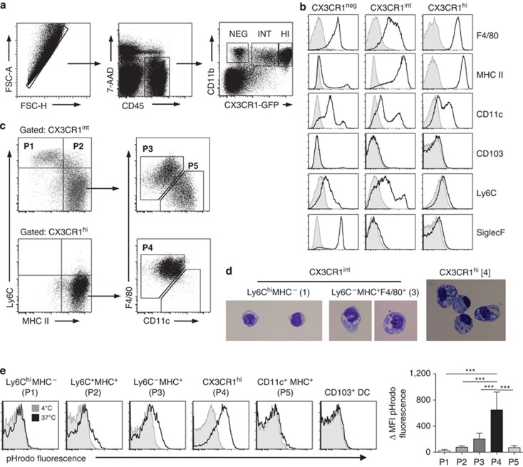
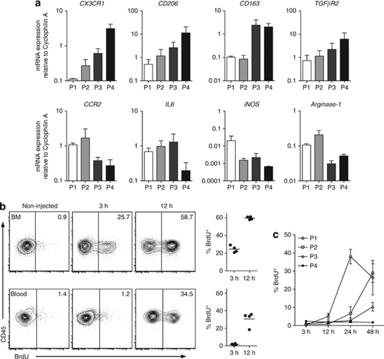
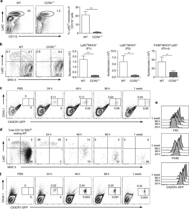
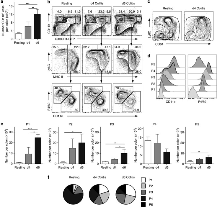
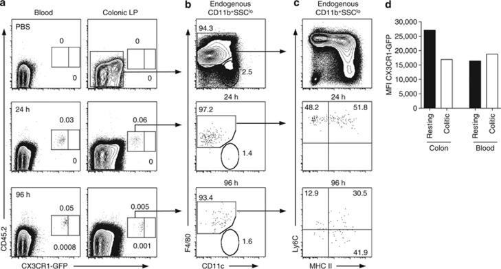
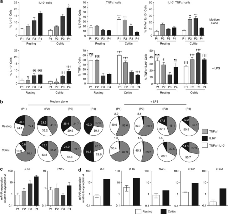
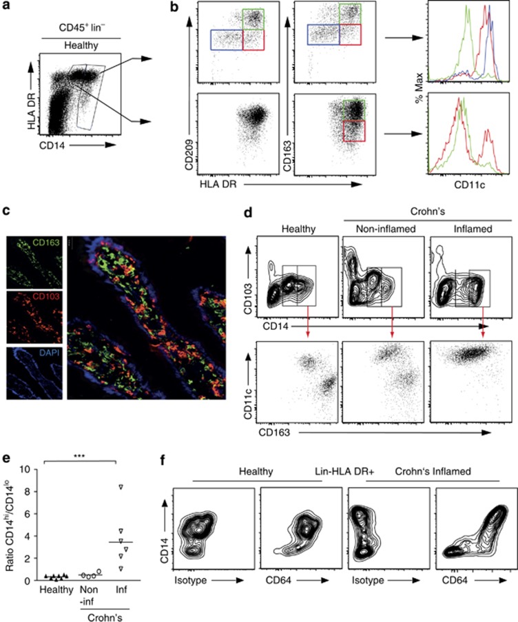
Macrophages (mφ) are essential for intestinal homeostasis and the pathology of inflammatory bowel disease (IBD), but it is unclear whether discrete mφ populations carry out these distinct functions or if resident mφ change during inflammation. We show here that most resident mφ in resting mouse colon express very high levels of CX3CR1, are avidly phagocytic and MHCII(hi), but are resistant to Toll-like receptor (TLR) stimulation, produce interleukin 10 constitutively, and express CD163 and CD206. A smaller population of CX3CR1(int) cells is present in resting colon and it expands during experimental colitis. Ly6C(hi)CCR2(+) monocytes can give rise to all mφ subsets in both healthy and inflamed colon and we show that the CX3CR1(int) pool represents a continuum in which newly arrived, recently divided monocytes develop into resident CX3CR1(hi) mφ. This process is arrested during experimental colitis, resulting in the accumulation of TLR-responsive pro-inflammatory mφ. Phenotypic analysis of human intestinal mφ indicates that analogous processes occur in the normal and Crohn's disease ileum. These studies show for the first time that resident and inflammatory mφ in the intestine represent alternative differentiation outcomes of the same precursor and targeting these events could offer routes for therapeutic intervention in IBD.


Figures

Figure 1
Figure 1
Phenotypic characterization of colonic mononuclear phagocytes. (a) Doublets were excluded from colonic digests from CX3CR1gfp/+ mice on the basis of FSC-A and FSC-H, and live leukocytes were selected as being 7-AAD CD45+. The resulting cells were then analyzed for CD11b and CX3CR1-green fluorescent protein (GFP) expression. (b) Histograms show the expression of the indicated cell surface markers on the CX3CR1, CX3CR1int, and CX3CR1hi subsets. (c) Expression of Ly6C, MHCII, F4/80, and CD11c on CX3CR1int (top panels) and CX3CR1hi CD11b+ cells (bottom panels). (d) Morphological assessment of fluorescence-activated cell-sorted P1, P3, and P4 cells (× 200). (e) Phagocytic activity of colonic MP cell subsets, as measured by the uptake of pHrodo E. coli bioparticles for 15 min at 37 °C (black lines) or at 4 °C as a control (shaded histograms). Representative pHrodo dye fluorescence of CX3CR1-based P1 to P5 and CD103+ DC. Mean ΔMFI (mean fluorescence intensity (MFI) of pHrodo dye fluorescence at 37 °C-MFI at 4 °C control) for populations 1 to 4 + 1 s.d. for 4–5 mice is shown. Representation of three individual experiments (***P<0.001).
Figure 2
Figure 2
Transcriptional and kinetic analysis of colonic CX3CR1-defined myeloid cells. (a) Quantitative reverse transcription PCR (qRT-PCR) quantitation of CX3CR1, CD163, CD206, TGFβR2, CCR2, interleukin 6 (IL6), and tumor necrosis factor α (TNFα) messenger RNA by fluorescence-activated cell sorted P1, P2, P3, and P4 from colonic mucosa. Results shown are mean expression relative to cyclophilin A using the 2−ΔC(t) method. Means of three independent experiments using pooled cells from 12 mice per experiment. (b, c) CX3CR1+/gfp mice received 1 mg 5-bromo-2-deoxyuridine (BrdU) intraperitoneally and 3, 12, 24, and 48 h later, BrdU uptake was assessed in Ly6Chi blood and bone marrow monocytes (CD11b+Ly6ChiCX3CR1int) (b) and colonic CX3CR1int (P1–P3) and CX3CR1hi (P4) populations (c). Results are means of four mice per group.
Figure 3
Figure 3
Recruitment of Ly6Chi monocytes to colon of CCR2 null mice. (a) Representative staining and mean percentage of CD115+ Ly6Chi monocytes within the CD11b+ fraction of blood leukocytes in wild-type (WT) or CCR2−/− mice. (b) Representative Ly6C and MHCII expression on live-gated CD45+ CD11b+SSClow colon leukocytes and the absolute numbers of P1, P2, and P3/P4 subsets from resting WT or CCR2−/− mice. Results representative of at least three individual experiments with 3–4 mice in each group. (**P<0.01 and ***P<0.001). (c) Donor-derived cells (CD45.1+/CD45.2+ CX3CR1+/gfp) within the live-gated CD45+ CD11b+ fraction of colonic LP cells of CCR2−/− mice, 24 h, 48 h, 96 h, and 7 days after transfer of 2 × 10⁶ fluorescence-activated cell-sorted Ly6Chi BM monocytes. (d) Expression of Ly6C, MHCII, and F4/80 on donor-derived cells isolated from recipient CCR2−/− colon at 24 h, 48 h, 96 h, and 7 days after transfer of Ly6Chi monocytes, compared with live-gated CD45+ CD11b+SSClo cells from wild-type (WT) colon (left panel). (e) Expression of CX3CR1-green fluorescent protein (GFP) by donor-derived cells in colon at 24 h, 48 h, 96 h, and 7 days after transfer. Gates for identifying CX3CR1int and CX3CR1hi cells were set using CD11b+ cells from resting CX3CR1+/gfp colon. (f) Donor-derived cells (CD45.1+/CD45.2+ CX3CR1+/gfp) among live-gated CD45+ CD11b+ cells in bloodstream of CCR2−/− mice, 24 h, 48 h, 96 h, and 7 days after transfer of Ly6Chi monocytes and in phosphate-buffered saline (PBS)-treated controls. Results are representative of two individual experiments with two recipient mice at each time point.
Figure 4
Figure 4
Effects of inflammation on composition of colonic myeloid cell populations. (a) CX3CR1+/gfp mice were fed 2% dextran sodium sulfate in the drinking water for 7 days to induce colitis and the absolute numbers of CD11b+ LP leukocytes per colon assessed at d4 and d6, and in resting mice. (b) Representative expression of CD11b and CX3CR1-green fluorescent protein (GFP) by live-gated CD45+ colonic cells (upper panels), expression of Ly6C and MHCII by CD11b+ CX3CR1int colonic cells (middle panels), and expression of F4/80 and CD11c among CX3CR1int Ly6C MHCII+ fraction (lower panels) in resting mice and on d4 and d6 of colitis. (c) Expression of Ly6C and CD64 by CX3CR1int CD11b+ cells in resting mice and on d4 of colitis. (d) Expression of CD11c and F4/80 by subsets P1–P5 on d4 of colitis. (e) Absolute numbers of CX3CR1-defined P1, P2, P3, P4, and P5 subsets of colonic CD11b+ cells during colitis. (f) Relative proportions of P1–P5 subsets among total CX3CR1+ cells in resting mice and on d4 and d6 of colitis. *P<0.05, **P<0.01, and ***P<0.001.
Figure 5
Figure 5
Effects of inflammation on recruitment of monocytes to colon. Monocyte recruitment into inflamed colon was studied by transferring 2 × 10⁶ fluorescence-activated cell sorted Ly6Chi bone marrow monocytes from CD45.1+/CD45.2+ CX3CR1+/gfp mice into CD45.1+ wild-type (WT) mice on d3 of dextran sodium sulfate (DSS) colitis. After a further 2 days of DSS, mice were returned to normal drinking water. (a) Donor-derived cells within the live-gated CD45+ CD11b+ fraction of colonic LP cells of colitic mice 24 and 96 h after transfer compared with PBS-injected controls. CX3CR1int and CX3CR1hi gates were set using donor-derived monocytes in blood, which remain CX3CR1int. (b) Expression of F4/80 and CD11c by donor-derived cells in inflamed colon 24 and 96 h after transfer, compared with endogenous CD45+ CD11b+SSClo cells in colon on d4 DSS. (c) Expression of Ly6C and MHCII by F4/80+ donor-derived cells in inflamed colon 24 and 96 h after transfer, compared with endogenous CD45+ CD11b+ F4/80+ SSClo cells in colon on d4 DSS. (d) Mean fluorescence intensity (MFI) of CX3CR1-green fluorescent protein (GFP) expression on donor-derived monocytes derived from the colon and blood of resting CCR2−/− mice or from WT mice taken on day 4 of DSS colitis, with two mice per group. Results are representative of two individual experiments. PBS, phosphate-buffered saline.
Figure 6
Figure 6
Functional characterization and Toll-like receptor (TLR) responsiveness of colonic CX3CR1-defined myeloid cells in healthy and inflamed intestine. (a) Colonic LP cells isolated from resting CX3CR1+/gfp mice, or from mice receiving 2% dextran sodium sulfate (DSS) for 4 days, were cultured for 4.5 h in medium (top panels) or with 1 μg ml−1 lipopolysaccharide (LPS) (lower panels). Interleukin 10 (IL10) and tumor necrosis factor α (TNFα) production was assessed by intracellular cytokine staining. Results shown are mean proportions of IL10+, TNFα+, and IL10+TNFα+ cells within P1, P2, P3, and P4 cells in each condition + 1 s.d. for 3–4 mice per group. (b) Proportions of total cytokine-producing cells within P1 to P4 that produce IL10 alone, TNFα alone, or both IL10 and TNFα in resting (top panels) or inflamed colon (lower panels) ± lipopolysaccharide (LPS). Data representative of two individual experiments. (*P<0.05, **P<0.01, ***P<0.001 and P<0.05, ¶¶P<0.01, ¶¶¶P<0.001 versus P1–P4 subsets from resting mice cultured in medium alone; P<0.05, ††P<0.01, †††P<0.001 versus P1–P4 subsets from colitic mice cultured in medium alone.) (c) Quantitation of messenger RNA (mRNA) for IL10 and TNFα by fluorescence-activated cell sorted subsets P1–P4 from resting colon. (d) Quantitation of mRNA for pro-inflammatory cytokines and TLR by P1 subset of colonic myeloid cells from resting and colitic mice. Results are shown as mean expression relative to cyclophilin A (CPA) using the 2−ΔC(t) and are from three independent experiments using pooled cells from 12 mice per experiment (resting mice) or one experiment using pooled cells from 10 mice fed DSS for 4 days.
Figure 7
Figure 7
Macrophage subsets in the human ileum. Lin (CD3, CD19, CD20, CD56, TCRαβ), live (PI) singlet cells were identified in digests of resected healthy ileum (n=8) and CD45+ cells purified by magnetic antibody cell sorting. (a) CD14hi and CD14lo cells were identified and HLA-DR expression assessed. (b) Subsets of CD209/CD163 DR (blue gate), CD209/CD163 DR+ (red gate), and CD209/CD163+ DR+ (green gate) cells were then identified among the CD14hi (top panels) and CD14lo cells (lower panels), and their expression of CD11c assessed. Plots are representative of eight (A) or three (B) individual samples. (c). Anatomical location of resident mφ in healthy human ileum. CD163+ mφ (green) are found immediately below the epithelial layer (46-diamidino-2-phenyl indole (DAPI)—blue) and are a discrete population from CD103+ DC and lymphocytes (red) (× 100–× 400 final magnification). (d) Representative plots identifying CD14hi and CD14lo CD103 cells among CD45+ Lin cells from samples of healthy ileum, non-inflamed, or inflamed areas of Crohn's disease ileum (top panels). CD11c and CD163 expression were then assessed on the CD14hi subsets from each sample (lower panels). Results representative of 4–8 individual samples. (e) Ratios of CD14hi:CD14lo CD103 mφ in healthy, non-inflamed, and inflamed Crohn's disease ileal samples. Each symbol represents an individual patient. (f) CD64 vs. CD14 expression by live Lin MHCII+ cells from normal and inflamed Crohn's disease human ileum. The left panels in each pair show CD64 isotype staining. ***P<0.001.

References

    1. Bain C.C., Mowat A.McI. Intestinal macrophages - specialised adaptation to a unique environment. Eur. J. Immunol. 2011;41,:2494–2498. - PubMed
    1. Geissmann F., Manz M.G., Jung S., Sieweke M.H., Merad M., Ley K. Development of monocytes, macrophages, and dendritic cells. Science. 2010;327,:656–661. - PMC - PubMed
    1. Hadis U., et al. Intestinal tolerance requires gut homing and expansion of foxP3+ regulatory T cells in the lamina propria. Immunity. 2011;34,:237–246. - PubMed
    1. Kayama H., et al. Intestinal CX3C chemokine receptor 1high (CX3CR1high) myeloid cells prevent T-cell-dependent colitis. Proc. Natl Acad. Sci. USA. 2012;109,:5010–5015. - PMC - PubMed
    1. MacDonald T.T., Monteleone I., Fantini M.C., Monteleone G. Regulation of homeostasis and inflammation in the intestine. Gastroenterology. 2011;140,:1768–1775. - PubMed

Publication types

MeSH terms