. 2012 Oct 9;107(8):1327-36.
doi: 10.1038/bjc.2012.410. Epub 2012 Sep 18.

Individuality in FGF1 expression significantly influences platinum resistance and progression-free survival in ovarian cancer

Affiliations

Individuality in FGF1 expression significantly influences platinum resistance and progression-free survival in ovarian cancer

G Smith et al. Br J Cancer. .

Abstract

Background: Ovarian cancer is frequently advanced at presentation when treatment is rarely curative. Response to first-line platinum-based chemotherapy significantly influences survival, but clinical response is unpredictable and is frequently limited by the development of drug-resistant disease.

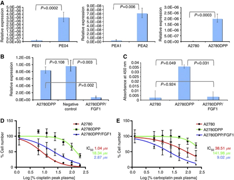
Methods: We used qRT-PCR analysis to assess intertumour differences in the expression of fibroblast growth factor 1 (FGF1) and additional candidate genes in human ovarian tumours (n=187), and correlated individuality in gene expression with tumour histology, chemotherapy response and survival. We used MTT assays to assess platinum chemosensitivity in drug-sensitive and drug-resistant ovarian cell lines.

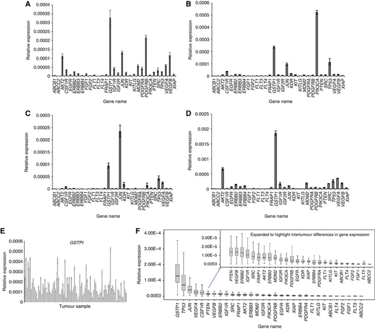
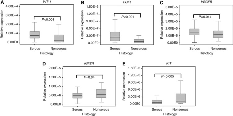
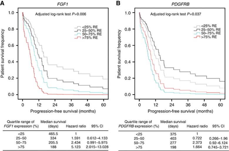
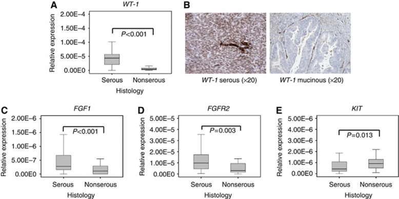
Results: Marked intertumour differences in gene expression were observed, with each tumour having a unique gene expression profile. Nine genes, including FGF1 (P=1.7 × 10(-5)) and FGFR2 (P=0.003), were differentially expressed in serous and nonserous tumours. MDM2 (P=0.032) and ERBB2 (P=0.064) expression was increased in platinum-sensitive patients, and FGF1 (adjusted log-rank test P=0.006), FGFR2 (P=0.04) and PDRFRB expression (P=0.037) significantly inversely influenced progression-free survival. Stable FGF1 gene knockdown in platinum-resistant A2780DPP cells re-sensitised cells to both cisplatin and carboplatin.

Conclusion: We show for the first time that FGF1 is differentially expressed in high-grade serous ovarian tumours, and that individuality in FGF1 expression significantly influences progression-free survival and response to platinum-based chemotherapy.


Figures

Figure 1
Figure 1
TLDA cards were used to compare the expression of 31 candidate genes, relative to the expression of the loading control 18S ribosomal RNA, as described in Materials and Methods. Gene expression was assessed in fresh-frozen ovarian tumours (n=96), and is represented as mean±s.d. of triplicate replicates. (A–D) Gene expression in 4 representative ovarian tumours; (E) intertumour variation in GSTP1 expression, where each bar represents one tumour; (F) summary of variation in relative gene expression (box plot median) and the extent of intertumour variability in the expression of each gene (box plot whiskers).
Figure 2
Figure 2
Association of gene expression with tumour histology. TLDA cards were used to compare gene expression in fresh-frozen ovarian tumours (n=96) as described in Materials and Methods, and tumours subclassified according to histology – serous tumours (n=59) and nonserous tumours (n=37, combined mucinous, endometrioid and clear-cell histologies). WT-1 (A), FGF1 (B) and VEGFB (C) were significantly overexpressed in serous tumours; IGF2R (D) and KIT (E) were significantly overexpressed in nonserous tumours. Significant differences in ABCC2, CSF1R, ERBB4, JUN and KDR gene expression were also observed (Supplementary Data Figure S2).
Figure 3
Figure 3
Correlation of (A) FGF1 and (B) PDGFRB expression with patient survival. Log-rank analysis was used to compare quartiles (0–25%, 25–50%, 50–75% and 75–100% of maximum expression) of FGF1 and PDGFRB expression with progression-free survival, assessed from the end of the first course of chemotherapy, as described in Materials and Methods. Survival was adjusted for patient histology, the extent of debulking surgery and tumour histology, stage and grade in multivariate analysis. Correlation of FGF1 expression with overall survival is illustrated in Supplementary Data Figure S3.
Figure 4
Figure 4
Validation of ovarian tumour histology associations. Single-gene Taqman probes were used to compare gene expression in FFPE ovarian tumours (n=91) as described in Materials and Methods, and tumours subclassified according to histology – serous tumours (n=58) and nonserous tumours (n=33, combined mucinous, endometrioid and clear-cell histologies). WT-1 (A), FGF1 (C) and FGFR2 (D) were significantly more highly expressed in serous tumours; increased WT-1 expression in serous tumours was confirmed by immunohistochemical analysis as described in Materials and Methods (B). In contrast, KIT (E) was significantly overexpressed in nonserous tumours.
Figure 5
Figure 5
FGF1 expression influences platinum chemosensitivity. qRT–PCR analysis was used to compare FGF1 expression in paired platinum-sensitive and -resistant ovarian cancer cell lines, as described in Materials and Methods (A). FGF1 expression was stably knocked down in A2780DPP cells and reduction in gene expression confirmed by qRT–PCR analysis (B) and quantitative immunoassay (C). MTT assays were used to compare chemosensitivity to (D) cisplatin and (E) carboplatin in drug-sensitive A2780 cells, drug-resistant A2780DPP cells and A2780DPP/FGF1 knockdown cells, as described in Materials and Methods.

References

    1. Al-Hussaini M, Stockman A, Foster H, McCluggage WG (2004) WT-1 assists in distinguishing ovarian from uterine serous carcinoma and in distinguishing between serous and endometrioid ovarian carcinoma. Histopathology 44(2): 109–115 - PubMed
    1. Banerjee S, Kaye S (2011) The role of targeted therapy in ovarian cancer. Eur J Cancer 47(Suppl 3): S116–S130 - PubMed
    1. Bartel F, Jung J, Bohnke A, Gradhand E, Zeng K, Thomssen C, Hauptmann S (2008) Both germ line and somatic genetics of the p53 pathway affect ovarian cancer incidence and survival. Clin Cancer Res 14(1): 89–96 - PubMed
    1. Birrer MJ, Johnson ME, Hao K, Wong KK, Park DC, Bell A, Welch WR, Berkowitz RS, Mok SC (2007) Whole genome oligonucleotide-based array comparative genomic hybridization analysis identified fibroblast growth factor 1 as a prognostic marker for advanced-stage serous ovarian adenocarcinomas. J Clin Oncol 25(16): 2281–2287 - PubMed
    1. Blackledge G, Lawton F, Redman C, Kelly K (1989) Response of patients in phase II studies of chemotherapy in ovarian cancer: implications for patient treatment and the design of phase II trials. Br J Cancer 59(4): 650–653 - PMC - PubMed

Publication types

MeSH terms