. 2013 Feb;45(2):159-68.
doi: 10.1055/s-0032-1321789. Epub 2012 Sep 18.

Expression profiles of the nuclear receptors and their transcriptional coregulators during differentiation of neural stem cells

Affiliations

Expression profiles of the nuclear receptors and their transcriptional coregulators during differentiation of neural stem cells

A Androutsellis-Theotokis et al. Horm Metab Res. 2013 Feb.

Abstract

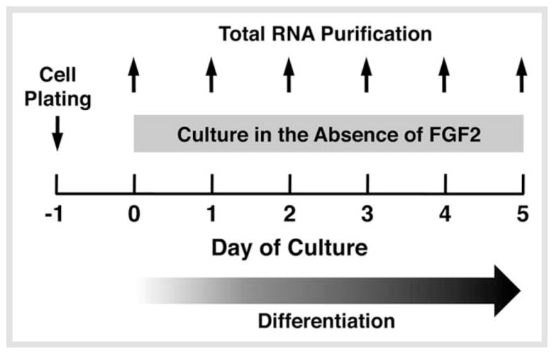
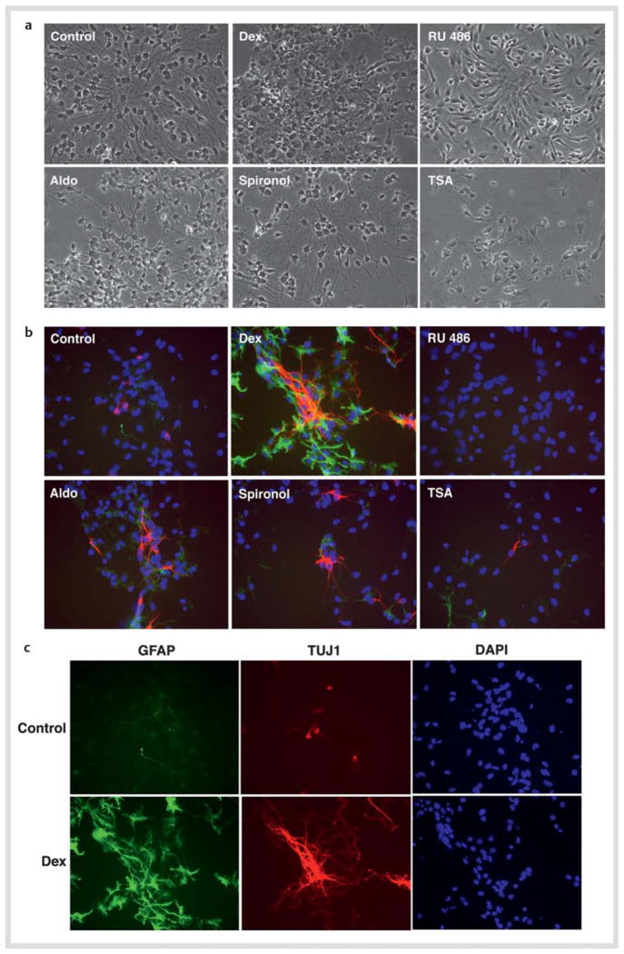
Neural stem cells (NSCs) are pluripotent precursors with the ability to proliferate and differentiate into 3 neural cell lineages, neurons, astrocytes and oligodendrocytes. Elucidation of the mechanisms underlying these biologic processes is essential for understanding both physiologic and pathologic neural development and regeneration after injury. Nuclear hormone receptors (NRs) and their transcriptional coregulators also play crucial roles in neural development, functions and fate. To identify key NRs and their transcriptional regulators in NSC differentiation, we examined mRNA expression of 49 NRs and many of their coregulators during differentiation (0-5 days) of mouse embryonic NSCs induced by withdrawal of fibroblast growth factor-2 (FGF2). 37 out of 49 NRs were expressed in NSCs before induction of differentiation, while receptors known to play major roles in neural development, such as THRα, RXRs, RORs, TRs, and COUP-TFs, were highly expressed. CAR, which plays important roles in xenobiotic metabolism, was also highly expressed. FGF2 withdrawal induced mRNA expression of RORγ, RXRγ, and MR by over 20-fold. Most of the transcriptional coregulators examined were expressed basally and throughout differentiation without major changes, while FGF2 withdrawal strongly induced mRNA expression of several histone deacetylases (HDACs), including HDAC11. Dexamethasone and aldosterone, respectively a synthetic glucocorticoid and natural mineralocorticoid, increased NSC numbers and induced differentiation into neurons and astrocytes. These results indicate that the NRs and their coregulators are present and/or change their expression during NSC differentiation, suggesting that they may influence development of the central nervous system in the absence or presence of their ligands.


Conflict of interest statement

Conflict of Interest

The authors do not have any conflict of interest.

Figures

Fig. 1
Fig. 1
Experimental procedures for evaluating NR/coregulator mRNA expression profiles upon differentiation of NSCs. NSCs were plated one day before FGF2 withdrawal. They were then cultured in the absence of FGF2 for 5 days. Total RNA was extracted from the cells daily for examining mRNA expression of NRs and their coregulators using the custom PCR arrays.
Fig. 2
Fig. 2
mRNA expression of 49 NRs and coregulators in NSCs at the baseline and fold changes after FGF2 withdrawal. a The mRNA of 37 NRs receptors and 35 coregulators is expressed in NSCs. Thirty seven NRs and all coregulators examined were expressed in mouse NSCs at baseline, under the criterion that the Ct value ≤ 35 is the lowest limit for expression (shown as a dashed line). Their Ct values are shown as open circles, while mean values are indicated with bold horizontal lines. mRNA expression profiles of NRs and coregulators at the baseline condition were used to create this Figure. b and c Fold increase b and decrease c of mRNA expression of NRs and coregulators in NSCs before and after FGF2 withdrawal. Fold changes in the C t values of expressed NRs and coregulators are shown as open circles, while their mean values are indicated with bold horizontal lines. Dashed line indicates the fold change of “1”. Maximum fold changes found in the mRNA expression profiles of NRs and coregulators upon FGF2 withdrawal were used to create this Figure.
Fig. 3
Fig. 3
mRNA and protein expression of selected NRs and HDAC11 in NSCs before and after FGF2 withdrawal. a, b, and c mRNA expression of selected NRs and HDAC11 in NSCs upon FGF2 withdrawal. mRNA expression of THRα, RORγ, COUP-TFI and ERRβ a, GR, MR, AR and PR b, and LRH1, PMC11, and HDAC11 c were examined with the SYBR Green-based real-time PCR using their specific primers listed in Table 1. Total RNA samples employed in Fig. 1 were used. Bars represent the mean ± S.E. values of fold mRNA expression upon FGF2 withdrawal compared with baseline (in the absence of FGF2 withdrawal). D: days after GFG2 withdrawal. *p < 0.05, **p < 0.01, n. s.: not significant, compared to baseline. d Protein expression of MR and HDAC11 in NSCs upon FGF2 withdrawal. NSCs before and after incubation for 5 days in the absence of FGF2 were immunostained with anti-MR or -HDAC11 antibody, and subsequently, by appropriate fluorescence-conjugated secondary antibodies. Cells were also stained with DAPI.
Fig. 4
Fig. 4
Dexamethasone and aldosterone increase NSC numbers, while TSA suppresses them. NSCs were cultured in the presence of 10−6 M of dexamethasone (Dex) and/or RU 486, 10−8 M of aldosterone (Aldo) and/or spironolactone (Spironol), or 10−6 M of trichostatin-A (TSA) in the presence of FGF2 for 2 days. Cells were then fixed and stained with DAPI and their images were taken or the numbers of NSCs were counted under fluorescence microscopy. Images of DAPI staining are shown in a, while percent increases of cell numbers compared to baseline (the cells before FGF2 withdrawal) are shown in b. Bars indicate mean ± S.E. values of % cell numbers compared to control. *p < 0.05, **p < 0.01, n. s.: not significant, compared to control.
Fig. 5
Fig. 5
Dexamethasone and aldosterone influence differentiation of NSCs. NSCs were cultured in the presence of 10−6 M of dexamethasone (Dex) or RU 486, 10−8 M of aldosterone (Aldo) or spironolactone (Spironol), or 10−6 M of trichostatin-A (TSA) for 2 days in the absence of FGF2, and were fixed for staining of GFAP (astrocytes: green) and TUJ1 (neurons: red). Nomarski images are shown in a, while images of immunostaining for GFAP and TUJ1 together with DAPI staining are shown in b. c demonstrates results of individual images of GFAP, TUJ1, and DAPI staining for the cells cultured in the presence or absence of dexamethasone.

References

    1. Burris TP. The nuclear receptor superfamily. In: Burris TP, McCabe ERB, editors. Nuclear receptors and genetic disease. London: Academic Press; 2001. pp. 1–58.
    1. Horn S, Heuer H. Thyroid hormone action during brain development: More questions than answers. Mol Cell Endocrinol. 2010;315:19–26. - PubMed
    1. Wolkowitz OM, Burke H, Epel ES, Reus VI. Glucocorticoids. Mood, memory, and mechanisms. Ann NY Acad Sci. 2009;1179:19–40. - PubMed
    1. Hughes ZA, Liu F, Marquis K, Muniz L, Pangalos MN, Ring RH, Whiteside GT, Brandon NJ. Estrogen receptor neurobiology and its potential for translation into broad spectrum therapeutics for CNS disorders. Curr Mol Pharmacol. 2009;2:215–236. - PubMed
    1. Kino T, Chrousos GP. Glucocorticoid effect on gene expression. In: Steckler T, Kalin NH, Reul JMHM, editors. Handbook on Stress and the Brain. Amsterdam: Elsevier BV; 2005. pp. 295–312.

Publication types

MeSH terms