. 2013 Mar;57(3):1250-61.
doi: 10.1002/hep.26086. Epub 2013 Feb 11.

Intestinal mucus-derived nanoparticle-mediated activation of Wnt/β-catenin signaling plays a role in induction of liver natural killer T cell anergy in mice

Affiliations

Intestinal mucus-derived nanoparticle-mediated activation of Wnt/β-catenin signaling plays a role in induction of liver natural killer T cell anergy in mice

Zhong-Bin Deng et al. Hepatology. 2013 Mar.

Erratum in

  • Hepatology. 2016 Apr;63(4):1404

Abstract

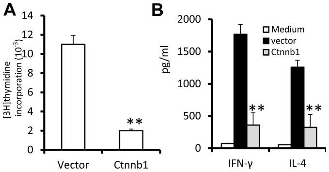
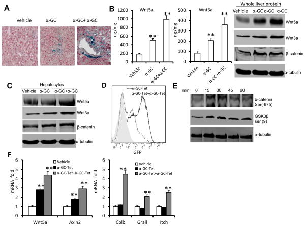
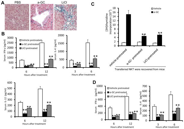
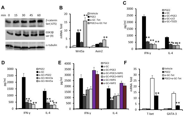
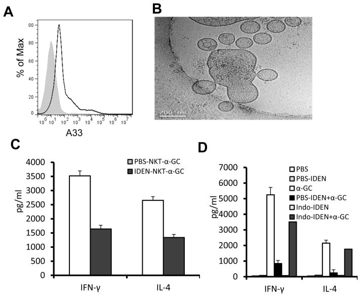
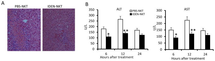
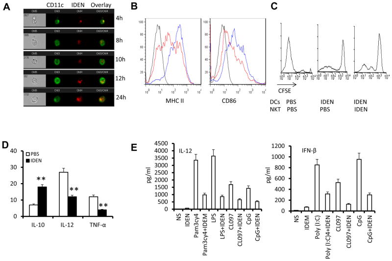
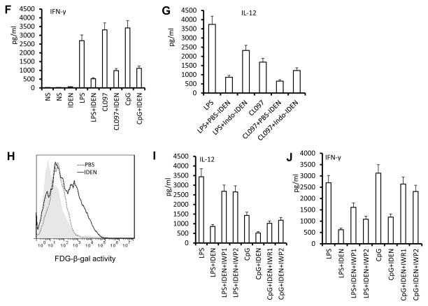
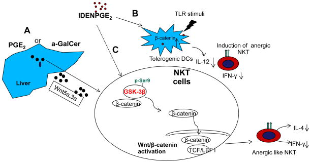
The Wnt/β-catenin pathway has been known to play a role in induction of immune tolerance, but its role in the induction and maintenance of natural killer T (NKT) cell anergy is unknown. We found that activation of the Wnt pathways in the liver microenvironment is important for induction of NKT cell anergy. We identified a number of stimuli triggering Wnt/β-catenin pathway activation, including exogenous NKT cell activator, glycolipid α-GalCer, and endogenous prostaglandin E2 (PGE2). Glycolipid α-GalCer treatment of mice induced the expression of wnt3a and wnt5a in the liver and subsequently resulted in a liver microenvironment that induced NKT cell anergy to α-GalCer restimulation. We also found that circulating PGE2 carried by nanoparticles is stable, and that these nanoparticles are A33(+) . A33(+) is a marker of intestinal epithelial cells, which suggests that the nanoparticles are derived from the intestine. Mice treated with PGE2 associated with intestinal mucus-derived exosome-like nanoparticles (IDENs) induced NKT cell anergy. PGE2 treatment leads to activation of the Wnt/β-catenin pathway by inactivation of glycogen synthase kinase 3β of NKT cells. IDEN-associated PGE2 also induces NKT cell anergy through modification of the ability of dendritic cells to induce interleukin-12 and interferon-β in the context of both glycolipid presentation and Toll-like receptor-mediated pathways.

Conclusion: These findings demonstrate that IDEN-associated PGE2 serves as an endogenous immune modulator between the liver and intestines and maintains liver NKT cell homeostasis. This finding has implications for development of NKT cell-based immunotherapies. (HEPATOLOGY 2013).


Figures

Figure 1
Figure 1. Stable expression of β-catenin gene induces anergy in liver NKT cells
Sorted β-catenin-GFP+ or control vector-GFP+ retroviral transduced NKT cells were co-cultured with BMDCs in the presence of α-GalCer. (A) Cell proliferation measured by 3H-thymidine incorporation assay; (B) Levels of IFN-γ and IL-4 in the supernatant 24 h after α-GalCer treatment of cultures. **p <0.01 (Student’s t-test). Data are the mean SEM of three experiments.
Figure 2
Figure 2. α-GalCer simulation leads to the activation of Wnt signaling in the liver
(A–C) TCF/LEF1-reporter mice were given a single-dose of α-GalCer (5μg, α-GC), multiple doses α-GalCer (5μg, every 3 d for 3 times, α-GC+ α-GC), or vehicle by intravenous injection and assessed 24 h after the last injection (n=7). (A) X-gal staining of sections of paraffin-embedded liver tissue. Magnification = ×20. (B) Expression of Wnt 3a and Wnt 5a in whole liver extracts from treated mice as assessed using an ELISA (left two panels) and immunoblot analysis (right panel). (C) Immunoblot analysis of proteins of primary hepatocytes co-cultured with liver leucocytes in the presence of α-GalCer for 24 h. (D) FACS analysis of GFP expression on Tcf-GFP-NKT hybridoma 1.2 after stimulation with a single dose of α-GalCer-tetramer (α-GC-Tet) or after restimulation with α-GalCer-tetramer (-α-GC-Tet+α-GC-Tet). (E) Immunoblot analysis of phosphorylated β-catenin ser (675) and GSK3β ser (9) in NKT cell hybridoma 1.2 stimulated with α-GalCer tetramer. (F) Changes in mRNA abundance in NKT cell hybridoma 1.2 treated with α-GalCer tetramer for 24 h, washed with PBS, and then restimulated with α-GalCer tetramer (α-GC-Tet+α-GC-Tet), or untreated NKT cell hybridoma 1.2 stimulated by α-GalCer (α-GC-Tet) or vehicle (vehicle) for 5h. Data are the mean ± SEM of three experiments (B,F) or representative of three experiments (A, B, right panel, C, D, E).
Figure 3
Figure 3. Activation of Wnt signaling in the liver led to NKT cell anergy
Recipient TCF/LEF1-reporter mice were treated with α-GalCer (3μg, i.v.), LiCl (200 mg/kg, i.p.) or vehicle as a control every three days for 12 days then irradiated (750 rads). After 5d, they were injected i.v. with enriched liver NKT cells (10 × 106 per mouse) from naïve C57BL/6 CD45.1+ mice. (A) X-gal staining of sections of paraffin-embedded liver tissue obtained 24 h after the last injection of LiCl. (B) Serum cytokine levels in the reconstituted mice at different time points after α-GalCer injection. (C) Proliferation of NKT cell recovered from recipient mice 3 days after cell transfer as described above and co-cultured with liver DCs from naïve mice in the presence of α-GalCer. (D) The recovered NKT cells were transferred into Rag1 KO mice for 9h, and the mice were then administered α-GalCer (intravenously); the serum cytokine levels assessed were examined at the indicated time points after the α-GalCer injection. *P < 0.05 and **P < 0.01 (Student’s t-test (B), one-tailed unpaired Student’s t-test (A, C, D). Data are the mean ± SEM of at least 3 experiments (n=5) (A, C,D), or are representative of at least three independent experiments (D).
Figure 4
Figure 4. PGE2 cross-talk with Wnt signaling leads to NKT cell anergy
(A) Immunoblot analysis of phosphorylated β-catenin ser (675) and GSK3β ser (9) in sorted liver NKT cells stimulated with PGE2. (B) Changes in mRNA abundance in NKT cell hybridoma 1.2 stimulated with α-GalCer-tetramer (α-GC-Tet) and/or PGE2 for 5 h. (C–E) Levels of IFN-γ and IL-4 in the supernatants of liver NKT cells co-cultured with BMDCs that had been pretreated with PGE2 or the following inhibitors/ligands for 3h and then stimulated with α-GalCer or vehicle (DMSO) for 24 h. (C) TDZD (10 μM), LiCl (10 mM); (D) Wnt3a (0.5 μg/ml), wnt5a (0.5 μg/ml); and/or (E) IWR1 (5 μM), IWP2 (5 μM). (F) Changes in mRNA abundance in NKT cell hybridoma 1.2 stimulated with α-GalCer-tetramer (α-GC-Tet) and/or PGE2 for 5 h. *P < 0.05 and **P < 0.01 (one-tailed unpaired Student’s t-test). Data (B–F) are the mean ± SEM of at least 4 experiments (n=5) or representative of three independent experiments (A).
Figure 5
Figure 5. PGE2 associated with intestinal mucus derived exosome-like nanoparticles (IDEN) induces liver NKT cell anergy
(A) FACS analysis was performed to determine expression of A33 on the surface of exosomes derived from peripheral blood. (B) Characterization of nanosized nanoparticles isolated from intestinal mucus. Intestine from B6 mice was used for isolation of nanosized nanoparticles by differential centrifugation as described previously(44). Sucrose-purified nanosized nanoparticles were examined by electron microscopy (Bar = 200 nm). (C) Cytokine production by liver NKT cells co-cultured with BMDCs in the presence of α-GalCer (100ng/ml) for 24h. NKT cells isolated from mice were i.v. injected along with IDEN or PBS every 2 days for 14 days. (D) Cytokine production by liver NKT cells from naïve mice cultured with BMDCs in the presence of PBS-IDEN, Indo-IDEN or PBS for 3h, and then stimulated by α-GalCer (100ng/ml) for 24h. *P < 0.05 and **P < 0.01 (Student’s t-test). Data are representative of three experiments (A, B) or are the mean ± SEM of five independent experiments (C, D).
Figure 6
Figure 6. Intestinal mucus derived exosome-like nanoparticles (IDEN)-protect mice against ConA induced hepatitis
Effects of adoptive transfer of NKT cells isolated from mice treated with IDEN (100μg/mouse) or vehicle control (PBS) given i.v. every 3 d for 15 d into irradiated NOD-SCID mice on ConA-induced liver damage (25 mg ConA/kg, by intravenous injection 2 d after the last injection of IDEN) (n = 10). (A) H&E-staining of liver sections. (B) Levels of serum ALT and AST.
Figure 7
Figure 7. Wnt signaling plays a role in Intestinal mucus-derived exosome-like microparticle (IDEN) induced NKT cell anergy in the context of both glycolipid presentation and TLR-mediated pathways
(A) CD11c+ DCs take up IDEN. Hepatic MNCs (5 × 10⁵) were cultured with PKH26-labeled IDEN (10 μg/ml) for 4-24 h at 37°C. IDEN positive cells are positive for: CD11c (green) and PKH26+ IDEN (red). At least 104 total events were collected for each sample. Data are representative of three independent experiments done in triplicate. (B–D) C57BL/6 mice were injected i.v. with PBS or IDEN (100 μg/mouse, every 2 d for 14 d. (B) Expression of MHC II and CD86 on CD11c+ DCs from the livers of mice pretreated with IDE (red) or PBS (blue) 24 h after being i.v. injected with α-GalCer (5μg/ml). (C) Liver NKT cells and DCs were sorted from mice pretreated with IDEN or PBS. The division of CFSE labeled liver NKT cells co-cultured with liver DCs for 3 d in the presence of α-GalCer was FACS analyzed. (D) Analysis of IL-10, IL-12 and TNF-α mRNA in DCs sorted from the livers of PBS or IDEN-treated mice 5 h after injection of α-GalCer. (E) Levels of IL-12 and IFN-β in supernatants of BMDCs cultured for 24 h in the presence of IDEN (50 μg/ml) with pam3cy4, LPS, poly(I:C), CL097 or CpG. (F) Presence of IFN-γ in supernatants of BMDCs cultured with sorted hepatic NKT cells in the presence of IDEN for 3 h, and then stimulated for 24 h with different TLR ligands indicated in the figure. (G) Levels of IL-12 in supernatants of BDMCs cultured for 24 h in the presence of IDEN (50 μg/ml) with LPS or CL097. IDEN were derived from mice treated with PBS or indomethacin. (H–J) IDEN mediated Wnt/β-catenin BMDC activation. (H) IDEN treatment leads to transactivation of TCF/LEF1 reporter. BMDCs from transgenic TCF/LEF1 reporter mice were treated by PBS or IDEN, and β-galactosidase activity was measured by flow cytometry with fluorescein di-β-D-galactosidase (FDG) as a substrate. Data are representative of three independent experiments. (I) Levels of IL-12 in supernatants of BCMCs cultured for 24 h in the presence of IWR1, IWP2 and/or IDEN (50 μg/ml) with LPS or CpG. (J) Presence of IFN-γ in supernatants of BMDCs co-cultured with sorted hepatic NKT cells in the presence of IWR1, IWP2 and/or IDEN for 3 h, and then stimulated by LPS or CpG for 24 h. *P < 0.05 and **P < 0.01 (Student’s t-test). Data are representative of three esperiments (A, B, C, H) or are the mean SEM of three independent experiments (D, E, F, G, I, J).
Figure 8
Figure 8
We propose three possible pathways that could lead to liver NKT cell anergy. (A). Repeated α-GalCer stimulation or PGE2 carried by IDEN leads to induction of Wnt ligands in the liver. Released Wnt 3a and Wnt 5a subsequently bind to liver NKT cells and activate β-catenin mediated TCF/LEF1 activation, which results in induction of NKT cell anergy. (B) Up take of IDEN-PGE2 by hepatic DCs results in activation of the β-catenin mediated pathway which then prevents the secretion of IL-12 induced by TLR stimuli. (c) IDEN-PGE2 could also directly bind to PGE2 receptors and subsequently activate the β-catenin mediated pathway by inactivation of GSK-3β.

References

    1. Venkataswamy MM, Porcelli SA. Lipid and glycolipid antigens of CD1d-restricted natural killer T cells. Seminars in immunology. 2010;22:68–78. - PMC - PubMed
    1. Kawano T, Cui J, Koezuka Y, Toura I, Kaneko Y, Motoki K, Ueno H, et al. CD1d-restricted and TCR-mediated activation of valpha14 NKT cells by glycosylceramides. Science. 1997;278:1626–1629. - PubMed
    1. Parekh VV, Wilson MT, Olivares-Villagomez D, Singh AK, Wu L, Wang CR, Joyce S, et al. Glycolipid antigen induces long-term natural killer T cell anergy in mice. The Journal of Clinical Investigation. 2005;115:2572–2583. - PMC - PubMed
    1. Crispe IN. The liver as a lymphoid organ. Annual review of immunology. 2009;27:147–163. - PubMed
    1. Crispe IN. Hepatic T cells and liver tolerance. Nature reviews Immunology. 2003;3:51–62. - PubMed

Publication types

MeSH terms