. 2012 Oct;139(20):3732-40.
doi: 10.1242/dev.083980.

The P granule component PGL-1 promotes the localization and silencing activity of the PUF protein FBF-2 in germline stem cells

Affiliations

The P granule component PGL-1 promotes the localization and silencing activity of the PUF protein FBF-2 in germline stem cells

Ekaterina Voronina et al. Development. 2012 Oct.

Abstract

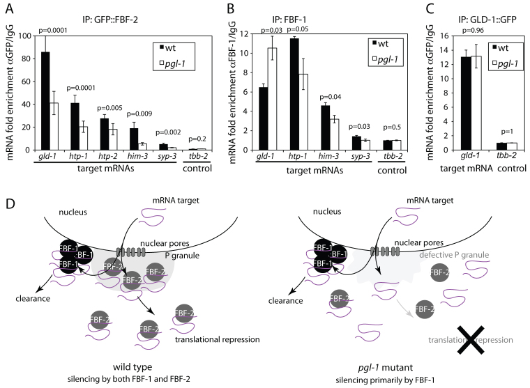
In the C. elegans germline, maintenance of undifferentiated stem cells depends on the PUF family RNA-binding proteins FBF-1 and FBF-2. FBF-1 and FBF-2 are 89% identical and are required redundantly to silence the expression of mRNAs that promote meiosis. Here we show that, despite their extensive sequence similarity, FBF-1 and FBF-2 have different effects on target mRNAs. FBF-1 promotes the degradation and/or transport of meiotic mRNAs out of the stem cell region, whereas FBF-2 prevents translation. FBF-2 activity depends on the P granule component PGL-1. PGL-1 is required to localize FBF-2 to perinuclear P granules and for efficient binding of FBF-2 to its mRNA targets. We conclude that multiple regulatory mechanisms converge on meiotic RNAs to ensure silencing in germline stem cells. Our findings also support the view that P granules facilitate mRNA silencing by providing an environment in which translational repressors can encounter their mRNA targets immediately upon exit from the nucleus.


Figures

Fig. 1.
Fig. 1.
FBF-1 and FBF-2 have distinct distributions in the distal gonad. (A) Diagram of the distal end of one gonadal arm. The gonad is a syncytium: each nucleus (circle) is surrounded by P granules (red) and partially enclosed by membranes open to a central cytoplasmic core (rachis). Signaling from the distal tip cell maintains cells at the distal end of the gonad in mitosis. In the transition zone, cells enter meiosis and initiate pairing of homologous chromosomes (‘crescent’ chromatin morphology). (B) Magnified view of the mitotic zone of wild-type and pgl-1(ct131) gonads double immunostained for the P granule component GLH-2 and FBF-1 or FBF-2. DNA is in blue. (C,E) Gonads of the indicated genotypes stained for FBF-2 or imaged live for GFP::FBF-2. FBF-2 localizes to aggregates in the mitotic zone of fbf-1 mutant gonads. Formation of the aggregates depends on pgl-1. (D,F) The percentage of germlines with FBF-2 or GFP::FBF-2 aggregates in the indicated genotypes. N, number of hermaphrodites scored. (G) Magnified view of the distal region of an fbf-1 mutant gonad co-stained for PGL-1 (OIC1D4 antibody) and FBF-2. The FBF-2 aggregate (arrow) is negative for PGL-1.
Fig. 2.
Fig. 2.
FBF-1 and FBF-2 localize to distinct perinuclear granules. (A,B) Deconvolved optical sections of wild-type mitotic zone nuclei (blue) co-stained for PGL-1 (red; K76 antibody) and FBF-1 or GFP::FBF-2 (green). Numbers refer to examples of different overlap patterns as shown in C. (C) Percentage granule colocalization as determined from three GFP::FBF-1 and five GFP::FBF-2 gonadal sections imaged as in A and B. Five gonadal sections co-stained with GLH-2 and PGL-1 (K76 antibody; supplementary material Fig. S2B) were quantified for colocalization as controls. Only FBF-1 and GFP::FBF-2 granules immediately adjacent to nuclei (‘perinuclear’) were counted.
Fig. 3
Fig. 3
fbf-1;pgl-1 mutants show the same range of phenotypes as fbf-1 fbf-2 mutants. (A) The percentage of sterile hermaphrodites of the indicated genotypes. Error bars indicate s.e.m. (from three to six experiments). N, number of hermaphrodites scored. (B,C) Full gonads of the indicated genotype stained with DAPI to reveal nuclei. The wild-type gonad contains all stages of germ cell differentiation including oocytes and sperm. By contrast, the fbf-1;pgl-1 germline contains primarily spermatogenic cells (dotted line). Excess sperm was observed in 42% of fbf-1;pgl-1 germlines. (D,F) Distal gonads of the indicated genotypes expressing a GFP::Histone H2B fusion under the control of the fog-1 3′ UTR (D) or immunostained for the synaptonemal complex proteins HTP-1 and HTP-2 (F) [the antibody recognizes both proteins (Martinez-Perez et al., 2008)]. Gonads are outlined; vertical dotted lines indicate the position of the transition zone as recognized by the ‘crescent-shaped’ chromatin. (E,G) The percentage of gonads of the indicated genotypes with GFP::H2B:fog-1 3′ UTR or HTP-1/2 expression extending to the distal end. fbf-1;pgl-1 gonads are divided into two groups: those that were fertile (had embryos, FER) and those that were sterile (no embryos, STE). Gonads were scored at the young adult stage before all cells entered meiosis. A minority of gonads in which all cells had already entered meiosis (no mitotic zone) were excluded from analysis. (H) Magnified view of nuclei in the pachytene region stained for DNA (blue) and HTP-1/2 (red); maximum intensity projection of confocal stack spanning the depth of the nucleus. HTP-1/2 forms aggregates (arrows) in fbf-1 fbf-2 and fbf-1;pgl-1 double mutants, but not in wild-type or fbf-1 fbf-2/mIn1 (balancer) controls. (I,J) Magnified view of DAPI-stained chromosomes in diakinesis stage oocytes; maximum intensity projection of confocal stack spanning the depth of the nucleus. At this stage, homologous chromosomes should be held together by chiasmata forming six distinct DNA masses (wild type, I). Nine chromosomal masses are visible in an fbf-1;pgl-1 oocyte (J) indicating that some homologs have failed to recombine. (K) The percentage of dead embryos produced by fertile hermaphrodites of the indicated genotypes. Error bars indicate s.e.m. (from two to seven experiments).
Fig. 4.
Fig. 4.
fbf-1 affects the distribution of meiotic mRNAs in the distal gonad. (A) Distal gonads of the indicated genotypes hybridized to fluorescent probes specific for gld-1, htp-1/2, him-3 and tbb-2 mRNAs. Germlines are outlined; vertical dotted lines indicate the transition zone as recognized by DAPI staining (not shown). (B) The percentage of germlines with aggregates of the indicated mRNAs as visualized by in situ hybridization as shown in A. N, the number of gonads scored for each genotype and mRNA. (C) fbf-1 mutant distal gonad double stained for GFP::FBF-2 and gld-1 mRNA. Arrow points to a cytoplasmic aggregate that is positive for both GFP::FBF-2 and gld-1 mRNA.
Fig. 5.
Fig. 5.
Efficient immunoprecipitation of meiotic mRNAs by GFP::FBF-2 requires pgl-1. (A-C) Enrichment of the indicated mRNAs in immunoprecipitates of GFP::FBF-2, FBF-1 and GLD-1::GFP in wild-type and pgl-1 extracts as determined by qPCR. The mean is calculated from two (FBF-1) or three (GFP::FBF-2, GLD-1::GFP) biological replicates. Error bars indicate s.e.m. P-values evaluating difference between wild type and pgl-1 are shown for each analyzed mRNA. Western blots used to equalize cDNA loading per amount of precipitated protein are shown in supplementary material Fig. S5A-C. Levels of analyzed mRNAs in the input lysates normalized to ama-1 mRNA are shown in supplementary material Fig. S5D. (D) Working model of cooperation between FBF-2 and PGL-1. In the mitotic zone, upon exit from the nucleus, mRNAs encoding meiotic proteins encounter FBF-2 in P granules or FBF-1 in other perinuclear granules. mRNAs bound by FBF-2 are maintained in the cytoplasm in translationally repressed complexes. mRNAs bound by FBF-1 are cleared from the cytoplasm by an unknown mechanism. In pgl-1 mutants, FBF-2 fails to localize to P granules and does not bind efficiently to its target mRNAs. FBF-1 is unaffected.

References

    1. Arur S., Ohmachi M., Nayak S., Hayes M., Miranda A., Hay A., Golden A., Schedl T. (2009). Multiple ERK substrates execute single biological processes in Caenorhabditis elegans germ-line development. Proc. Natl. Acad. Sci. USA 106, 4776-4781 - PMC - PubMed
    1. Bernstein D., Hook B., Hajarnavis A., Opperman L., Wickens M. (2005). Binding specificity and mRNA targets of a C. elegans PUF protein, FBF-1. RNA 11, 447-458 - PMC - PubMed
    1. Brenner S. (1974). The genetics of Caenorhabditis elegans. Genetics 77, 71-94 - PMC - PubMed
    1. Cinquin O., Crittenden S. L., Morgan D. E., Kimble J. (2010). Progression from a stem cell-like state to early differentiation in the C. elegans germ line. Proc. Natl. Acad. Sci. USA 107, 2048-2053 - PMC - PubMed
    1. Crittenden S. L., Bernstein D. S., Bachorik J. L., Thompson B. E., Gallegos M., Petcherski A. G., Moulder G., Barstead R., Wickens M., Kimble J. (2002). A conserved RNA-binding protein controls germline stem cells in Caenorhabditis elegans. Nature 417, 660-663 - PubMed

Publication types

MeSH terms

LinkOut - more resources