

CT Diagnosis of a Thoracic Aort Aneurysm with Type B Aortic Dissection Clinically Misdiagnosed as Acute Pulmonary Embolism
- PMID: 22991520
- PMCID: PMC3444002
- DOI: 10.1155/2012/720394
CT Diagnosis of a Thoracic Aort Aneurysm with Type B Aortic Dissection Clinically Misdiagnosed as Acute Pulmonary Embolism
Abstract
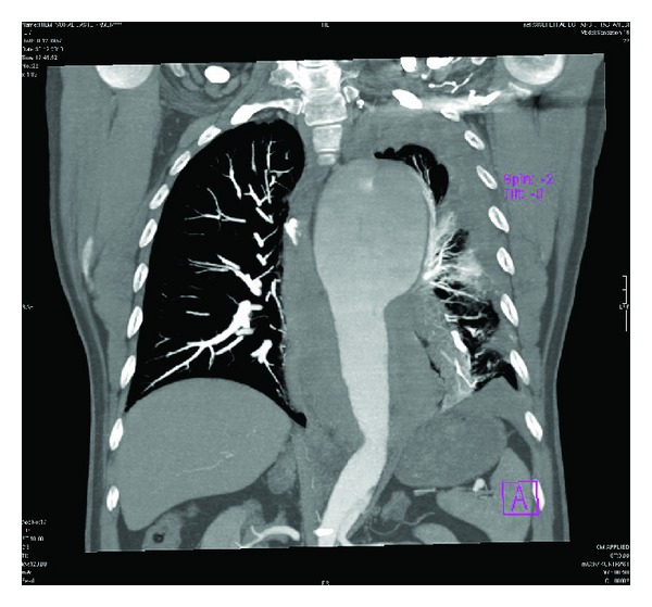
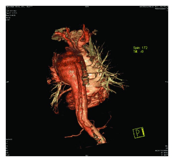
A 54-year-old man was admitted to the emergency department, presenting with an acute onset of chest pain and severe respiratory distress symptoms. He was medicated with intravenous analgesia and antihypertensive drugs. The patient was subjected to a chest X-ray which revealed a prominent widening of the mediastinum and pleural effusion on the left side. In laboratory tests-d-dimer level was highly elevated. The patient was clinically interpreted as having an acute pulmonary embolism and referred to the radiology clinic to perform a computed tomography (CT) examination. Contrast-enhanced CT demonstrated that there was no abnormality related to the pulmonary vasculature, but a huge thoracic aorta aneurysm measuring 11 × 8.1 × 7.7 cm in diameter was detected. Accompanying the aneurysm, an intimal flap was also present in the proximal descending thoracic aorta, distal to the origin of the left subclavian artery and extending into the bifurcation level. The patient was therefore diagnosed as having a type B aortic dissection as well. Once these serious conditions were detected, he was immediately transferred to a cardiovascular thoracic surgery hospital for endovascular repairment operation.
Figures


References
-
- Bhalla S, West OC. CT of nontraumatic thoracic aortic emergencies. Seminars in Ultrasound, CT and MRI. 2005;26(5):281–304. - PubMed
-
- Mehta RH, Suzuki T, Hagan PG, et al. Predicting death in patients with acute type A aortic dissection. Circulation. 2002;105(2):200–206. - PubMed
-
- Nienaber CA, Fattori R, Mehta RH, et al. Gender-related differences in acute aortic dissection. Circulation. 2004;109(24):3014–3021. - PubMed
-
- Nienaber CA, Fattori R. Aortic diseases—do we need MR techniques? Herz. 2000;25(4):331–341. - PubMed
-
- Urbania TH, Hope MD, Huffaker SD, Reddy GP. Role of computed tomography in the evaluation of acute chest pain. Journal of Cardiovascular Computed Tomography. 2009;3(1):S13–S22. - PubMed
LinkOut - more resources
Full Text Sources
