

Beam Hardening Artifacts: Comparison between Two Cone Beam Computed Tomography Scanners
- PMID: 22991636
- PMCID: PMC3445314
- DOI: 10.5681/joddd.2012.011
Beam Hardening Artifacts: Comparison between Two Cone Beam Computed Tomography Scanners
Abstract
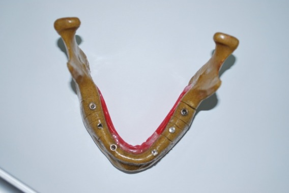
Background and aims: At present, cone beam computed tomography (CBCT) has become a substitute for computed tomography (CT) in dental procedures. The metallic materials used in dentistry can produce artifacts due to the beam hard-ening phenomenon. These artifacts decrease the quality of images. In the present study, the number of artifacts as a result of beam hardening in the images of dental implants was compared between two NewTom VG and Planmeca Promax 3D Max CBCT machines.
Materials and methods: An implant drilling model was used in the present study. The implants (Dentis) were placed in the canine, premolar and molar areas. Scanning procedures were carried out by two CBCT machines. The corresponding sections (coronal and axial) of the implants were evaluated by two radiologists. The number of artifacts in each image was determined using the scale provided. Mann-Whitney U test was used for two-by-two comparisons at a significance level of P<0.05.
Results: There were statistically significant differences in beam hardening artifacts in axial and coronal sections between the two x-ray machines (P<0.001), with a higher quality in the images produced by the NewTom VG.
Conclusion: Given the higher quality of the images produced by the NewTom VG x-ray machine, it is recommended for imaging of patients with extensive restorations, multiple prostheses or previous implant treatments.
Keywords: Artifacts; beam hardening; cone beam computed tomography.

References
-
- chindasombatjareon j, kakimoto n, murakami s, maeda y, furukawa s. quantitative analysis of metallic artifacts caused by dental metals: comparison of cone-beam and multi-detector row ct scanners. oral radiol . 2011;27:114–20.
-
- ludlow jb, davis-ludlow le, brooks sl, howerton wb. dosimetry of 3 cbct devices for oral and maxillofacial radiology: cb mercuray, newtom 3g and i-cat. dentomaxillofac radiol . 2006;35:219–26. - PubMed
-
- ludllow jb, ivanovic m. comparative dosimetry of dental cbct devices and 64-slice ct for oral and maxillofacial radiology. oral surg oral med oral pathol . 2008;106:930–38. - PubMed
-
- schulze rk, berndt d, d'hoedt b. on cone-beam computed tomography artifacts induced by titanium implants. clin oral implants res . 2010;21:100–7. - PubMed
-
- white sc, pharoah mj. oral radiology: principles and interpretation. 6th ed. st. louis: mosby; 2009. 235
LinkOut - more resources
Full Text Sources