. 2012 Dec 1;448(2):213-20.
doi: 10.1042/BJ20121365.

Assessing the range of kinase autoinhibition mechanisms in the insulin receptor family

Affiliations

Assessing the range of kinase autoinhibition mechanisms in the insulin receptor family

Stephen C Artim et al. Biochem J. .

Abstract

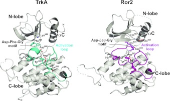
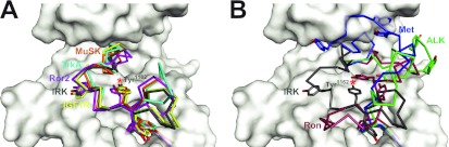
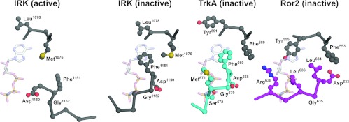
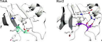
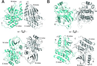
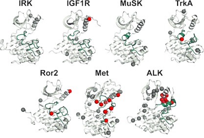
To investigate the range of autoinhibitory mechanisms used by TKDs (tyrosine kinase domains) from the insulin receptor family of RTKs (receptor tyrosine kinases), we determined crystal structures of TKDs from TrkA (tropomyosin receptor kinase A, a nerve growth factor receptor) and Ror2 (receptor tyrosine kinase-like orphan receptor 2, an unconventional Wnt receptor). TrkA autoinhibition closely resembles that seen for the insulin receptor, relying on projection of an activation loop tyrosine residue into the substrate-binding site and occlusion of the ATP-binding site by the activation loop. Ror2 employs similar mechanisms, but the unusual replacement of the phenylalanine residue in its Asp-Phe-Gly motif with leucine necessitates occlusion of the ATP-binding site by other means. The unusual Asp-Leu-Gly motif in Ror2 is displaced compared with other inactive kinases, allowing the activation loop to interact directly with the TKD's αC helix, in another mode of autoinhibition that is characteristic of the other extreme of this receptor family: ALK (anaplastic lymphoma kinase) and Met. These findings provide insight into the expected range of activating mutations in these TKDs in cancer. We also describe symmetrical dimers of the inactive TrkA TKD resembling those found in other RTKs, possibly reflecting an arrangement of kinase domains in a pre-formed TrkA dimer.


Figures

Figure 1
Figure 1. Structures of TrkA and Ror2 TKDs
Structures of inactive TKDs from TrkA (left) and Ror2 (right) in cartoon representation. The N-lobe and C-lobe are labelled, as is the catalytically important αC helix. The activation loops are labelled and highlighted in cyan (TrkA) and magenta (Ror2). Side chains in the DFG motif of TrkA (Asp668-Phe669-Gly670) and the equivalently located DLG motif in Ror2 (Asp633-Leu634-Gly635) are shown, and tyrosine residues from the YXXXYY motif are shown in stick representation. Also shown is the invariant lysine residue in strand β3 (Lys544 in TrkA, Lys507 in Ror2), which forms a salt bridge with αC Glu524 in Ror2 (but not TrkA). The catalytic base aspartate (Asp650 in TrkA, Asp615 in Ror2) is also shown and labelled. Molecule A was used for Ror2 TKD. The N- and C-terminal residues of the modelled structure are marked where visible in the orientation shown.
Figure 2
Figure 2. Comparison of activation loop configurations in IRK family kinases
Known inactive conformation structures of IRK family kinases (in addition to TrkA and Ror2) were overlaid on that of the insulin receptor (PDB code 1IRK [13]; grey, with the TKD shown in surface representation), and their activation loops coloured. (A) The activation loops of MuSK (PDB code 1LUF [15]; orange), TrkA (cyan), Ror2 molecule A (magenta) and IGF1R (PDB code 1M7N [14]; yellow) align well with that of IRK. (B) The activation loops of Ron (PDB code 3PLS [17]; red), Met (PDB code 2G15 [16]; blue) and ALK (PDB code 3L9P [8]; green) are more widely variable and are compared with that of IRK (grey). Tyrosine side chains in the YXXXYY motif are shown, and the substrate-mimicking tyrosine residue (Tyr1162 in IRK) is marked with a red asterisk.
Figure 3
Figure 3. ATP-binding-site occlusion by the activation loop in inactive IRK, TrkA and Ror2
Structures of the inactive TKDs from IRK [13], TrkA and Ror2 were overlaid on the structure of active IRK (PDB code 1IR3) determined with bound peptide substrate and a non-hydrolysable ATP analogue AMP-PNP (adenosine 5′-[β,γ-imido]triphosphate) [41]. Close-up views of residues surrounding the binding site for AMP-PNP (shown from the 1IR3 structure) are shown for inactive IRK (activation loop coloured grey), TrkA (activation loop coloured cyan) and Ror2 (activation loop coloured magenta). Whereas the 1IR3 (active) structure readily accommodates AMP-PNP, the nucleotide-binding site is occluded directly by the activation loop in inactive IRK, TrkA and Ror2, with backbone and side-chain clashes with the phosphate groups. In addition, the DFG motif phenylalanine residue (Phe1151 in IRK and Phe669 in TrkA) blocks the binding site for the adenine ring of AMP-PNP. This inhibitory interaction is not maintained in Ror2, which has a phenylalanine-to-leucine substitution in the DFG motif (yielding DLG), and Tyr555 from elsewhere in the TKD takes an a similar role.
Figure 4
Figure 4. DFG motif conformation and activation loop–αC interactions in TrkA and Ror2
Left: close-up of the DFG motif region in TrkA TKD, with the DFG motif itself coloured cyan. Note the absence of salt bridge between the β3 invariant lysine residue (Lys544) and αC glutamate residue (Glu560), for which complete side-chain density was not seen. Van der Waal's contacts between the Phe589 and Leu564 side chains are likely to contribute to stabilization of the αC position. Right: close-up of the DFG motif equivalent in Ror2 TKD (coloured magenta), which has the sequence DLG, with Leu634 replacing the normal phenylalanine residue at this position in other kinases. Note that the β3 invariant lysine residue (Lys507) forms a salt bridge with the αC helix glutamate residue (Glu524) even in the inactive Ror2 kinase (as also seen in ALK and Met), although the displaced αC helix position prevents Lys507 from contributing to an active-like ATP-binding site. The DLG motif of Ror2 TKD in the activation loop also interacts directly with the αC helix, thus stabilizing the inactive state through a mode that more closely resembles autoinhibition in Met, ALK or EGFR. Arg528 in the αC helix interacts directly with both backbone and side chain of Asp633 in the DLG motif to stabilize this conformation. Mutations at Arg528 might be predicted to activate Ror2.
Figure 5
Figure 5. Possible inactive TrkA TKD dimers
Two orthogonal views of crystallographic dimers of TrkA TKDs. One TrkA TKD is coloured cyan, and the other grey. (A) A dimer that is also found in an inhibitor-bound TrkC TKD crystal structure (PDB code 3V5Q [33]) and in inactive FGFR1 TKD structures. The αC helix, which is responsible for the majority of the interactions is labelled, as is the αE helix, the C-terminus of which also makes contributions to the dimer interface. The N- and C-termini of the TKD are marked. Note the projection of the N-terminus towards the TKD active site, a feature that is also seen in the TrkC crystallographic dimer from 3V5Q. (B) a dimer that is also found in a recently released TrkA TKD structure (PDB code 4F0I [34]), which involves the ‘hinge’ region, kinase insert domain and strand β1. This dimer could help to maintain the inactive configuration through restraints on the hinge region, for example. No evidence for TrkA TKD dimerization in solution was obtained in analytical ultracentrifugation studies (not shown). Tyrosine residues in the YXXXYY motif within the activation loop are coloured black, to highlight the fact that they are quite distant in these dimers from the active site of the neighbouring TKD that may transphosphorylate them. The tyrosine trans-autophosphorylation events that lead to TrkA activation could not occur in the context of these inactive dimers, but would require their reorganization upon extracellular ligand binding or association with additional receptor molecules.
Figure 6
Figure 6. Somatic mutations in IRK family kinases in cancer
IRK family TKDs in their inactive conformation (PDB codes listed in Figure 2) are shown in the same orientation as in Figure 1, with the activation loop coloured cyan. Sites at which mutations have been reported in cancer patients, in the literature or COSMIC (http://www.sanger.ac.uk/genetics/CGP/cosmic/) database [40], are represented as spheres, coloured red if the mutations lie in the activation loop or αC helix. Note that the region of contact between the activation loop and αC helix is the site of many mutations in Met and ALK. TKDs without this autoinhibitory feature have fewer known mutations to date, and fewer in the activation loop or αC. These analyses identify a vulnerability in ALK and Met for oncogenic mutations as described in the text. Ron is not included because no mutations in this TKD have yet been described. IRK is included for comparison.

References

    1. Dancey J. E., Bedard P. L., Onetto N., Hudson T. J. The genetic basis for cancer treatment decisions. Cell. 2012;148:409–420. - PubMed
    1. Yauch R. L., Settleman J. Recent advances in pathway-targeted cancer drug therapies emerging from cancer genome analysis. Curr. Opin. Genet. Dev. 2012;22:45–49. - PubMed
    1. Lemmon M. A., Schlessinger J. Cell signaling by receptor tyrosine kinases. Cell. 2010;141:1117–1134. - PMC - PubMed
    1. Zhang X., Gureasko J., Shen K., Cole P. A., Kuriyan J. An allosteric mechanism for activation of the kinase domain of epidermal growth factor receptor. Cell. 2006;125:1137–1149. - PubMed
    1. Sharma S. V., Bell D. W., Settleman J., Haber D. A. Epidermal growth factor receptor mutations in lung cancer. Nat. Rev. Cancer. 2007;7:169–181. - PubMed

Publication types

MeSH terms