. 2013 Jan;83(1):72-83.
doi: 10.1038/ki.2012.328. Epub 2012 Sep 19.

In vivo multiphoton imaging of mitochondrial structure and function during acute kidney injury

Affiliations

In vivo multiphoton imaging of mitochondrial structure and function during acute kidney injury

Andrew M Hall et al. Kidney Int. 2013 Jan.

Abstract

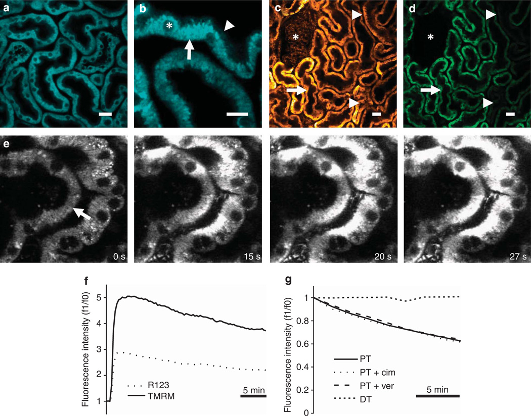
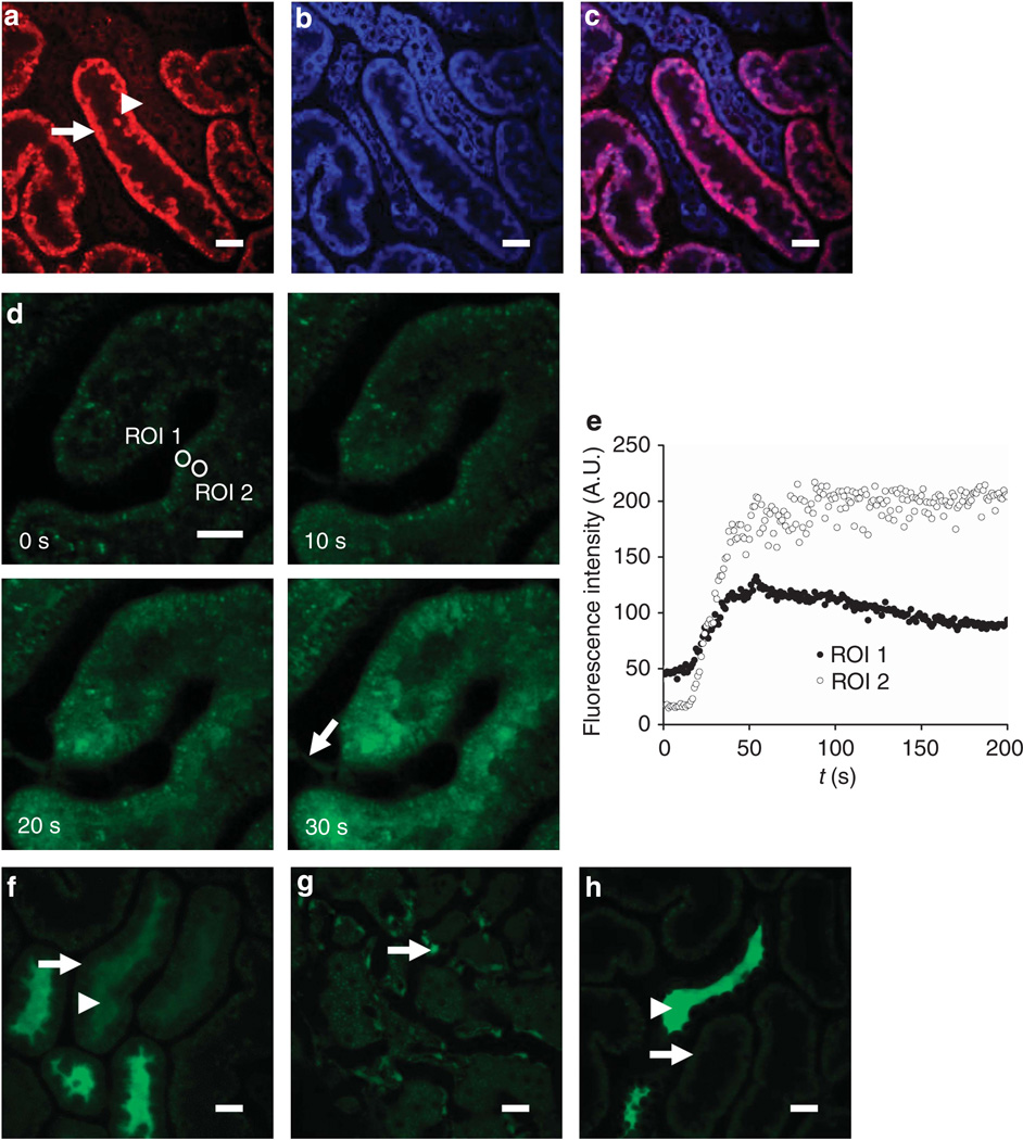
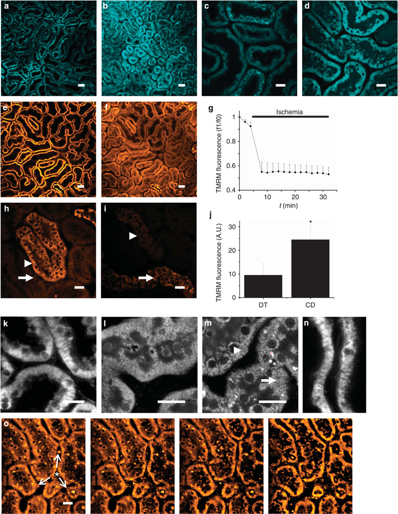
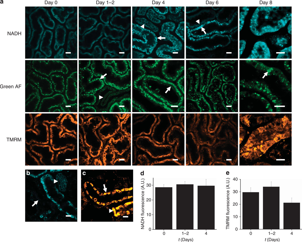
Mitochondrial dysfunction has been implicated in the pathogenesis of acute kidney injury due to ischemia and toxic drugs. Methods for imaging mitochondrial function in cells using confocal microscopy are well established; more recently, it was shown that these techniques can be utilized in ex vivo kidney tissue using multiphoton microscopy. We extended this approach in vivo and found that kidney mitochondrial structure and function can be imaged in anesthetized rodents using multiphoton excitation of endogenous and exogenous fluorophores. Mitochondrial nicotinamide adenine dinucleotide increased markedly in rat kidneys in response to ischemia. Following intravenous injection, the mitochondrial membrane potential-dependent dye TMRM was taken up by proximal tubules; in response to ischemia, the membrane potential dissipated rapidly and mitochondria became shortened and fragmented in proximal tubules. In contrast, the mitochondrial membrane potential and structure were better maintained in distal tubules. Changes in mitochondrial structure, nicotinamide adenine dinucleotide, and membrane potential were found in the proximal, but not distal, tubules after gentamicin exposure. These changes were sporadic, highly variable among animals, and were preceded by changes in non-mitochondrial structures. Thus, real-time changes in mitochondrial structure and function can be imaged in rodent kidneys in vivo using multiphoton excitation of endogenous and exogenous fluorophores in response to ischemia-reperfusion injury or drug toxicity.


Figures

Figure 1
Figure 1. In vivo imaging of mitochondrial nicotinamide adenine dinucleotide (NADH) and membrane potential in the kidney
(a, b) Mitochondrial NADH was visible at 720 nm excitation in both mouse (a) and rat (b) kidneys and showed a characteristic basolateral mitochondrial distribution in tubular cells (arrow), with very little fluorescence signal observed in non-mitochondrial structures including the apical brush border (arrowhead) or cell nuclei (asterisk). (c–g) The mitochondrial membrane potential (Δψm)–dependent dyes tetramethyl rhodamine methyl ester (TMRM) and rhodamine 123 loaded into rodent kidney tubules and localized to the mitochondria; TMRM loaded well into rat proximal tubules (PTs—arrow), distal tubules (DTs—arrowheads), and glomeruli (asterisk) following intravenous injection (c), whereas rhodamine 123 loaded well into PTs (arrow), but not DTs (arrowheads) or glomeruli (asterisk) (d). Uptake of TMRM into tubules occurred initially from the basolateral side (arrow) (e); images depicted were acquired shortly after an intravenous injection of the dye into a mouse. Representative traces are depicted showing the rapid increase and subsequent slow decrease in fluorescence that occurred in rat PTs following intravenous injection of TMRM and rhodamine 123 (f). Representative traces are depicted showing that the decline of TMRM fluorescence in rat PTs was not prevented by prior intravenous injection of either cimetidine or verapamil (g); no decline in TMRM fluorescence was observed in DTs. Bars = 20 µm in (a, b) and 40 µm in (c, d).
Figure 2
Figure 2. In vivo imaging of reactive oxygen species (ROS) production and glutathione in the kidney
(a–c) Imaging of ROS production. Following intravenous injection of the reactive ROS-sensitive dye dihydroethidium (HEt) in rats, the fluorescence signal was higher in proximal tubules (PTs—arrow) than in adjacent distal tubules (DTs—arrowhead) (a); simultaneous excitation of mitochondrial nicotinamide adenine dinucleotide (NADH) (blue) (b) showed that the two signals colocalized (c). (d–h) Imaging of intracellular glutathione using monochlorobimane (MCB). Following intravenous injection of MCB in rats, an increase in fluorescence signal intensity was observed in PTs, which originated at the basolateral aspect of cells and spread apically over time (d, e), with simultaneous uptake into endothelial cells (arrow). MCB was subsequently rapidly excreted from PT cells (arrow) into the tubular lumen (arrowhead) (f); the image depicted was acquired 8min post injection. Although MCB was excreted by PTs, the signal remained stable in endothelial cells (arrow) (g); the image depicted was acquired 18 min post injection. After 20 min, the fluorescence signal had disappeared from PTs (arrow) but was visible in DT lumens (arrowhead) (h). Bars = 20 µm in all images.
Figure 3
Figure 3. Real-time in vivo imaging of mitochondrial structure and function in the kidney during ischemia reperfusion
(a–d) Resting nicotinamide adenine dinucleotide (NADH) signal in rat renal cortical tubules (a) increased markedly in response to ischemia (b); the image depicted was acquired 2min post occlusion of the renal artery; higher resolution images acquired pre- (c) and post-ischemia (d) confirmed that the signal change was localized to the mitochondria. (e–g) In rat proximal tubules (PTs) loaded with tetramethyl rhodamine methyl ester (TMRM), resting mitochondrial membrane potential (Δψm) (e) dissipated rapidly in response to ischemia (f); the image depicted was acquired 2min post ischemia; data depicted in (g) are mean (±s.e.m.) mitochondrial TMRM fluorescence intensity in PTs of three separate experiments. Δψm was better maintained during ischemia in distal tubules (DTs—arrowhead) than in PTs (arrow) (h); the image depicted was acquired 9min post ischemia. After 30 min of ischemia, Δψm was better maintained in collecting ducts (arrow) than in DTs (arrowhead) (i); data depicted in (j) are mean (±s.e.m.) of mitochondrial TMRM fluorescence of three separate experiments (*P<0.05). (k–n) Changes in mitochondrial morphology during ischemia and reperfusion. Images depicted show the following: normal elongated mitochondria in PTs loaded with TMRM (k); NADH signal in a PT 10 min post ischemia demonstrating widespread mitochondrial shortening and fragmentation (l); NADH signal 30 min post ischemia demonstrating normal mitochondrial morphology in a DT (arrowhead) adjacent to a PT (arrow) (m); fragmented mitochondria 20min post reperfusion in a PT loaded with TMRM (n). The signal intensity gain was adjusted in images (k–n) to optimally visualize mitochondrial structure. (o) Repolarization of mitochondria immediately post reperfusion in ischemic PTs preloaded with TMRM; images depicted were acquired 50s apart and demonstrate a spreading wave of repolarization (arrows) from a central blood vessel (asterisk), with reuptake of TMRM from the cytosolic compartment into the mitochondria within cells. Bars = 40 µm in (a, b) and (e, f) and 20 µm in all other images.
Figure 4
Figure 4. In vivo imaging of gentamicin toxicity in the kidney
(a–c) Representative images are depicted displaying the time course of intracellular changes in gentamicin toxicity. After 1–2 days of exposure, bright structures were visible in the proximal tubules (PTs) in the green autofluorescence (AF) signal (arrow), most likely representing enlarged lysosomes, which were not visible in the distal tubules (DTs—arrowhead); after 4 days, abnormalities were also noted in the PT brush border (arrow). Mitochondrial nicotinamide adenine dinucleotide (NADH) signals remained normal in tubules at 1–2 days; after 4 days, sporadic areas of dysmorphic mitochondria and increased NADH signal were observed in PTs (arrow), but not in DTs (arrowhead). However, the majority of mitochondria in PTs loaded with the mitochondrial membrane potential (Δψm)–dependent dye tetramethyl rhodamine methyl ester (TMRM) appeared well energized up to 4 days. After 6 days, abnormalities in NADH signal were more widespread in PTs, but remained highly variable, with damaged tubules (arrow) appearing directly adjacent to normal-looking tubules (arrowhead); a high degree of variability was also observed in mitochondrial TMRM signal intensity. After 8 days, mitochondria in the surviving PTs were grossly dysmorphic, as seen in the NADH image, and massively enlarged lysosomes were visible in the green AF signal (arrow); there were also marked variations in mitochondrial TMRM signal between adjacent cells within surviving PTs. After 8 days of gentamicin exposure, widespread necrosis occurred in PTs, leading to the appearance of ghost tubules devoid of living cells (arrow) (b); in contrast, mitochondrial NADH (b) and TMRM (c) signals remained normal in DTs (arrowhead) and collecting ducts (arrow). (d, e) Data depicted show mean mitochondrial NADH (d) and TMRM (e) fluorescence intensity (±s.e.m.) after exposure to gentamicin; n = 4 animals in each group. Bars = 20 mm in all images.

References

    1. Duchen MR, Szabadkai G. Roles of mitochondria in human disease. Essays Biochem. 2010;47:115–137. - PubMed
    1. Plotnikov EY, Kazachenko AV, Vyssokikh MY, et al. The role of mitochondria in oxidative and nitrosative stress during ischemia/reperfusion in the rat kidney. Kidney Int. 2007;72:1493–1502. - PubMed
    1. Brooks C, Wei Q, Cho SG, et al. Regulation of mitochondrial dynamics in acute kidney injury in cell culture and rodent models. J Clin Invest. 2009;119:1275–1285. - PMC - PubMed
    1. Burke TJ, Wilson DR, Levi M, et al. Role of mitochondria in ischemic acute renal failure. Clin Exp Dial Apheresis. 1983;7:49–61. - PubMed
    1. Sun Z, Zhang X, Ito K, et al. Amelioration of oxidative mitochondrial DNA damage and deletion after renal ischemic injury by the KATP channel opener diazoxide. Am J Physiol Renal Physiol. 2008;294:F491–F498. - PubMed

Publication types

MeSH terms

LinkOut - more resources