. 2012 Nov;160(3):1342-56.
doi: 10.1104/pp.112.205955. Epub 2012 Sep 19.

A novel approach to dissect the abscission process in Arabidopsis

Affiliations

A novel approach to dissect the abscission process in Arabidopsis

Zinnia Haydee González-Carranza et al. Plant Physiol. 2012 Nov.

Abstract

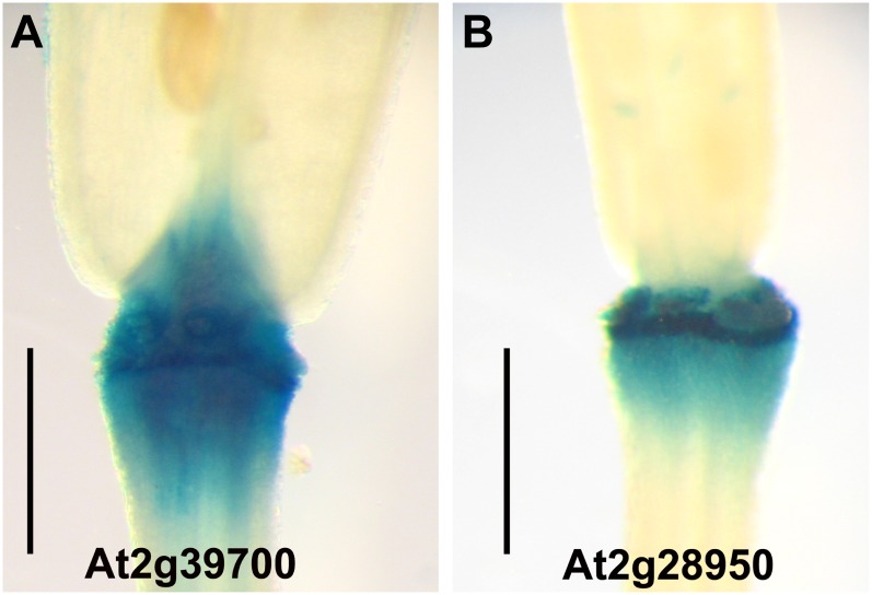
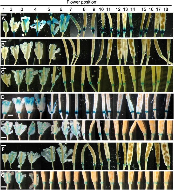
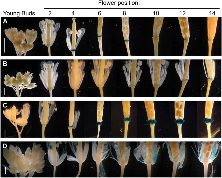
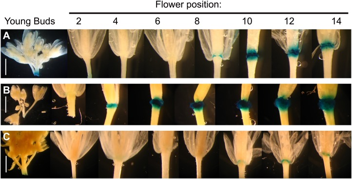
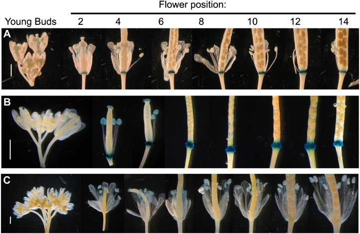
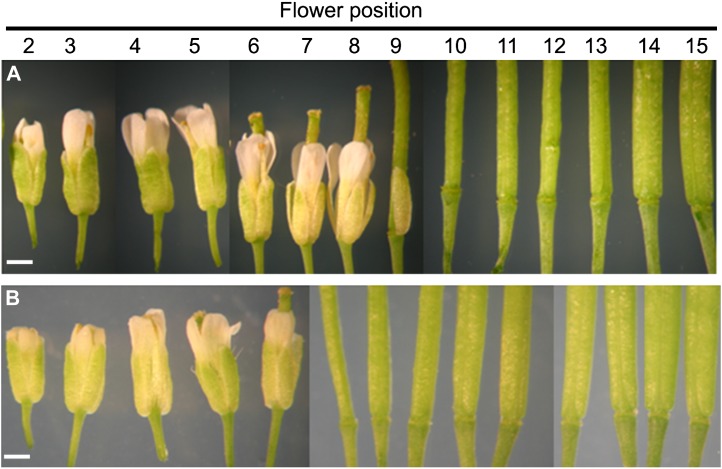
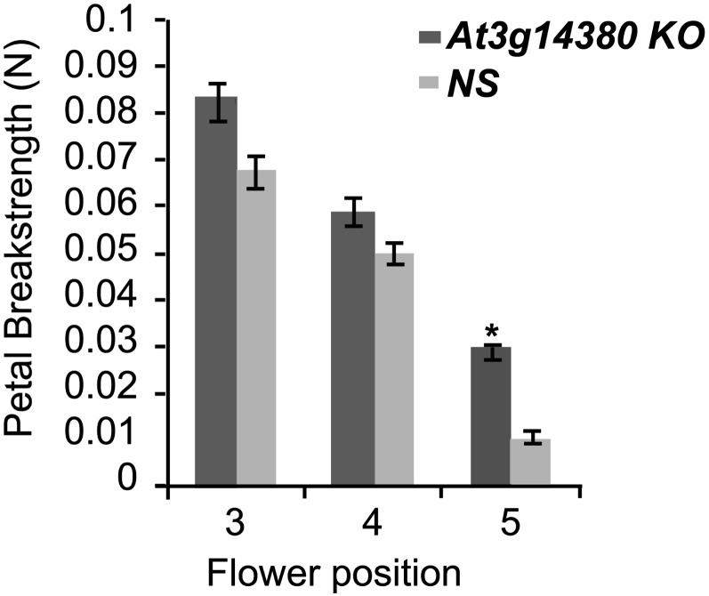
Abscission is the consequence of a specialized layer of cells undergoing a complex series of molecular and biochemical events. Analysis of the specific molecular changes associated with abscission is hampered by contamination from neighboring nonseparating tissues. Moreover, studies of abscission frequently involve the examination of events that take place in isolated segments of tissue exposed to nonphysiological concentrations of ethylene or indole-3-acetic acid for protracted periods (more than 24 h) of time. To resolve these problems, we have adopted the use of a transgenic line of Arabidopsis (Arabidopsis thaliana) where the promoter of an abscission-specific polygalacturonase gene (At2g41850/ARABIDOPSIS DEHISCENCE ZONE POLYGALACTURONASE2) has been fused to a green fluorescent protein reporter. RNA was extracted from green fluorescent protein-tagged cells, released from abscising floral organs, and used to generate a complementary DNA library. This library was used to probe a microarray, and a population of abscission-related transcripts was studied in detail. Seven novel abscission-related genes were identified, four of which encode proteins of unknown function. Reverse transcription-polymerase chain reaction analyses and promoter fusions to the β-glucuronidase reporter gene confirmed the expression of these genes in the abscission zone and revealed other places of expression during seedling development. Three of these genes were studied further by crossing reporter lines to the abscission mutants inflorescence deficient in abscission (ida) and blade-on-petiole1 (bop1)/bop2 and an IDA-overexpressing line. Phenotypic analysis of an At3g14380 transfer DNA insertion line indicates that this gene plays a functional role in floral organ shedding. This strategy has enabled us to uncover new genes involved in abscission, and their possible contribution to the process is discussed.


Figures

Figure 1.
Figure 1.
GUS expression in Arabidopsis floral AZs from ProAt2g39700::GUS (AtEXP4) and ProAt2g28950::GUS (AtEXP6). Flower position 8 is shown, where position 1 is the flower with the first visible petal. Bars = 1 mm.
Figure 2.
Figure 2.
RT-PCR analysis of the expression of At1g64405 (A), At2g23630 (B), At2g44010 (C), At3g14380 (D), At3g53040 (E), At3g56350 (F), At5g50540 (G), and AT5G44200 (H; cap-binding protein) as a housekeeping gene. RNA was extracted from Col-0 wild-type roots, rosette leaves, cauline leaves, buds, flowers, stems, siliques, and yellow siliques. For primer information, see Supplemental Table S1.
Figure 3.
Figure 3.
Flower and silique expression from ProAt1g64405::GUS (A), ProAt2g23630::GUS (B), ProAt2g44010::GUS (C), ProAt3g14380::GUS (D), ProAt3g53040::GUS (E), ProAt3g56350::GUS (F), and ProAt5g50540::GUS (G). Time course of expression is shown from floral development positions 1 to 18, where position 1 is the first flower where petals are visible to the eye. Tissue was GUS stained for a period of 6 h (A and D) or 12 h (B, C, and E–G) of incubation in GUS substrate. Bars = 1 mm.
Figure 4.
Figure 4.
Flower and silique expression from ProAt2g41850::GUS (A), ProAt2g41850::GUS crossed with ida mutant (B), ProAt2g41850::GUS crossed with Pro35S::IDA (C), and ProAt2g41850::GUS crossed with bop1/bop2 mutant (D). Time course of expression is shown from young buds to flower position 14, where position 1 is the first flower where petals are visible to the eye. Tissue was GUS stained for a period of 12 h of incubation in GUS substrate. Bars = 1 mm.
Figure 5.
Figure 5.
Flower and silique expression from ProAt1g64405::GUS crossed with ida mutant (A), ProAt1g64405::GUS crossed with Pro35S::IDA (B), and ProAt1g64405::GUS crossed with bop1/bop2 mutant (C). Time course of expression is shown from young buds to flower position 14, where position 1 is the first flower where petals are visible to the eye. Tissue was GUS stained for a period of 6 h of incubation in GUS substrate. Bars = 1 mm.
Figure 6.
Figure 6.
Flower and silique expression from ProAt3g14380::GUS crossed with ida mutant (A), ProAt3g14380::GUS crossed with Pro35S::IDA (B), and ProAt3g14380::GUS crossed with bop1/bop2 mutant (C). Time course of expression is shown from young buds to flower position 14, where position 1 is the first flower where petals are visible to the eye. Tissue was GUS stained for a period of 12 h of incubation in GUS substrate. Bars = 1 mm.
Figure 7.
Figure 7.
Developmental time course of flowers from 4-week-old plants. Flowers shown represent positions 2 to 15, where position 1 is the first flower where petals are visible to the eye. A, At3g14380 KO plants. B, At3g14380 NS plants (SALK_03248). Bars = 1 mm. [See online article for color version of this figure.]
Figure 8.
Figure 8.
Break-strength analysis of petals from flowers excised from 4-week-old plants of At3g15380 KO-L3 wild type and NS-L1 (SALK_03248) at positions 3, 4, and 5, where position 1 is the first flower where petals are visible to the eye. Four flowers from each stage were used in a Lloyds LF plus machine to measure the force required to separate the petal from the pedicel of the flower. Measurements were statistically analyzed by ANOVA. The asterisk indicates a difference in the mean at P < 0.002 (n = 12).

Similar articles

Cited by

References

    1. Adamczyk BJ, Lehti-Shiu MD, Fernandez DE. (2007) The MADS domain factors AGL15 and AGL18 act redundantly as repressors of the floral transition in Arabidopsis. Plant J 50: 1007–1019 - PubMed
    1. Aharoni A, Dixit S, Jetter R, Thoenes E, van Arkel G, Pereira A. (2004) The SHINE clade of AP2 domain transcription factors activates wax biosynthesis, alters cuticle properties, and confers drought tolerance when overexpressed in Arabidopsis. Plant Cell 16: 2463–2480 - PMC - PubMed
    1. Alonso JM, Stepanova AN, Leisse TJ, Kim CJ, Chen H, Shinn P, Stevenson DK, Zimmerman J, Barajas P, Cheuk R, et al. (2003) Genome-wide insertional mutagenesis of Arabidopsis thaliana. Science 301: 653–657 - PubMed
    1. Battaglia M, Olvera-Carrillo Y, Garciarrubio A, Campos F, Covarrubias AA. (2008) The enigmatic LEA proteins and other hydrophilins. Plant Physiol 148: 6–24 - PMC - PubMed
    1. Belfield EJ, Ruperti B, Roberts JA, McQueen-Mason S. (2005) Changes in expansin activity and gene expression during ethylene-promoted leaflet abscission in Sambucus nigra. J Exp Bot 56: 817–823 - PubMed

Publication types