

Telomeres, tethers and trypanosomes
- PMID: 22992703
- PMCID: PMC3515529
- DOI: 10.4161/nucl.22167
Telomeres, tethers and trypanosomes
Abstract
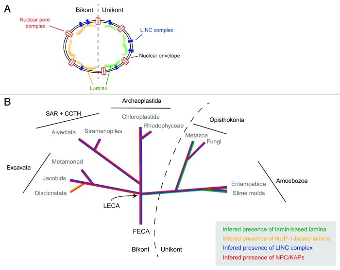
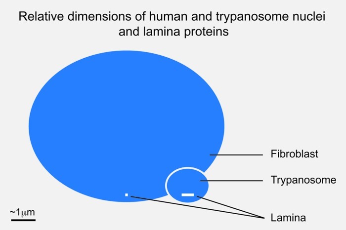
Temporal and spatial organization of the nucleus is critical for the control of transcription, mRNA processing and the assembly of ribosomes. This includes the occupancy of specific territories by mammalian chromosomes, the presence of subnuclear compartments such as the nucleolus and Cajal bodies and the division of chromatin between active and inactive states. These latter are commonly associated with the location of DNA within euchromatin and heterochromatin respectively; critically these distinctions arise through modifications to chromatin-associated proteins, including histones, as well as the preferential localization of heterochromatin at the nuclear periphery. Most research on nuclear organization has focused on metazoa and fungi; however, recent technical advances have made more divergent eukaryotes accessible to study, with some surprising results. For example, the organization of heterochromatin is mediated in metazoan nuclei in large part by lamins, the prototypical intermediate filament proteins. Despite the presence of heterochromatin, detected both biochemically and by EM in most eukaryotic organisms, until this year lamins were thought to be restricted to metazoan taxa, and the proteins comprising the lamina in other lineages were unknown. Recent work indicates the presence of lamin orthologs in amoeba, while trypanosomatids possess a large coiled-coil protein, NUP-1, that performs functions analogous to lamins. These data indicate that the presence of a nuclear lamina is substantially more widespread than previously thought, with major implications for the evolution of eukaryotic gene expression mechanisms. We discuss these and other recent findings on the organization of nuclei in diverse organisms, and the implications of these findings for the evolutionary origin of eukaryotes.
Figures

Comment on
- DuBois KN, Alsford S, Holden JM, Buisson J, Swiderski M, Bart JM, Ratushny AV, Wan Y, Bastin P, Barry JD, Navarro M, Horn D, Aitchison JD, Rout MP, Field MC. NUP-1 Is a large coiled-coil nucleoskeletal protein in trypanosomes with lamin-like functions. PLoS Biol. 2012;10:e1001287. doi: 10.1371/journal.pbio.1001287.
Similar articles
-
Co-dependence between trypanosome nuclear lamina components in nuclear stability and control of gene expression.Nucleic Acids Res. 2016 Dec 15;44(22):10554-10570. doi: 10.1093/nar/gkw751. Epub 2016 Sep 12. Nucleic Acids Res. 2016. PMID: 27625397 Free PMC article.
-
NUP-1 Is a large coiled-coil nucleoskeletal protein in trypanosomes with lamin-like functions.PLoS Biol. 2012;10(3):e1001287. doi: 10.1371/journal.pbio.1001287. Epub 2012 Mar 27. PLoS Biol. 2012. PMID: 22479148 Free PMC article.
-
Deviating from the norm: Nuclear organisation in trypanosomes.Curr Opin Cell Biol. 2023 Dec;85:102234. doi: 10.1016/j.ceb.2023.102234. Epub 2023 Sep 2. Curr Opin Cell Biol. 2023. PMID: 37666024 Review.
-
A hub-and-spoke nuclear lamina architecture in trypanosomes.J Cell Sci. 2021 Jun 15;134(12):jcs251264. doi: 10.1242/jcs.251264. Epub 2021 Jun 21. J Cell Sci. 2021. PMID: 34151975 Free PMC article.
-
Lamins as structural nuclear elements through evolution.Curr Opin Cell Biol. 2023 Dec;85:102267. doi: 10.1016/j.ceb.2023.102267. Epub 2023 Oct 21. Curr Opin Cell Biol. 2023. PMID: 37871500 Free PMC article. Review.
Cited by
-
The plant LINC complex at the nuclear envelope.Chromosome Res. 2014 Jun;22(2):241-52. doi: 10.1007/s10577-014-9419-7. Chromosome Res. 2014. PMID: 24801343 Review.
-
Evolution of the nucleus.Curr Opin Cell Biol. 2014 Jun;28(100):8-15. doi: 10.1016/j.ceb.2014.01.004. Epub 2014 Feb 6. Curr Opin Cell Biol. 2014. PMID: 24508984 Free PMC article. Review.
-
Co-dependence between trypanosome nuclear lamina components in nuclear stability and control of gene expression.Nucleic Acids Res. 2016 Dec 15;44(22):10554-10570. doi: 10.1093/nar/gkw751. Epub 2016 Sep 12. Nucleic Acids Res. 2016. PMID: 27625397 Free PMC article.
-
The biology and polymer physics underlying large-scale chromosome organization.Traffic. 2018 Feb;19(2):87-104. doi: 10.1111/tra.12539. Epub 2017 Dec 3. Traffic. 2018. PMID: 29105235 Free PMC article. Review.
-
Antigenic variation in African trypanosomes: the importance of chromosomal and nuclear context in VSG expression control.Cell Microbiol. 2013 Dec;15(12):1984-93. doi: 10.1111/cmi.12215. Epub 2013 Oct 10. Cell Microbiol. 2013. PMID: 24047558 Free PMC article. Review.
References
Publication types
MeSH terms
Substances
Grants and funding
LinkOut - more resources
Full Text Sources